Module #8 (Neurulation)
1/138
There's no tags or description
Looks like no tags are added yet.
Name | Mastery | Learn | Test | Matching | Spaced | Call with Kai |
|---|
No analytics yet
Send a link to your students to track their progress
139 Terms
What are the three big derivatives of the ectoderm?
Surface ectoderm (epidermis → most external)
Neural crest
Neural plate/neural tube
A structure that is transitory and extremely multi-potent, is also a derivative of the ectoderm
Neural crest
Neural tube is derived from what structure, and is also a derivative of the ectoderm
Neural plate
Most external layer of the embryo
Ectoderm
The ectoderm will receive _ to become and make the decisions to be surface, neural crest, neural plate/neural tube
different signals
Most important signal that the ectoderm can receive, a homolog in vertebrates of this gene called DPP (Decapentaplegic), characterized by having 10 different mutations
BMP
In a drosophila, in the ventral side there will be the expression of what that will inhibit DPP?
Dorsal → in the region where Dorsal is not present, DPP is going to be present
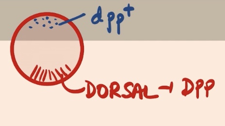
DPP is going to specify the
Surface ectoderm (epidermis → skin) in invertebrates (drosophila)
High elevated _ induces the formation of the surface ectoderm (epidermis) in vertebrates
BMP
Derivatives of the surface ectoderm (epidermis)?
1) Epidermis
2) Hair
3) Nails
4) Sebaceous Glands
5) Olfactory Epithelium
6) Lens + Cornea
7) Mouth Epithelium (example → tooth enamel)
In terms of the Neural tube, what do we have to specify it?
No BMP whatsoever
Derivatives of the Neural Crest?
1) Facial Cartilage
2) Dentine (from teeth)
3) Pigmented Cells (melanocytes
4) Adrenal Medula
5) Peripheral Nervous System (all nerve cells, all neurons that are outside of the CNS are neural crest derivatives)
Derivatives of the Neural Tube?
1) Brain
2) Spinal Cord
Brain + Spinal Cord = CNS
3) Motor Neurons
4) Retina (part of the eye)
Gives rise to many different derivatives that are essential for shape and different characteristics of very typical vertebrate characteristics?
Neural Crest
The morphological diversity of the face of different animals (including us) is going to be derived from differences in
facial cartilage → derives from the neural crest
Need to view this process of generating these three different classes of ectoderm as something that is
actually happening during gastrulation and the process of formation of the neural tube
Process of Neurulation is the
formation of the neural tube
The stage of the animal that is going through neurulation is called a
Neurula
Neurulation has how many different stages?
3, Early Neurula → Late Neurula
Emphasizes that this process of Neurulation is going to happen at the same time that
gastrulation is happening, not at the same time → but gastrulation has not finished yet when neurulation start (do not think of finish one to get to another)
So in a part of the embryo gastrulation can still be occurring, and in a different part of the embryo
neurulation is now happening
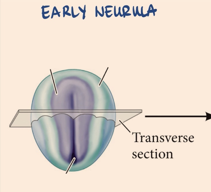
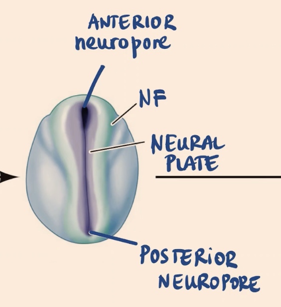
What can we see in the Early Neurula?
Surface ectoderm → most external layer
Central part of the ectoderm → Neural plate
Neural folds
Have orientation → Anterior & Posterior sides
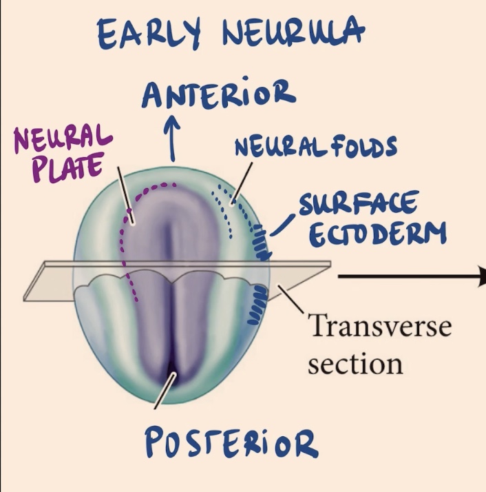
In the early Neurula, the border between the neural plate and surface ectoderm is the
Neural folds
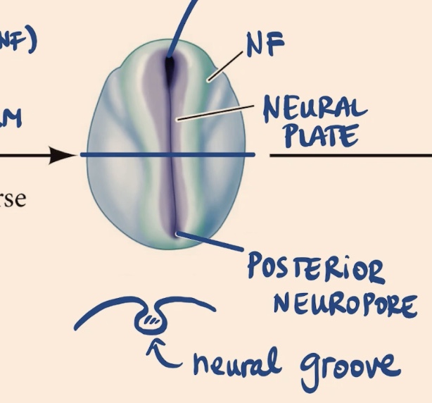
In the second stage of Neurulation we’ll see
Neural folds (NF) → now folded in
Neural plate → now getting inside the embryo
Openings of the neurula → Anterior and Posterior Neuropore
In the second stage of Neurulation we’ll see that the elements are
getting inside of the embryo → moving into the internal part of the embryo
Openings during the second stage of neurulation are referred to as
either the Anterior or Posterior Neuropores
If you were to cut the Neurula at the second stage of Neurulation, waht would we see inside? What is the structure inside called?
We’ll see that there is a little bit of a canal inside, and this structure inside is called the neural groove
What if we were to section this animal and take a look at it during neurulation, what will we see in the Early Neurula?
Most inside layer → Endoderm (orange/yellow color)
Mesoderm (red color)
Ectoderm → most external layer
Can see Neural Plate
Surface ectoderm → most external side
Notochord (mesodermal derivative)
Archenteron
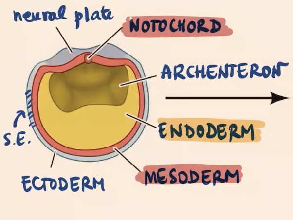
Most external side of a early Neurula sectioned during Neurulation is
the Surface Ectoderm
Process of neurulation culminates with the formation of the
Neural tube → once that is completely closed
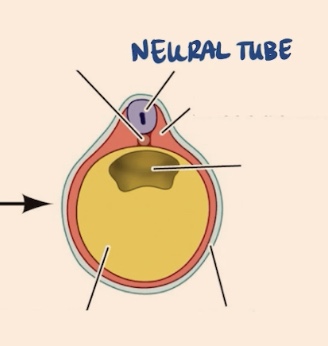
The external layer of cells in the Neurula (early) are now forming a tube and now entering the
embryo to close this tube
BMP
Bone Morphogenetic Proteins
Paracrine signaling means that
cells are signaling other cells in that process
Autocrine
I release factors and signal myself
Paracrine
Im going to signal cells next to me
BMP is involved in
Cell proliferation, Apoptosis (programed cell death), & Cell migration
This BMP signaling is part of a much larger family of factors that is called
TGF β Family → from transforming growth factor β family
TGF β Family
Big class of signaling molecules → paracrine molecules, that are extremely important for many diseases, many developmental processes
Have different receptors, these are different
proteins
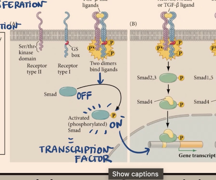
For BMP signaling we have different receptors, which ones?
Receptor type I
Receptor type II
However when those receptors (Receptors I and II) receive what ligand, then what will happen?
Receive the TGF β like ligands → they are going to form heterodimers (two dimers bind the ligand) → going to find that one copy or several copies of each of these receptors are going to form a complex
Which receptor involved in BMP signaling has a kinase domain?
Receptor Type II → meaning it can phosphorylate other proteins
So since Receptor II can a kinase domain, when those heterodimers form they are going to cross
phosphorylate each other
Due to the cross phosphorylation of those two dimers two one another, they are also going to phosphorylate what protein, that is in the cytoplasm?
Smad (now phosphorylated)
Now since Smad is phosphorylated, it is
active → without phosphorylation it is off
Once Smad is phosphorylated it is on and considered an
transcription factor
Smad as a transcription factor will go into the _, and bind to a bunch of different _
nucleus, genes → the promotors and promote transcription or repress the expression of certain genes
Strategies to over-activate or inhibit this pathway of Smad?
Lose it experiment → introduce a form of Smad that cannot be phosphorylated
Mutate the kinase specific domain → not going to transmit the signal (even if you send the ligand)
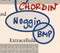
Another occurring inhibitor in BMP signaling pathway, that is an inhibitor that is an extra cellular protein that is called
Noggin
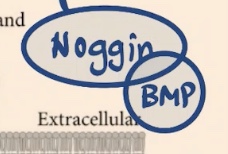
Noggin (a secreted protein) that will sequester what?
BMP → sort of like imagining releasing a bunch of different receptors that will compete with everything else → and you will not get BMP to bind to those receptors
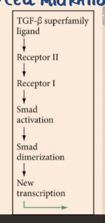
So depending on the ligand: Activin, Nodal, or TGF-β → it will activate a bunch of
receptors → result in phosphorylation of Smad’s that are called Smad 2,3 → then those Smad will recruit another factor (Smad4) -. go to nucleus and promote/repress transcription
BMP binds a different ligand → phosphorylating
Smad 1,5 → recruit Smad 4 and then go into the nucleus
Wants us to know that this pathway (TGF β), whether it is active in Nodal, TGF β itself works through these TGF β receptors
that are sending kinase receptors that are going to phosphorylate the Smad, once phosphorylated it is what is going to take care of regulating gene expression
Another secreted protein like Noggin, that is going to sequester BMP and is going to serve as an inhibitor of the pathway
Chordin
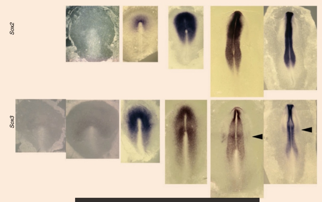
What are we seeing here?
All in-situ hybridization from this kind of like time course of development → all embryos at different stages
Sox2 and Sox3 are
Transcription factors
Sox (2,3) are going to encode for
proteins that will bind to DNA → they have a DNA binding domain → and are going to either promote the expression of certain genes or repress the expression of other genes
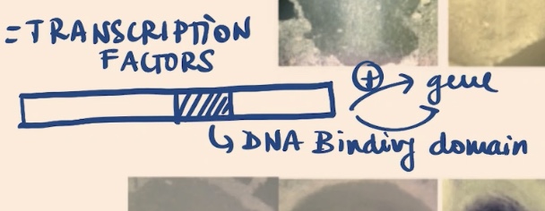
In particular, Sox factors are going to activate the expression of _, and at the same time they’re also going to inhibit _
neural plate gene programs, BMP signaling
When he says inhibit BMP signaling, please remember that they are TFs, their only way of inhibiting BMP signaling is
repressing the expression of something (could be receptor or ligands) → do not think of Sox2 interacting directly with BMP (talking at the expressional level)
If you reduce the expression of BMP, essentially what you are doing is modifying the
fate of these cells so that they are not epidermal cells
Remember the external ectoderm essentially had really high levels of
BMP
The process by which the neural plate, invaginates and forms a neural tube
Primary Neurulation
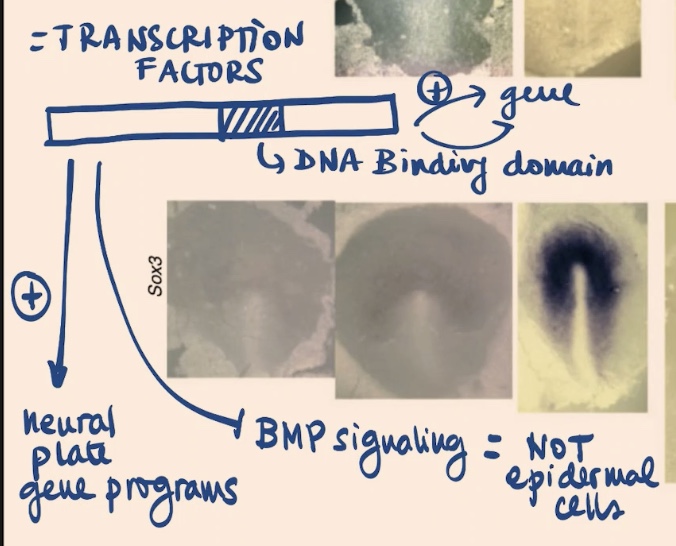
Other mechanism of Neurulation that involve ingression?
Secondary Neurulation
Secondary Neurulation (Ingression)
Where the neural plate is not folded and then goes as a tube → but there are cells in the ectoderm that are going to ingress → experience epithelial to mesenchymal transition → and now here, a part of these cells that derive from this layer (top blue)
In Secondary Neurulation, the cells that come from that ingression of the ectoderm will reconsolidate themselves (going to condense) and form a
tube where there is no gap inside
Once you have those condensates in Secondary neurulation you need to
cavitate → so there is this process of cavitation
Two mechanisms that form the Neural tube?
Primary Neurulation → involves the invagination of the neural plate
Secondary Neurulation (called Ingression) → and consists in those cells from the ectoderm moving loose in attachment → forming condensates and later on → cavitating
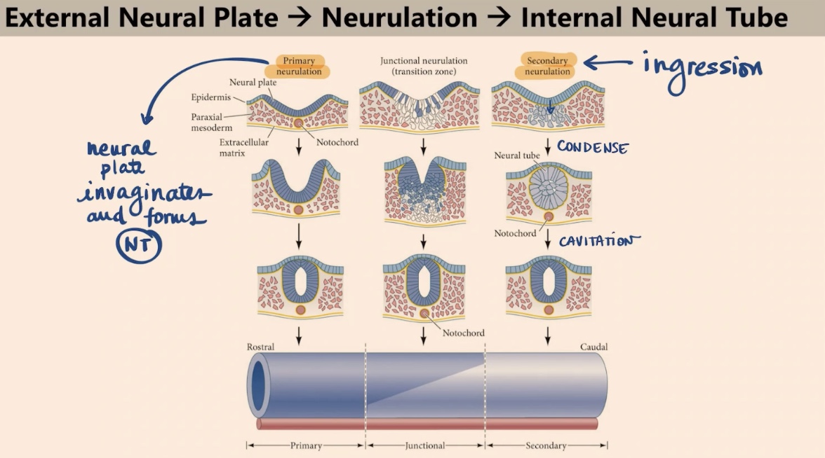
Both of those mechanisms that form the Neural tube (Primary and Secondary (Ingression) Neurulation), happen in every
embryo that we have been looking at, it’s just that they have differences in the location of the way these cells or this neural tube is formed
The Neurulation that is occurring in the anterior part of the Neural tube
Primary Neurulation
The secondary neurulation is what is happened in the _ of the neural tube
posterior part
Junctional Neurulation
A transition zone where the neural plate is in part forming this neural groove (invaginating), but there are also some cells that are undergoing epithelial to mesonchymal transition
In fish there is barely any, in chickens
secondary neurulation, most is primary. In chickens there is both primary, secondary neurulation and a really well characterize junctional neurulation → really depends on the species
Axis similar to Anterior → Posterior axis?
Rostral → Caudal
Rostral → your face
Caudal → tail
Mechanisms of Primary Neurulation involved?
Formation of the hinges
One layer of cells to forming a multilayer structure: where there is ectoderm, neural tube, notochord (mesoderm derivative)
First step of Primary Neurulation (Neurulation 1) is called? And has what?
This step is called Elongation
Have 2 layers → surface ectoderm and neural plate
Progenitors of the neural crest
In between those two layers of the first step of Neurulation we will find what?
The progenitors of the neural crest
Elongation is the
growth in the anterior → posterior axis
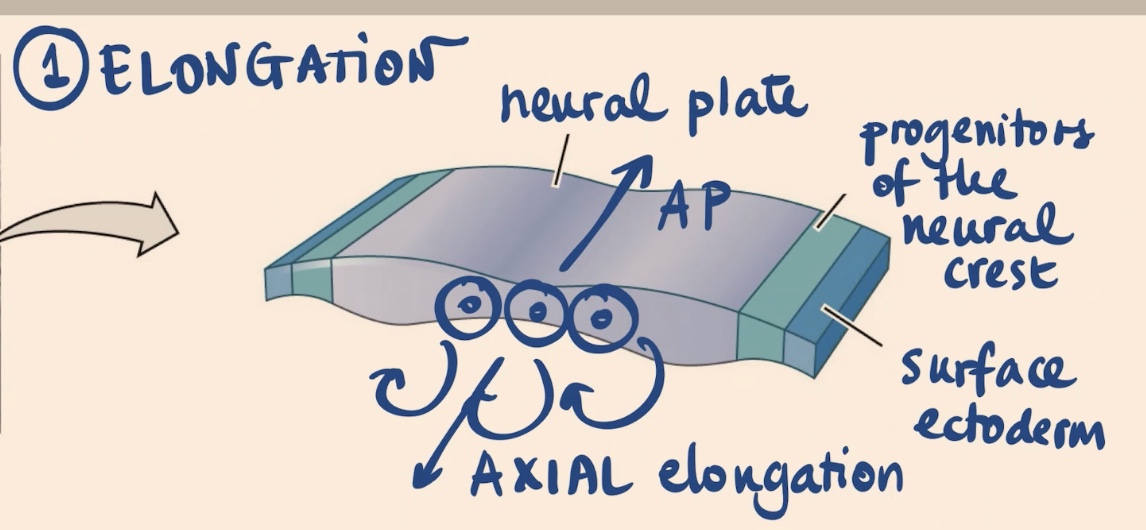
Elongation is driven by the fact that cells in the neural plate are .., also what is the name of this process?
actively dividing → with a particular orientation (towards AP axis), embryo going from a sphere to more complex structure (elongating the head and the tail). Process is Axial elongation.
Elongation will happen for a while in the embryo, but we need that neural plate to move inside the embryo → through a second step of Neurulation 1 called
Folding
Folding step of Nuerulation 1 has what?
A notochord (mesodermal derivative)
Medial hinge

Notochord (a mesodermal derivative) is going to be signaling to the _, and going to induce the differentiation of certain cells in the neural plate to become this _
neural plate, medial hinge (think of it at those cells (blob of blue), becoming more stiff)
Is going to essentially serve as the axis of the folding
The medial hinge → note also that there is a given movement of the embryo continuing to grow in that direction towards the midline
The notochord is going to attach itself to that medial hinge and going to induce this process of
invagination
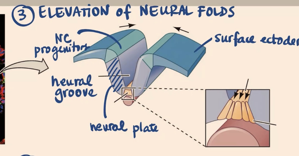
3rd step of Neuralation (Neuruation 2) is going to be
the Elevation of Neural Folds

In Elevation of Neural Folds, the _ is dragging towards the ventral side (the _ ). The _ is growing and pushing the neural folds towards the midline. And now we see what?
Notochord, Neural Tube, Surface Ectoderm. Now we see the Neural groove that has formed.
Step 4 of Neurulation (Neuruation) is _, which also has the formation of
Convergence, 2 new hinges → medial hinge & dorsal-lateral hinge points
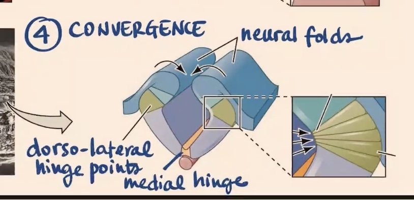
In convergence, the structures here now have kind of like a significant entity in the more dorsal part called the
Neural Folds
What happens in the last step (5th step) of Neurulation (Neuralation 3)?
Closure/Fusion
Closure/Fusion step of Neuralation? We can now see the _ , _ which is now closed. During the process, the cells of the _ are going to be located in most dorsal side of the tube → but will migrate
Surface ectoderm (completely outside now), Neural tube, Neural crest
When the cells of the Neural crest are located in most dorsal side of the tube, they are going to migrate and experience this process?
Process of epithelial to mesenchymal transition → and they are going to go very far, they will go and form the cartilage in your face, they will go and form all the melanocytes in your body
Exam question → Is the Neural crest a transient or a permanent structure?
Always a transient structure, but the cells that derive from the neural crest are permanent and they are one of the substrates of variability in vertebrate evolution
Mentioned that one of the layers, the genetic layers that are most important to animal evolution was the _, some people believe that the _ is sort of an updated mesoderm on steroids
Mesoderm, Neural crest (new source of variations for vertebrates)
Note that in terms of what we are learning about Neurulation it is talking about it in terms of
vertebrates
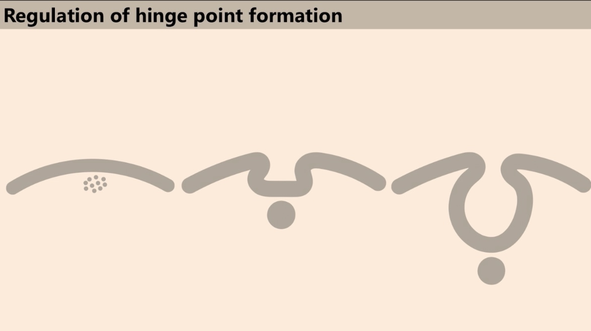
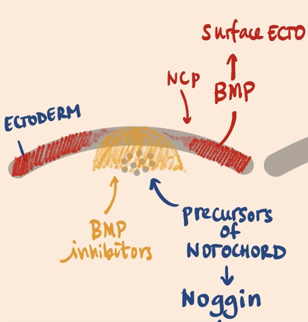
Added this slide, what is it? What is the goal?
Drawings of sections of embryos at different times → in a way pre-neurulation and then during neurulation. Describe the interplay of two signaling pathways in regulating the formation of the hinge points.
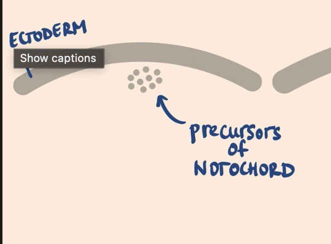
In the early embryo (pre-neurulation we see)
the Ectoderm and the precursors of The Notochord
_ is essential for the surface ectoderm, and in the absence of it, it was very important to form the _
BMP, Neural plate
Those precursors of the notochord are going to express (release), what two factors? By releasing these factors, one area of the Ectoderm will be exposed to these high levels of the _
Noggin + Chordin, BMP pathway inhibitors
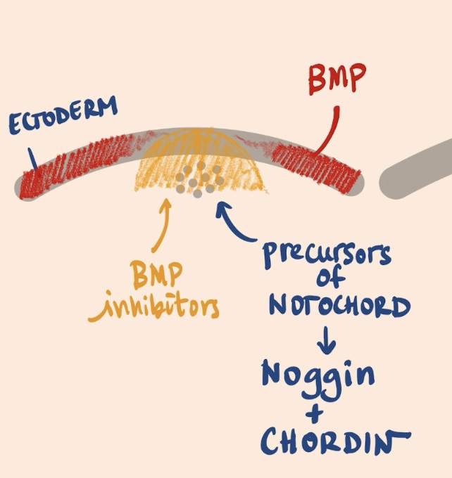
Remember that we have those gradients. Also why are Noggin + Chordin not morphogens?
Not since they are not inducing a response → they are inhibitors of the pathway, even though they are diffusing and creating a gradient →
Area that is super strong with BMP
Then kind of gradient where we have lower and lower amounts of BMP
Since there are areas that is super strong with BMP, then kind of gradient where we have lower and lower amounts of BMP. This means that really high levels of BMP are going to give rise to the _, and intermediate level where we are going have the _
Surface ectoderm, Neural crest progenitors