Cell ultrastructure 2.1.1.
1/73
There's no tags or description
Looks like no tags are added yet.
Name | Mastery | Learn | Test | Matching | Spaced | Call with Kai |
|---|
No analytics yet
Send a link to your students to track their progress
74 Terms
define prokaryotes
cell with no membrane bound nucleus or organelles
bacteria- prokaryotic or eukaryotic
example of a prokaryotic cell
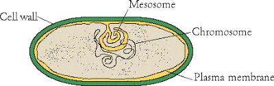
organelles in a bacteria cell (8)
cell surface membrane, mesomome, peptidoglycan cell wall, capsule, (naked) circular DNA, ribosomes, bacterial flagellum, plasmid
bacterial ribosomes
70S, free in cytoplasm, not membrane bound
bacterial mesomome
increases surface area for aerobic respiration
bacterial capsule
slimy layer outside cell wall, allows adhesion
define eukaryotes
cells with nucleus and membrane bound organelles, can be unicellular or multicellular
kingdoms made up of eukaryotic cells
animal, plant, fungi, protoctista
advantages of compartmentalisation
so enzymes and substrates stay in specific space, increases efficiency of reactions, increases likelihood of collisions, provides optimal conditions (e.g. pH) for specific reactions
organelles in animal cells (14)
nucleus, nucleolus, chromatin, nuclear envelope, nuclear pores, smooth endoplasmic reticulum, rough endoplasmic reticulum, centriole, ribosomes, golgi apparatus, lysosome, mitochondrion, plasma membrane, cytoplasm/cytosol
organelles in plant cells not in animal cells (3)
cellulose cell wall, chloroplasts, permanent vacuole
cytosol
cytoplasm= cytosol+all organelles (except nucleus), jelly-like liquid, contains cytoskeleton, site of many chemical reactions, surrounded by cell surface membrane
features and roles of the cell surface membrane
a.k.a plasma membrane, partially permeable, boundary between cell and its environment, site for chemical reactions and cell-cell signalling, dynamic structure e.g. phagocytosis, surface area can be increased by villi and microvilli
nucleus components (4)
nuclear envelope, nuclear pores, nucleolus, chromatin
nuclear envelope
double membrane, encloses DNA, protects it from cytoplasmic enzymes
nuclear pores
gaps in nuclear envelope, entry point for: regulatory proteins, nucleotides, steroid hormones, exit point for mRNA (DNA cannot leave nucleus)
nucleolus
site of ribosomes production
what is chromatin
DNA associates with histones (proteins)- {heterochromatin and euchromatin}
what happens to DNA before cell divides
DNA condenses into chromosomes, rest of the time appears as ‘grainy chromatin’
what are ribosomes and what is their role
site of protein synthesis, very large macromolecule made up of protein and ribosomal RNA, can be free in cytoplasm or on surface of RER, not membrane bound
ribosome sizes
80S in eukaryotes (large), 70S in prokaryotes (small)
endoplasmic reticulum
network of flattened sacs-cisternae- originating from outer membrane of nucleus/continuous with nuclear envelope, interior of cisternae called lumen, membranes of cisternae bud off into vesicles
vesicle
a small sac formed by a membrane (containing chemical from the lumen of the reticulum)
Rough endoplasmic reticulum
site of protein synthesis (on the ribosomes), and protein transport, ribosomes on surface (rough), newly formed polypeptides from ribosomes enter lumen of RER and are packaged into transport vesicles to go to the Golgi
what is the role of the smooth endoplasmic reticulum
site of lipid and carbohydrate synthesis, no ribosomes on it
roles of the golgi apparatus
sit of protein+ lipid modification and secretion, enter via vesicles
modification in the Golgi
-happens inside the lumen
-include: addition of a carbohydrate chain to proteins to make glycoproteins, and to lipids to make glycolipids
prosthetic groups of proteins added e.g. haemoglobin
final products are pinched off the end of the cisternae inside a secretory vesicle
secretion in the Golgi
vesicles transport products to their destination, inside or outside of cells
what are lysosomes
a vesicle with a specific function, formed by Golgi, remain in cell, contain hydrolytic (destructive) enzyme- lysozyme, degrades unwanted substances in the cell
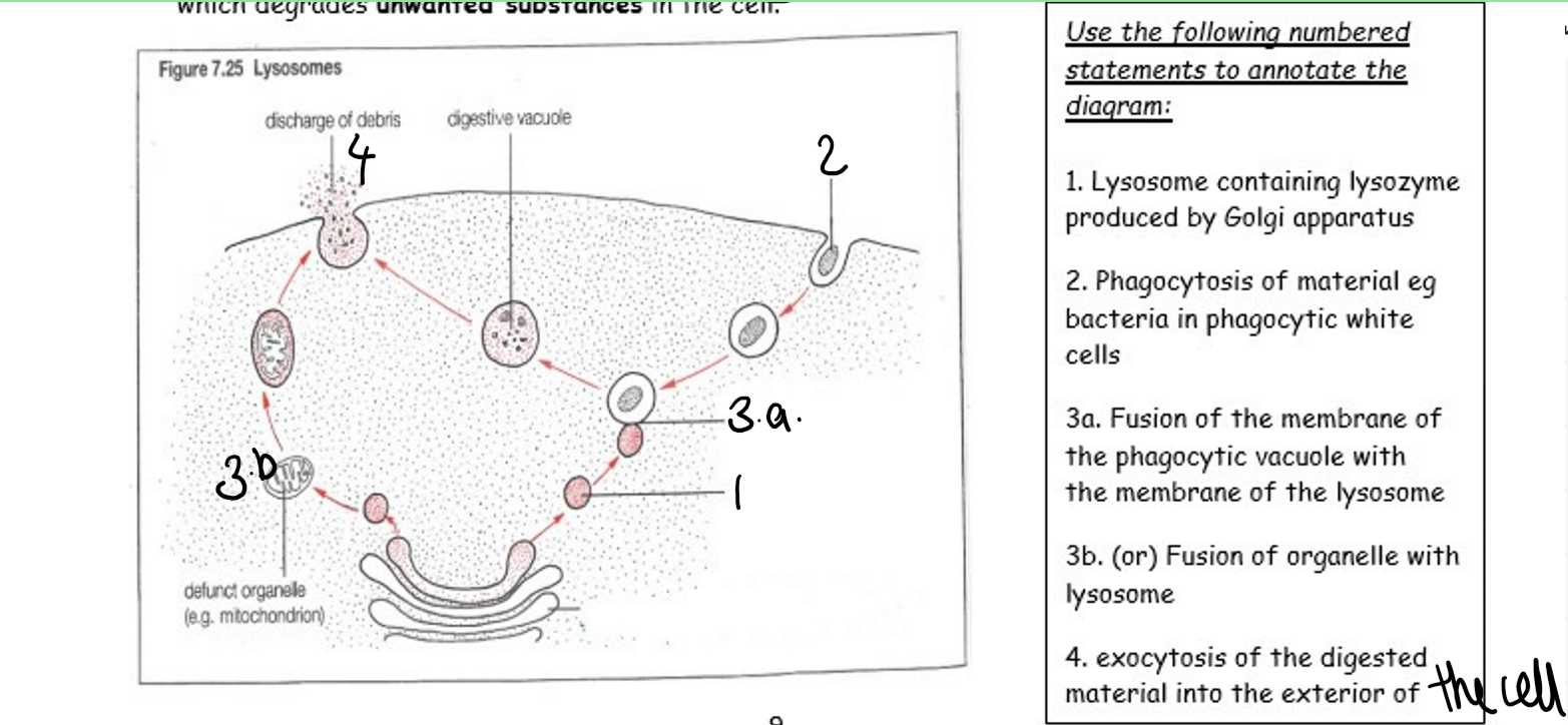
what are extracellular proteins
proteins made in the cell and secreted in order to carry out their functions outside the cell
examples of extracellular proteins
digestive enzymes, hormones, antibodies
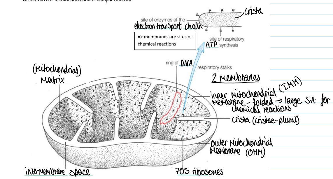
features and function of mitochondria
site for aerobic respiration, double membrane, has it’s own ribosomes and DNA, there are mitochondria in metabolically active cells (e.g. white blood cells, liver cells, neurones)
mitochondria structure diagram
aerobic respiration stages
glycolysis, link reaction and Krebs cycle, oxidative phosphorylation
where does glycolysis in aerobic respiration occur
1st stage. occurs in cytoplasm outside mitochondria
where does the link reaction and Krebs cycle in aerobic respiration occur
2nd stage, occurs in the matrix of mitochondria
aerobic respiration oxidative phosphorylation
3rd stage: electron transport chain and chemiosmosis, occurs on the inner mitochondrial membrane
evolution of mitochondria
aerobic bacteria:
same size ribosomes (70S)
same size ring of circular DNA
double membrane
size and shape of organelle
what is the cytoskeleton
filamentous structures which allow:
mechanical strength, support, stability and shape
transport within cells
movement of cells
made up of microfilaments, microtubules, intermediate filaments
intermediate filaments function
mechanical strength: help cell resist compression forces
support and stability for cell shape: holding organelles in place e.g. anchorage of nucleus
are a fixed length for stability so are not involved in cell movement
microtubules structure
cylindrical, polymerised, globular tubulin proteins, scaffolding like tracks
microfilaments structure
contractile actin fibres, change length with addition/ removal of subunits, contraction and polymerisation lead to change in length of filaments, subunits
microfilaments subunits
added at different rates to each end of fibres, not symmetrical, must be correct orientation to be added, at minus end must change shape before being added, not at plus end, added at a faster rate at plus end, filament increases in length faster in one direction, subunits added/removed controlled by concentration of subunits in cytoplasm at either end, at certain conditions subunits added at one end and removed at other, increasing length at one end at edge of cell causes cell to move in that direction
microtubules functions overview
mechanical strength
transport within cells:
a) during protein synthesis
b) during mitosis
cell movement
microtubules mechanical strength
help cell resist compression forces
microtubules transport within cell a) during protein synthesis
transport of mRNA from nucleus to ribosomes
transport of polypeptide within lumen of RER
movement of transport vesicles from RER to golgi apparatus
movement of vesicles between cisternae of golgi
movement of secretory vesicles from golgi to plasma membrane
microtubules transport within cells b) during mitosis
centrioles to allow attachment points for the spindle fibres at the poles
spindle fibres to allow movement of chromosomes to opposite poles
microtubules cell movement
flagellum to propel the cell forward e.g. sperm
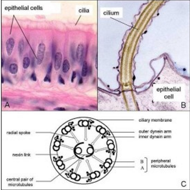
cilia which produce wafting motion to move substances outside the cell e.g. mucus
centrioles
formed by microtubules, involved in cell division, anchor point for spindle fibres
spindle fibres
formed by microtubules, form a centrosome with centrioles
flagella and cilia
formed by microtubules enclosed by a membrane, 9-2 structure, eukaryotic flagellum different to prokaryotic
microfilaments function
cell movement:
cytokinesis to divide the cell into two after mitosis
movement of plasma membrane e.g. phagocytosis, exocytosis
function of chloroplasts
site of photosynthesis
chloroplast structure
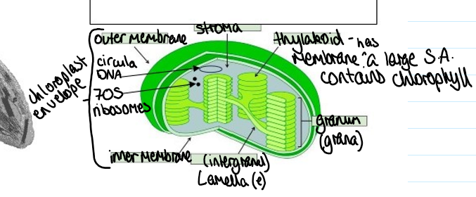
evolution of chloroplast
aerobic bacteria:
double membrane
70S ribosome
circular DNA
1st stage of photosynthesis
light dependent reactions: light energy → chemical energy
2nd stage of photosynthesis
light independent reactions: CO2 is fixed into sugar
membrane of the large permanent central vacuole
tonoplast
roles of the large permanent central vacuole
storage, maintaining turgor pressure
storage role of vacuole
contains:
waste (later removed by leaf fall)
pigments- anthocyanins (to attract pollinators and animals for seed dispersal)
cell sap: fluid containing sugars, amino acids, salts
maintaining turgor pressure role of vacuole
water enters vacuole
pressure against cell wall
turgor (pressure) creates
turgid cells support the plant
what is the cellulose cell wall made of and where makes it?
cellulose fibres: formed in SER, secreted by Golgi apparatus by secretory vesicles
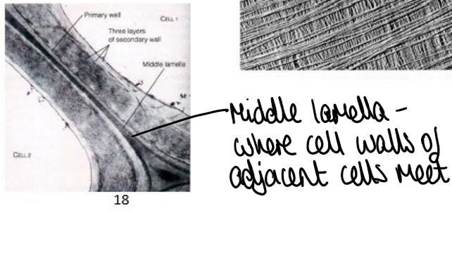
where cell walls of adjacent cells meet?
middle lamella
functions of cellulose cell wall
mechanical support and strength (increased by presence of lignin in wood)
osmotic support
osmotic support in cellulose cell wall
cellulose has high tensile strength, when water enters the cell, the cell wall resists expansion, prevents cell from bursting and internal pressure created (turgor pressure), cell becomes turgid, plant tissue supported
cellulose cell wall permeability?
highly permeable to water, allows easy passage of water through the roots and leaves
components of cellulose cell wall
lignin (wood), cutin (waxy cuticle), suberin (roots)- all make cell wall less permeable to water
how cellulose cell wall determines shape of leaf?
orientation of cellulose fibres determine the shape of the plant cell and therefore the shape of the leaf
what is the fungi cell wall made of
made of chitin
plasmodesmata
small gaps in cell wall and middle lamella where the cytoplasm of adjacent cell meet
what is cell fractionation
a method used to isolate/ separate organelles
cell fractionation method
done in ice-cold solution that has same solute concentration as the inside of the cells
homogenate is spun at increasingly high speeds in a centrifuge- called cell fractionation
define homogenisation
cellular organelles being isolated by breaking up the cells
how cell fractionation works
cell fragments, including organelles, move down the centrifuge tube depending on their mass and the speed of the centrifuge. heavier organelles need a lower centrifugal speed to compact the organelles into a pellet