HHP 210: Module 4 - Lecture 1
1/23
Earn XP
Description and Tags
Heart
Name | Mastery | Learn | Test | Matching | Spaced | Call with Kai |
|---|
No analytics yet
Send a link to your students to track their progress
24 Terms
What is the function of the heart?
Pump blood to lungs and body
Where is the location of the heart?
Mediastinum: middle of chest
2nd and 6th intercostal spaces
Apex: left and anterior
Base: superior and posterior
What is the fibrous pericardium?
Outside sac of dense connective tissue
What is the serous pericardium?
Two layers
Parietal: fibrous pericardium
Visceral: heart muscle
Layers of the heart? outer to inner
Epicardium: visceral serous pericardium
Myocardium: muscle
Endocardium: epithelium and connective tissue
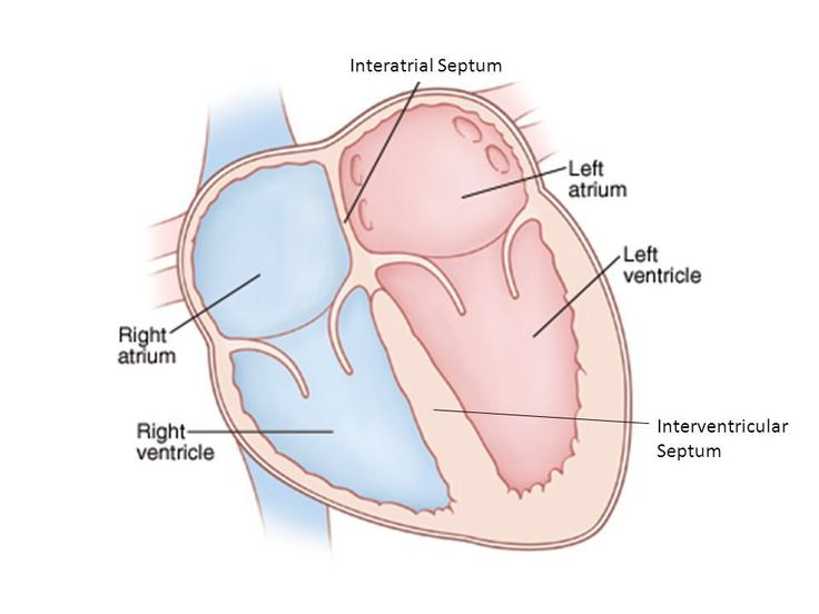
Chambers of the Heart? Auricles, trabecula carnae, and papillary muscles
Atria: right and left
Receive blood
Thin walls
Auricles: increase atrial volume, located at the top of each ventricle
Ventricle: right and left
Pump blood
Thick walls
Trabecula carnae: improve blood flow and contraction
Papillary muscles: proper closure of both valves
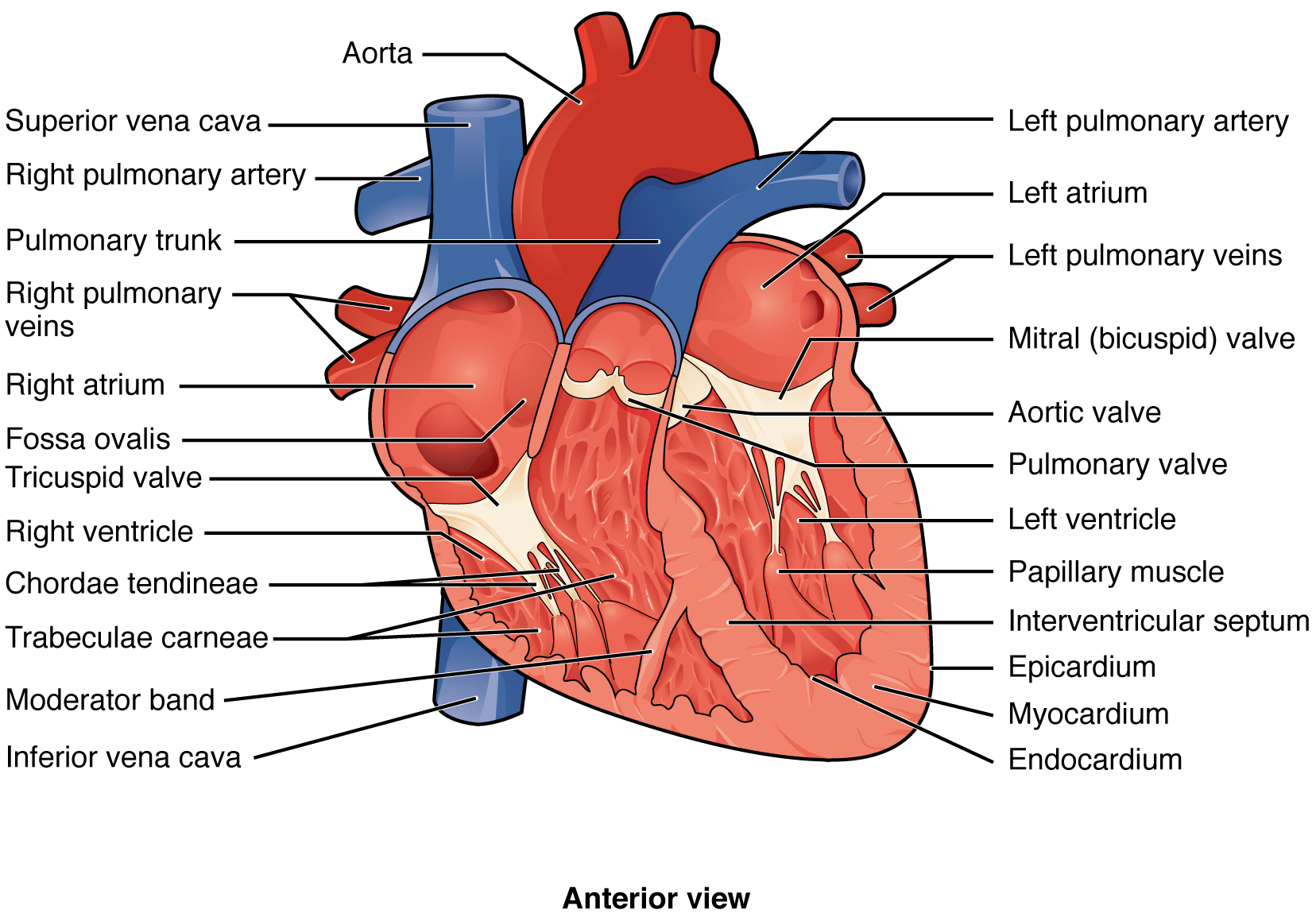
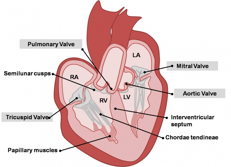
Septums of the heart
Division of left and right sides
Interventricular
Interatrial
Fossa ovalis?
Hole during fetus that allowed blood to pass
Major Vessels: Veins - to heart
Brings blood back to the heart
Right atrium: superior and inferior vena cava
Left atrium: right and left pulmonary veins
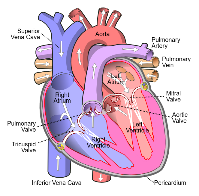
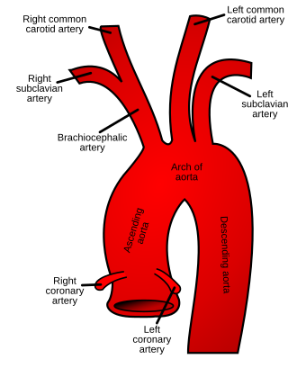
Major Vessels: Arteries; to lungs vs body; 3 major branches
Carry blood away from the heart
To lungs: pulmonary trunk, right & left pulmonary artery
Comes from right ventricle
To body:
Aorta (from left ventricle)
Ascending
Arch
3 Major Branches
Brachiocephalic
Right common carotid (superior)
Right Subclavian (lateral)
Left common carotid
Left subclavian
Descending: thoracic and abdominal cavity
Heart valves? Atrioventricular valves; chordae tendinae (2)
Prevent blood flow back into atrias, and made of cusps of connective tissues
Chordae tendinae: strings attached
Papillary muscle: tug and release
Tricuspid: right
Biscuspid/Mitral: left
-When one is open, the other is closed
-Located posterior

Heart valves? Semilunar valves (2)
Pulmonary: prevent blood flow back into right ventricle
Aortic: prevent blood flow back into left ventricle
Tricuspid w/o chordae tendinae
Heart sounds? Systole, diastole, lubb, dubb, average resting heart rate
Systole: contracting ventricles
Diastole: resting/expanding
Lubb: av valves closing
Dubb: semilunar valves closing
Avg resting heart rate: 40-80bpm
Order of how blood is pumped through the heart: deoxygenated vs oxygenated
Starting w/ deoxygenated
Superior and inferior vena cava
Right atrium
Tricuspid valve
Right ventricle
Pulmonary valve
Pulmonary artery
Left and right lungs
[now oxygenated]
Left and right pulmonary veins
Left atrium
Bicuspid valve
Left ventricle
Aortic valve
Aorta
To rest of the body
![<p>Starting w/ deoxygenated</p><ol><li><p>Superior and inferior vena cava</p></li><li><p>Right atrium</p></li><li><p>Tricuspid valve</p></li><li><p>Right ventricle</p></li><li><p>Pulmonary valve</p></li><li><p>Pulmonary artery</p></li><li><p>Left and right lungs</p></li></ol><p>[now oxygenated]</p><ol start="8"><li><p>Left and right pulmonary veins</p></li><li><p>Left atrium</p></li><li><p>Bicuspid valve</p></li><li><p>Left ventricle</p></li><li><p>Aortic valve</p></li><li><p>Aorta</p></li><li><p>To rest of the body</p></li></ol><p></p>](https://assets.knowt.com/user-attachments/0c4d5271-56d3-42be-93fd-20a3a909c02e.png)
Coronary vessels?