CT L2 - Light, Eye & Retina
1/22
There's no tags or description
Looks like no tags are added yet.
Name | Mastery | Learn | Test | Matching | Spaced | Call with Kai |
|---|
No analytics yet
Send a link to your students to track their progress
23 Terms
Three ways LIGHT travels:
Absorption
Refraction
Reflection
Vision in Simple Organisms vs Complex
Simple organisms (e.g. earthworm/euglena) may only be able to detect light levels rather than form images with their eyes. More complex organisms have image-forming eyes
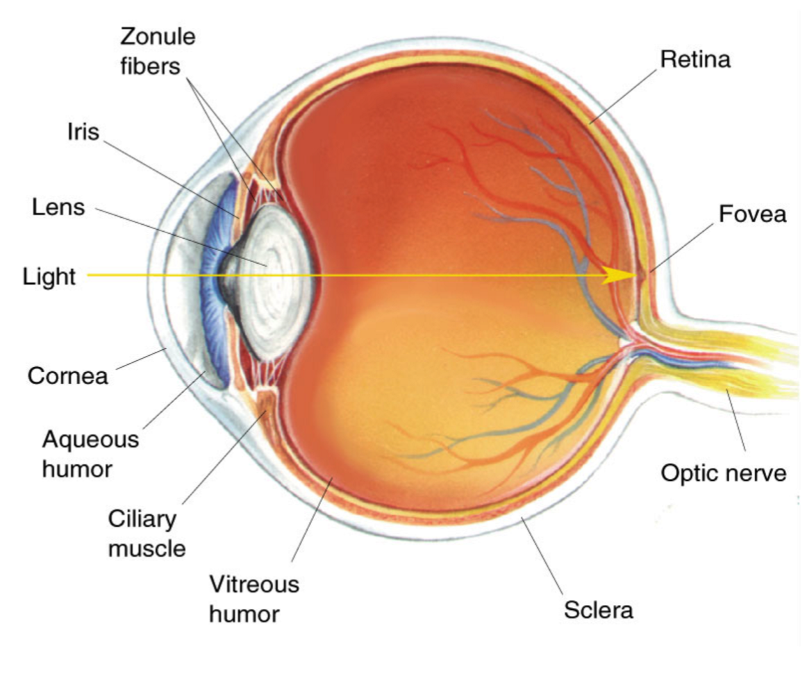
Anatomy of the Human Eye:
Retina = where light is focused, location of photoreceptors
Lens = can change shape - able to bend light rays in order to focus on far vs near objects
Fovea = tiny, pit, with high detail acuity + colour-sensitive
How do images form on the retina?
Each point in visual space is reflected onto the retina (retina forms a visual map)
Two Types of Photoreceptors in the Retina
RODS (approx. 100 million) = for low-light vision (SCOTOPIC) - detect light but have low detail, poor spatial resolution, high temporal resolution
CONES (approx. 6 million) = for daylight vision (PHOTOPIC) - detect colour and detail, high spatial resolution, poor temporal resolution
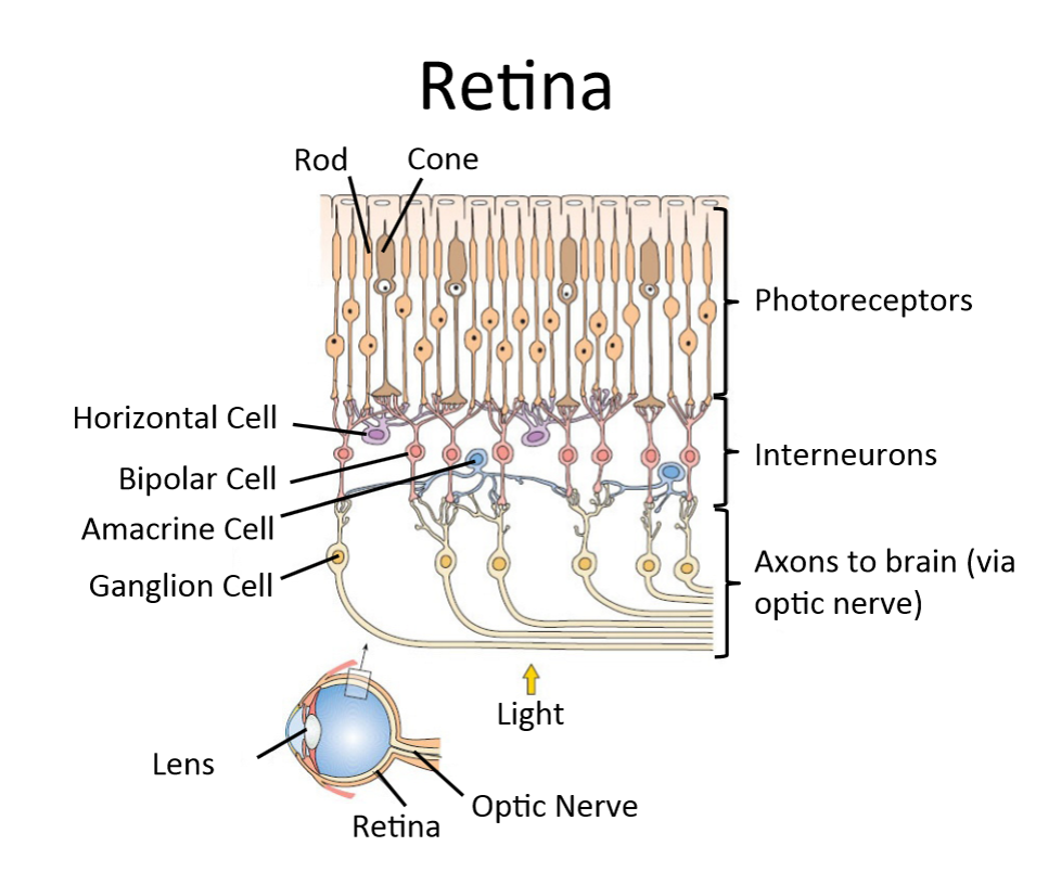
Other Structures in the Retina
Interneurons (horizontal, bipolar and amacrine cells) = allow for horizontal connectivity across the retina
Ganglion Cell = long axon to send info out of the retina to optic nerve
Optic nerve = bundle of ganglion axons send info directly to brain
The Duplex Theory of Vision
humans have two distinct visual systems in one eye: the ROD system and CONE system
The ROD System in the Retina - how it it wired + the impact?
Multiple photoreceptors connect to a single ganglion cell = because it pools information. it is much more light sensitive HOWEVER this means the brain can not tell which photoreceptor the light was detected by = LOW spatial resolution
The CONE system in the Retina - how it it wired + the impact?
one-to-one wiring = one photoreceptor for each ganglion cell = HIGH SPATIAL resolution but lowers sensitivity as light must hit that specific receptor to be detected
The Distribution of Photoreceptors
Only cones in the FOVEA, no rods = centre of visual field where you can focus on detail best
As you move to the periphery, the cones reduce and there are more rods.
What is the ‘blind spot’?
The ganglion cells have long axons, which leave the eye - the point at which they leave the eye has no photoreceptors causing a blind spot (located just to the side of the fovea) - brain typically "fills in" using the other eye's input.
What is foveal blindness (+ phenomenon)?
The Star Phenomenon: if you see a star in your periphery and then look at it directly and it disappears = this is because is bright enough to trigger the sensitive rods in your periphery is too dim to trigger the cones/lack of rods in your fovea
Lateral Inhibition Definition
that stimulating one part of the eye inhibited the activity of neighbouring parts used to sharpen edges of objects
Who discovered Lateral Inhibition and HOW?
Hartline (1938) used the horseshoe crab (Limulus polyphemus) because its compound eye has large, accessible nerve fibers that are easy to record from
Receptive Field Definition:
A specific region of sensory space where an appropriate stimulus leads to a response in a neuron
What is horizontal connectivity?
connections between lateral interneurons in the retina - allow for lateral inhibition
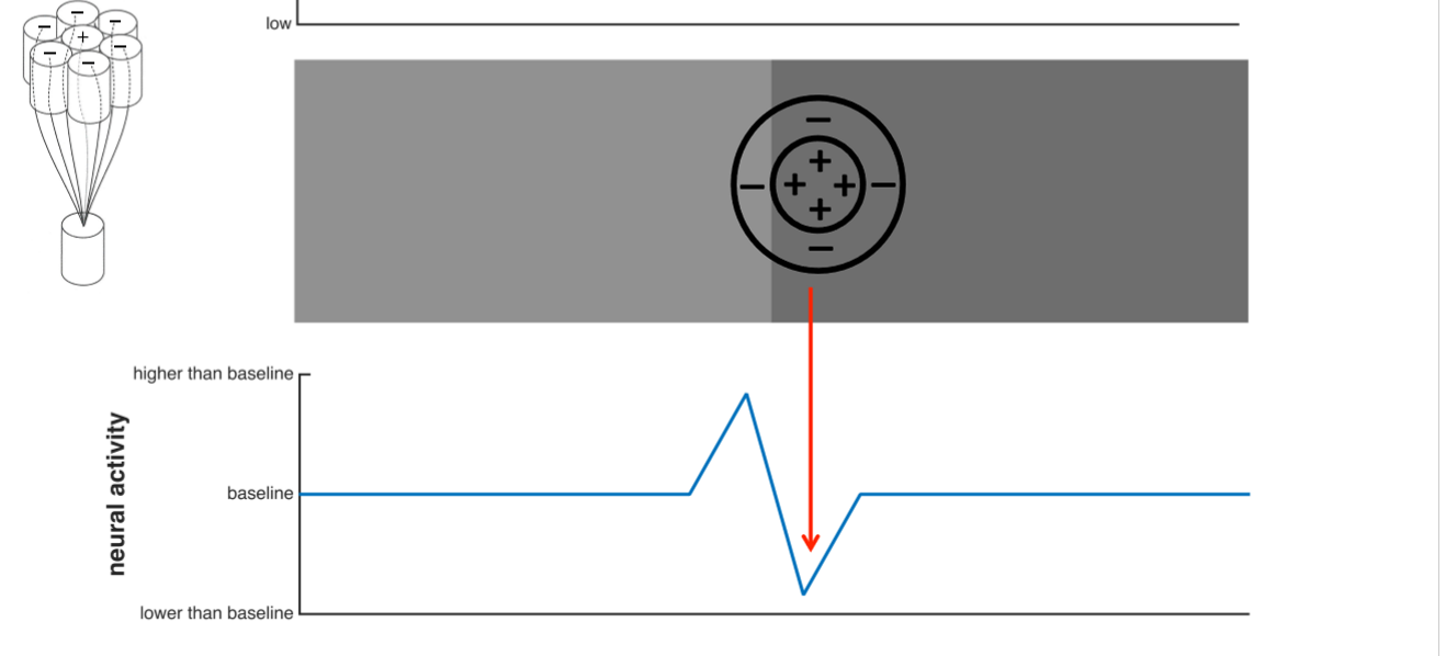
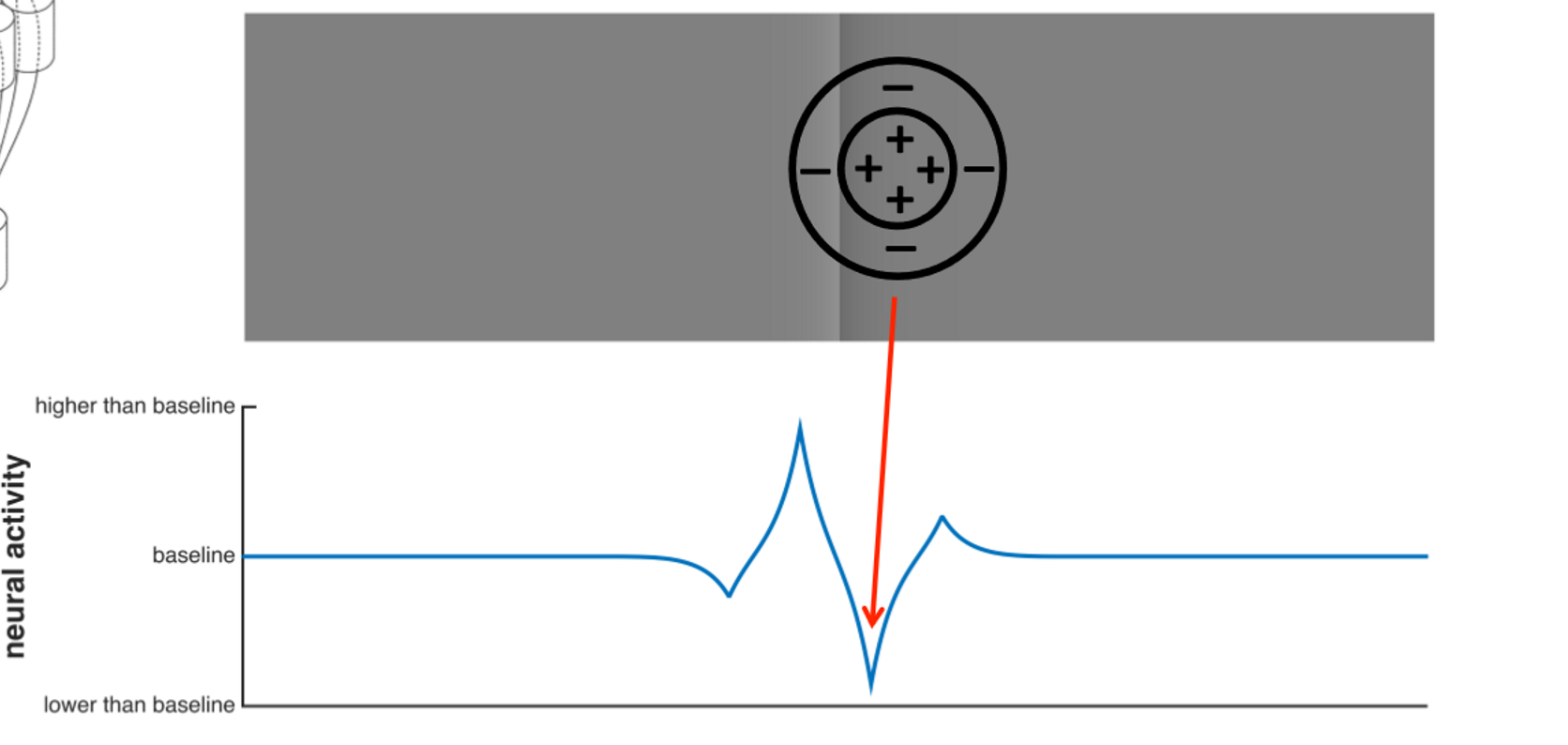
What is Centre-Surround Receptive Field?
Excited by light in the centre (+), inhibited by light in the surround (-)
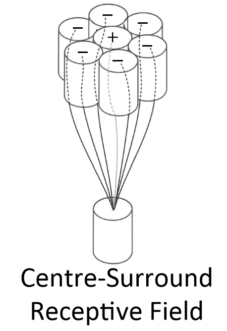
Why is the ganglion cell like a ballot box?
the ganglion cell adds up positive (facilitatory - centre-field) and negative (inhibitory - surround) "votes" from photoreceptors in the receptive field. If the sum reaches a threshold, it fires.
How does inhibition work in receptive fields/ ganglion cells?
The surround signals are inhibited by horizontal cells (aka lateral inhibition)
Why is the Centre-Surround Mechanism Necessary?
the retina only reports local differences (contrast), rather than absolute light levels. = efficient code for identifying the structure and edges of objects in the environment - only records changes rather than every individual pixel (similar to how a jpeg file works)
How does Edge Detection work (using the receptive fields)
As the eye moves across an edge (from light to dark) the inhibitory edge area of receptive field recieves the colour change info first. eg. darker area causes reduction in negative votes from surround - therefore overall neural acitvity increases above baseline (less inhibition). Then the centre passes from light to dark and the other side of the receptive field is the only part in the light. Therfore inhibition increases again causing a ‘BLIP’.
The Cornsweet Illusion (Craik-O’brien)
Because ganglion cells only care about local changes, they ignore the slow ramp and only react to the sharp contrast at the center.
The Illusion: The ganglion cell generates the exact same neural signature for this illusory edge as it does for a real one. - same BLIP effect
Result: The brain receives the signal "Edge: Light on left, Dark on right" and proceeds to "fill in" the entire left side as lighter and the right as darker.
Brain Evidence for Cornsweet Effect
Anderson, Dakin and Rees (2009) = The magnitude of the fMRI signal in the Lateral Geniculate Nucleus (LGN) for the illusory Cornsweet edge is strikingly similar to the signal produced by a physical edge = the LGN is part of the early visual system suggesting edge detection is low level and MONOCULAR process