Cell structure ALL
1/61
There's no tags or description
Looks like no tags are added yet.
Name | Mastery | Learn | Test | Matching | Spaced | Call with Kai |
|---|
No analytics yet
Send a link to your students to track their progress
62 Terms
What is cell theory?
all living organisms consist of cells
cells are formed by division of pre-existing cells
cells contain DNA that acts as instructions for growth
How long do temporary mounts last?
A few hours
How are permanent specimens prepared for microscopy?
specimen is dehydrated
fixed in wax
thinly sliced
How are dry mounts prepared?
solid specimens can be viewed as a whole
or cut into very thin slices (sectioning)
placed on slide and covered with a coverslip
How are wet mount prepared?
specimens suspended in liquid
coverslip placed at an angle to avoid bubbles
eg, aquatic life, living organisms
How are squash slides prepared?
wet mount prepared and coverslip pressed on
sample is squashed between two slides
How are smear slides prepared?
edge of slide used to smear a sample along another
coverslip then placed on top
eg. blood slide
What is the purpose of staining samples in microscopy?
Provides contrast - different components within a cell take up stains to different degrees
How is magnification calculated?
Image size ÷ actual size
What do positively charged dyes stain?
Cell components
What do negatively charged dyes stain?
Stain outside of the cells - the background
Why use a differential stain?
provides contrast
only some organelles take up stain
Why is gram staining used?
Separates bacteria into two groups:
gram positive
gram negative
What is the definition of magnification?
the number of times larger an image is compared with the real size of the object
What is the definition of resolution?
the ability to distinguish between two separate points
What are the two types of electron microscopes?
Transmission electron microscope
Scanning electron microscope
How does a transmission electron microscope work (+ magnification and resolution)?
beam of electrons passes through the sample
electrons are detected by a sensor
imaged is 2D
magnification x500 000
resolution = 0.5nm
How does a scanning electron microscope work (+ magnification and resolution)?
scans the surfaces of the sample
electrons are reflected
sensor detects electrons and an image is created
produces a 3D image
magnification x500 000
resolution = 3-10nm
How does a light microscope work (+ magnification and resolution)?
specimen can be viewed whilst still alive
can be stained to add contrast
color can be seen
light is shone through specimen then passes through two lenses
lenses magnify the image
magnification x1500
resolution = 200nm
What are the disadvantages of electron microscopes?
only produce black and white images
specimen must be in a vacuum - must not be living
specimen must be dehydrated
very expensive
very large
What is an artifact?
a visible structural detail caused by processing the specimen and is not a feature of the specimen eg. air bubbles trapped under a slide when using a light microscope
What is an eyepiece graticule?
small ruler that is inserted into the eyepiece of a microscope
scale is arbitrary
the eyepiece graticule remains constant at all magnifications
must be calibrated using a stage micrometer
What is a stage micrometer?
used to calibrate eyepiece graticule
it is 1cm long and is divided into 100 division
1 division = 100 micrometers
What type of organelles do prokaryotic cells contain?
They contain only non-membrane bound organelles.
How do prokaryotic cells store their genetic information?
As a single looped chromosome in cytoplasm
What is the size of a prokaryotic cell compared to a eukaryotic cell?
Much smaller (1-10 micrometers vs 10-100)
Are prokaryotes typically multi or uni-cellular?
Typically uni-cellular
How do prokaryotic cells divide?
Binary fission
Describe the structure of the nucleus
Largest organelle, surrounded by double membrane called nuclear envelope
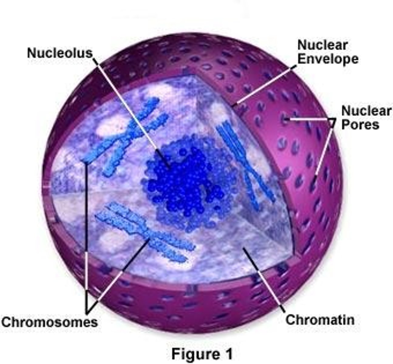
What is the function of the nucleus?
Contains genetic material, makes RNA and ribosomes, controls the cell's activities
Describe the structure of the mitochondria
Surrounded by double membrane, inner membrane folded to form cristae. Contains DNA and ribosomes.
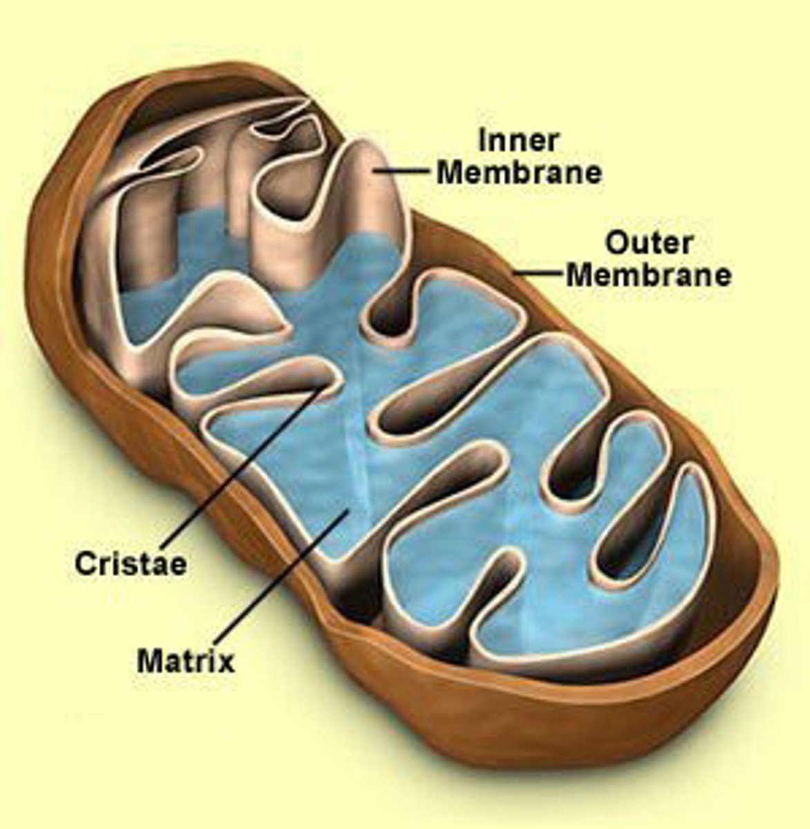
What is the function of mitochondria?
Site of aerobic respiration and produces ATP
Describe the structure of the rough endoplasmic reticulum
Flattened membrane sacs called cisternae with ribosomes attached
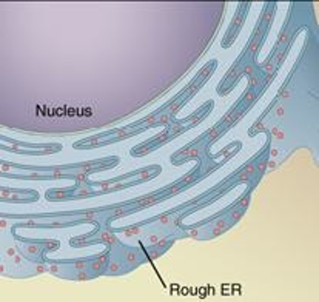
What is the function of the rough ER?
Site of protein synthesis and then transports proteins
Describe the structure of the smooth ER
Flattened membrane sacs called cisternae NO RIBOSOMES
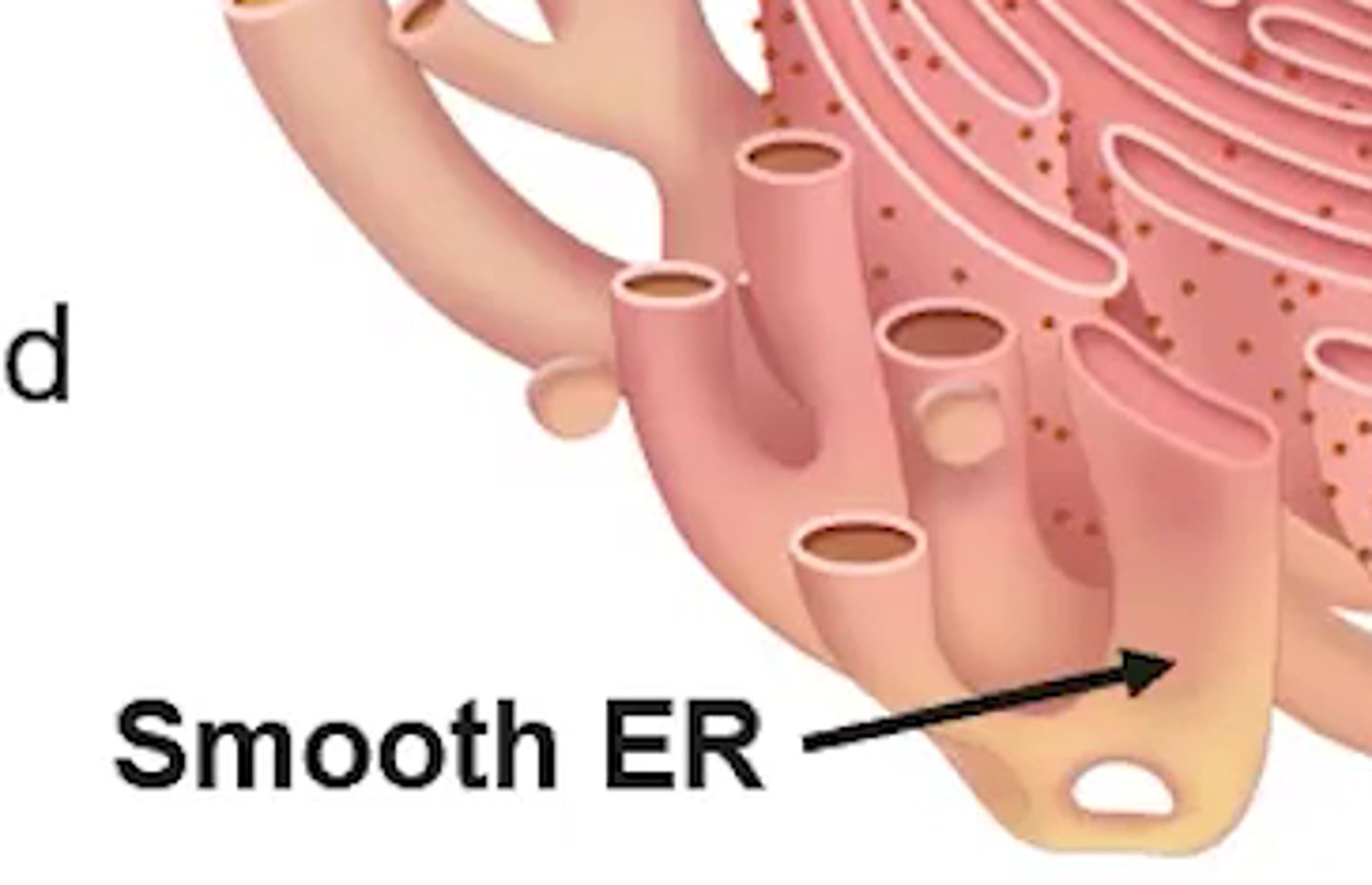
What is the function of the smooth ER?
Lipid and steroid synthesis
Describe the structure of the golgi apparatus
Stack of membrane bound sacs
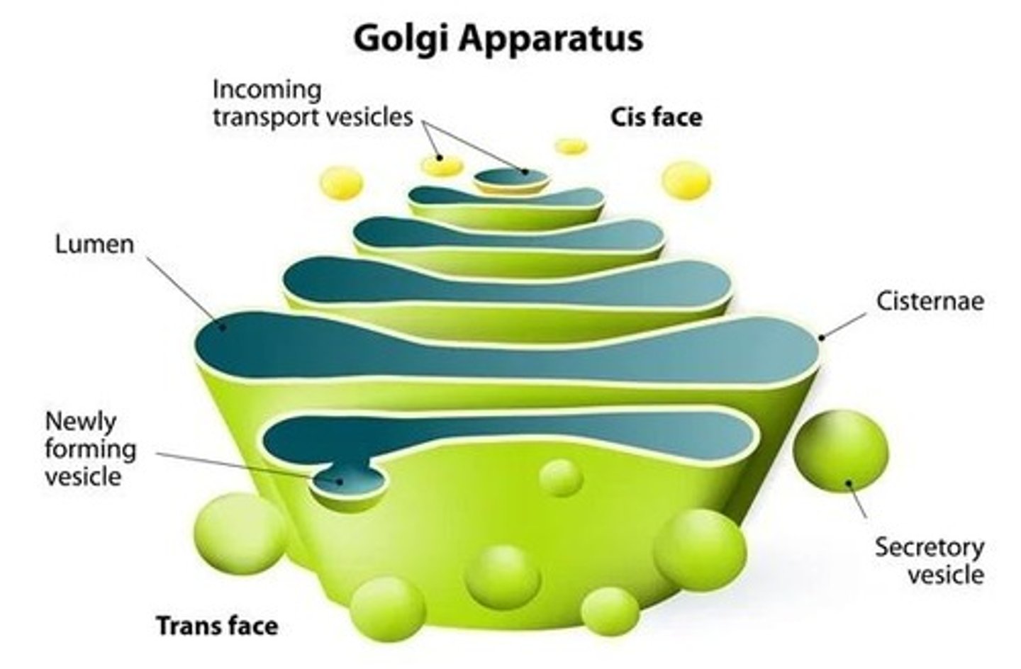
What is the function of the golgi apparatus?
Modifies proteins (adding sugar or lipid molecules) and folding proteins into 3D shapes. Packages proteins into vesicles for transport.
What are vesicles?
Membrane sacs used to transport materials
Describe the structure of a chloroplast
Surrounded by a double membrane with a highly folded inner membrane embedded with chlorophyll. Also contain DNA and ribosomes
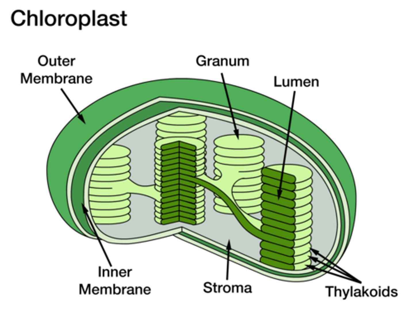
Describe the structure of the inside of a chloroplast
Contains a matrix of stroma which has a system of membranes running through it. These are stacked in places to form grana (contain chlorophyll). Stroma contains circular DNA, ribosomes and starch grains.
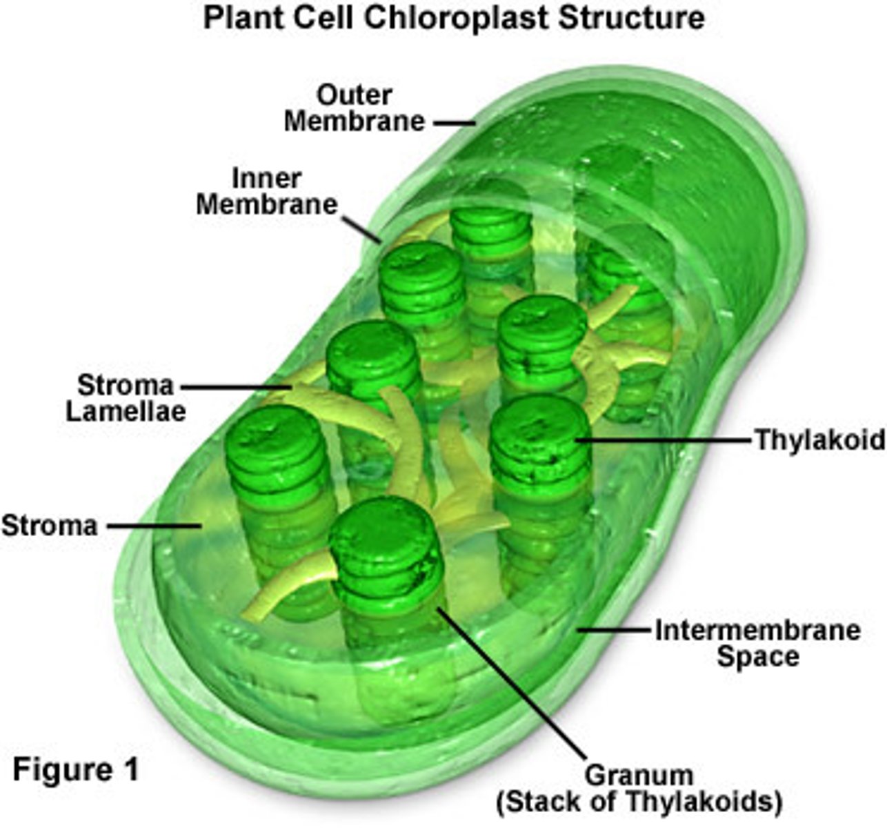
What is the function of the chloroplast?
Site of photosynthesis
Describe the structure of a lysosome
Spherical sacs containing powerful digestive enzymes
What is the function of a lysosome?
Destroys worn out organelles and digests the contents of vacuoles formed by phagocytosis
Describe the structure of a vacuole
Bound by a single membrane called a tonoplast. Contains cell sap.
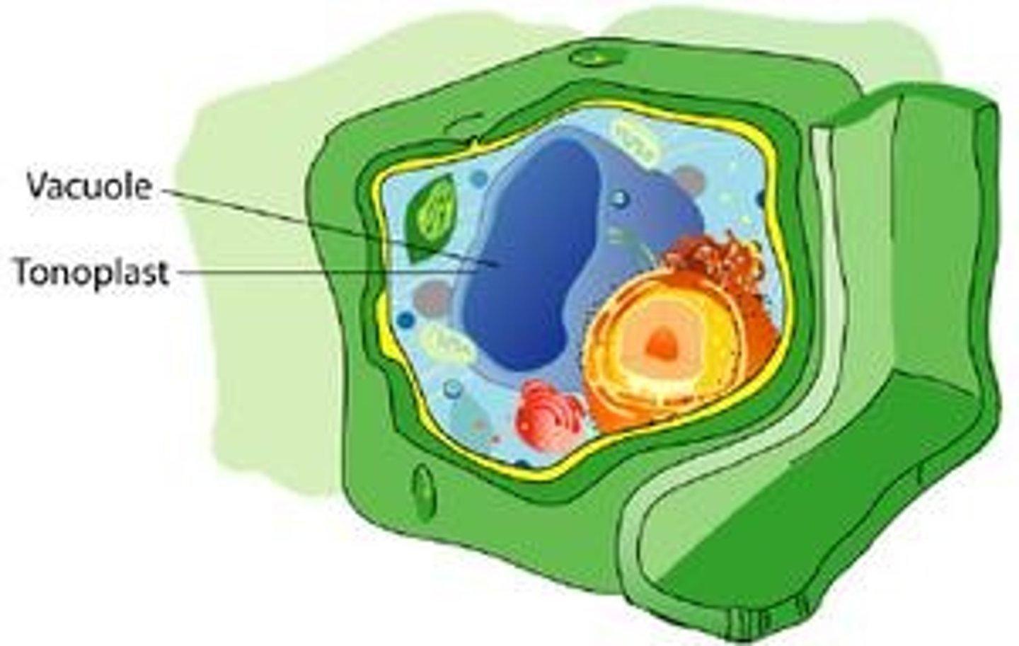
What are the main components of cell sap?
Mineral salts, pigments, organic acids and other substances.
What is the function of a vacuole?
Stores waste products and cell sap. Changes in volume affect turgidity of the cell.
Describe the structure of a ribosome
Not membrane bound. Made of protein and RNA
What is the function of a ribosome?
Protein synthesis
Describe the structure of a centriole
Made of two bundles of protein. Each cylinder made up of nine fibres.
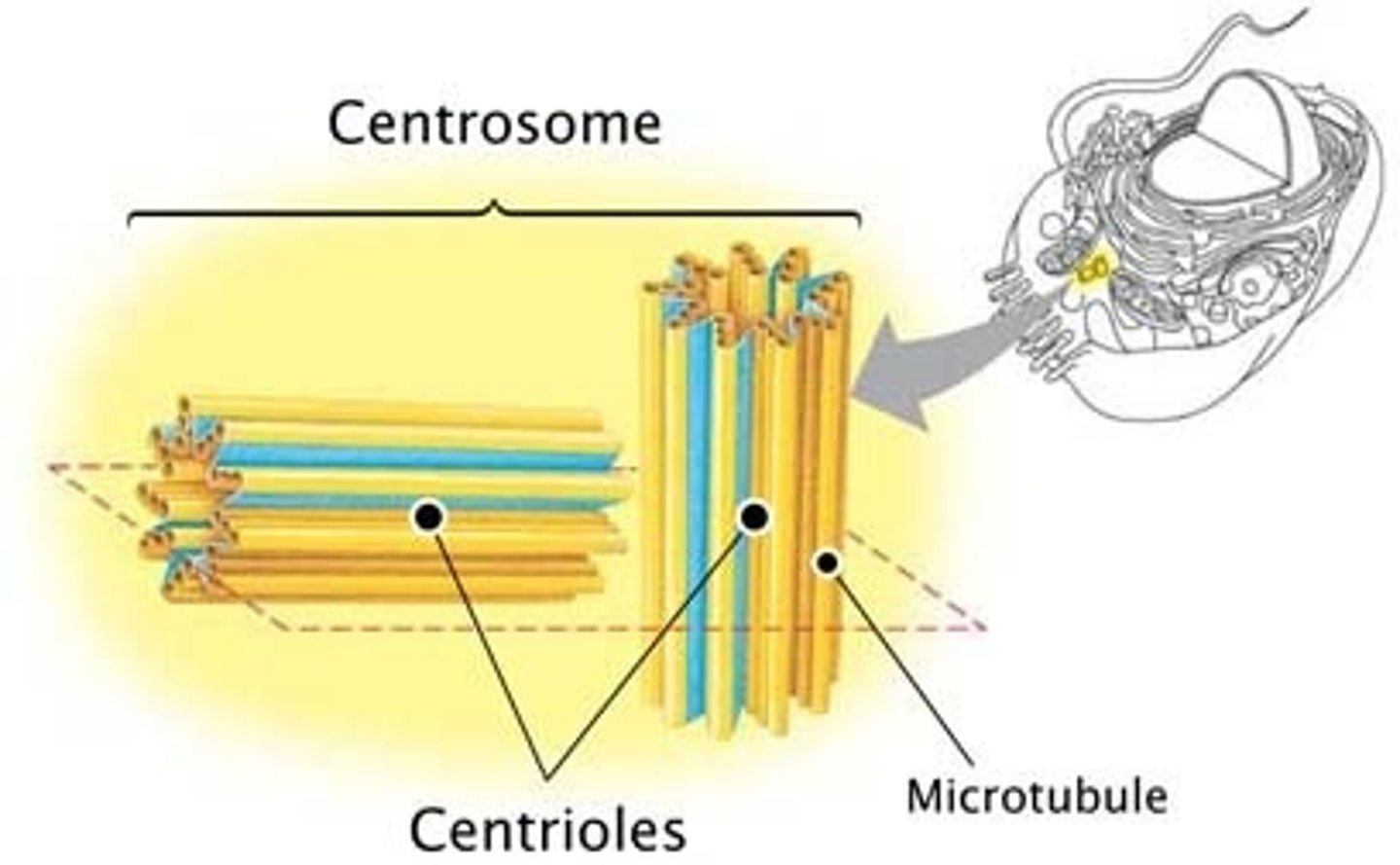
What is the function of a centriole?
Form spindle fibres and are involved in formation cilia and flagella.
Describe the structure of a cell wall
Made of cellulose and is fully permeable
What is the function of a cell wall?
Provides mechanical support and protection. Prevents cell from bursting.
What are the functions of the cytoskeleton?
Establishes cell shape, provide mechanical strength, locomotion, chromosome separation in mitosis and meiosis, movement of organelles within a cell
What are the 3 components of the cell cytoskeleton?
Microtubules, microfilaments and intermediate fibres
What is the size and function of microtubules?
25nm in diameter. Globular tubulin proteins polymerise to form tubes. Involved in transport of organelles. Make up the spindle fibres during cell division.
What is the size and function of microfillaments?
7nm in diameter. Contractile fibres formed from the protein actin. Responsible for cell movement and cell contraction during cytokinesis.
What is the size and function of intermediate fibres?
8-10nm in diameter. Give mechanical strength to cells and help maintain their integrity.
What are undulipodia and cilia + what are they made up of?
Hair like extensions that stick out from the surface of cells. Made of 9 pairs of microtubules in a circle w/ two more in the middle (9+2 arrangement)
What are the functions of undulipodia and cilia?
Move liquid past the surface of the cell. For single cells this enables them to swim. For anchored cells this moves liquid over the surface of the cell.
What are flagella made of + where are they present?
Only present in bacterial cells. Made of a spiral of flagellin protein attached by a hook to a protein disc than can rotate.
What is an advantage of being a eukaryote?
Organelles allow for division of labour. Each organelle is specialised to carry out a specific role.