hyperplasias and benign tumors of the jaws
1/102
Earn XP
Description and Tags
lecture given 5/21/2026
Name | Mastery | Learn | Test | Matching | Spaced | Call with Kai |
|---|
No analytics yet
Send a link to your students to track their progress
103 Terms
hyperplasias
torus palatinus, torus mandibularis, hyperostosis, dense bone island
odontogenic tumors
odontogenic epithelial tumors- ameloblastoma, calcifying epithelial odontogenic tumor
mixed odontogenic tumors
odontoma, ameloblastic fibroma, ameloblastic fibro-odontoma, adenomatoid odontogenic tumor
mesenchymal tumors (odontogenic ectomesenchyme)
odontogenic myxoma, benign cementoma
nonodontogenic benign tumors
benign tumors of neural origin- neurilemoma, neuroma, neurofibromatosis
mesodermal tumors
osteoma, gardner’s syndrome, central hemangioma, ossifying fibroma
torus palatinus
palatal torus
bony protuberance at the midline of the palate
located on the hard palate
well defined, convex, or lobulated
homogenously radiopaque
torus mandibularis
mandibular torus
bony protuberance on the lingual aspect of the mandible close to premolar
lingual and bilateral
sharply demarcated
homogenously radiopaque
what is this?
torus palatinus
what is this? besides baby luna
torus mandibularis
hyperostosis
small region of osseous hyperplasia
most commonly on the buccal surface of the maxilla
located on the maxillary alveolar process superimposed on teeth
well or poorly defined
radiopaque and homogenous
dense bone island
aka enostosis, periapical idiopathic osteosclerosis
localized growth of compact bone
located on mandible > maxilla, premolar to molar region
well defined, no capsule
radiopaque
may resorb root- not common but possible
what is this?
hyperostosis
what is this?
dense bone island
general characteristics of benign tumors
abnormal new growth of cells
non-malignant/non-cancerous tumor
localized, and does not spread to other parts of the body
if left untreated, some can grow large and lead to serious disease because of their size
can impinge upon and damage adjacent structures
what radiograph techniques are ideal for examining benign tumors?
intra orals, occlusal, panoramic radiographs
additional imaging CBCT, CT, MRI
what are the radiographic features of benign tumors?
odontogenic lesions- alveolar process, above IAN
vascular & neural lesion- within IAN
cartilaginous tumors- mandibular condyle
smooth, well defined, corticated
internal structure is radiolucent or radiopaque, or mixed, curved septa
can cause displacement of adjacent structures
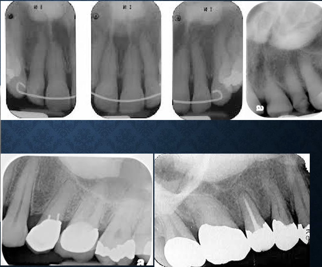
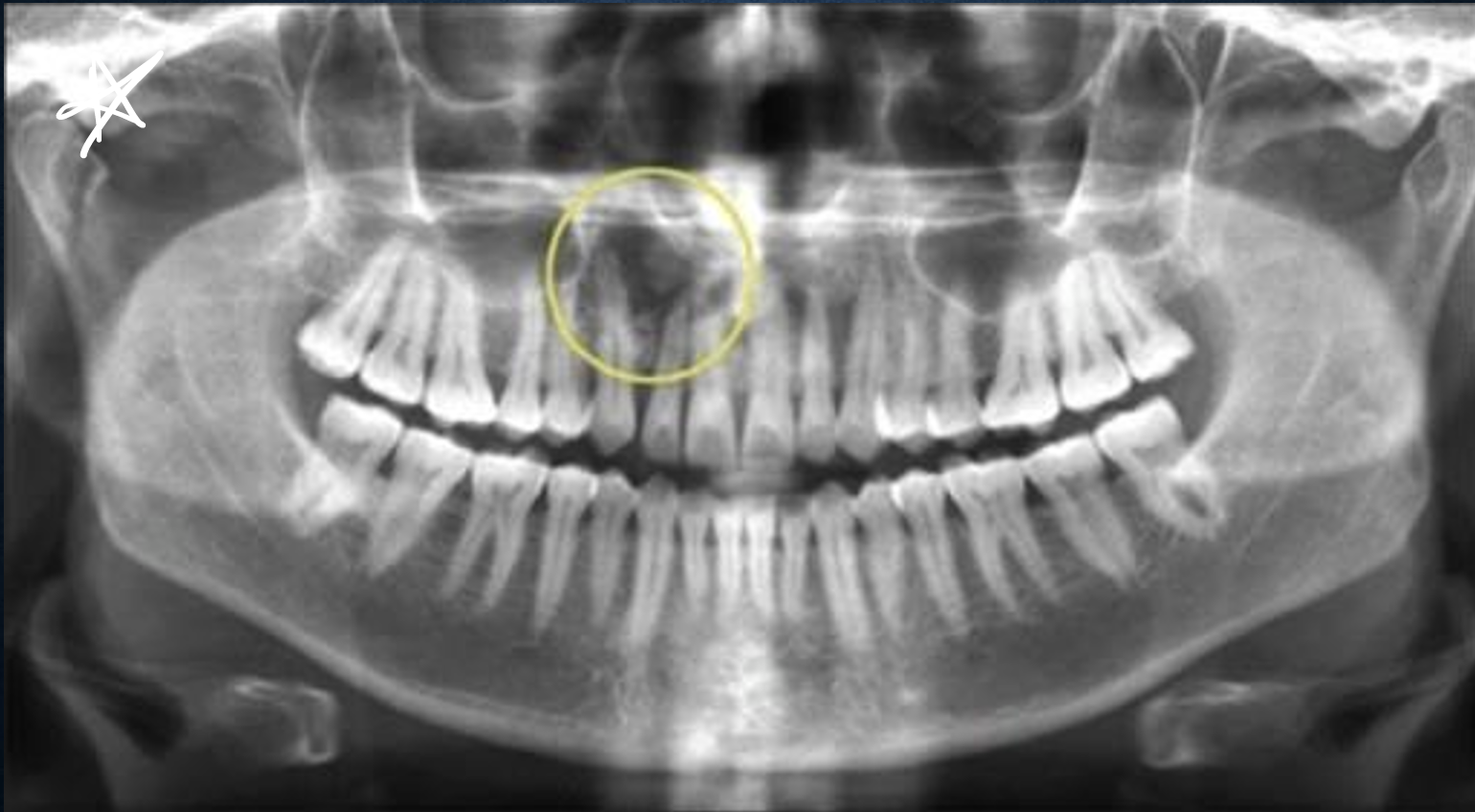
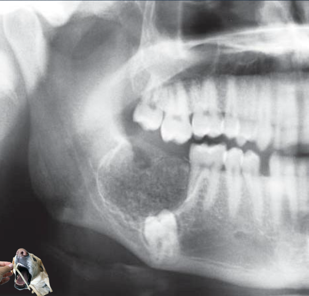
ameloblastoma
true neoplasm of the odontogenic epithelium
divided into multicystic, unicystic (mural), and desmoplpastic types
men>women, age 20-50, slow growing, symptoms occur early on, increasing facial asymmetry
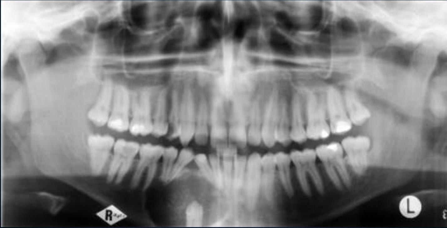
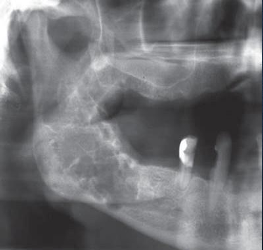
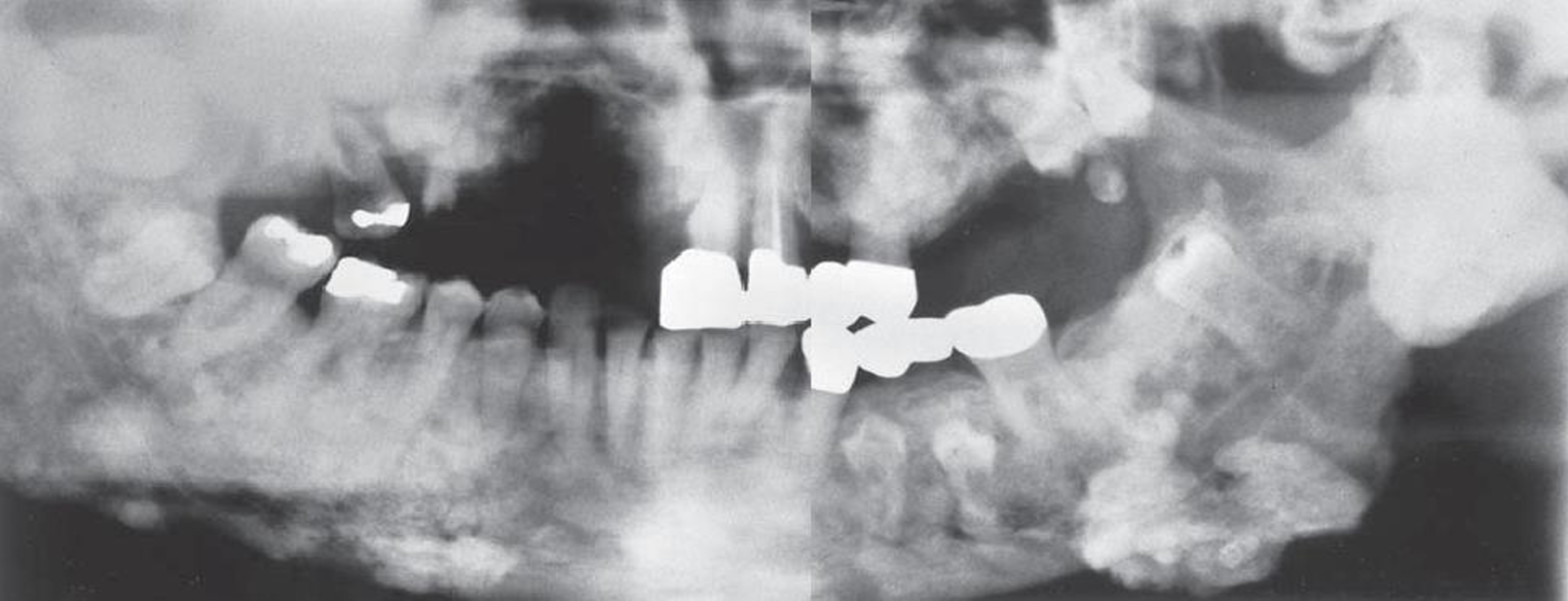
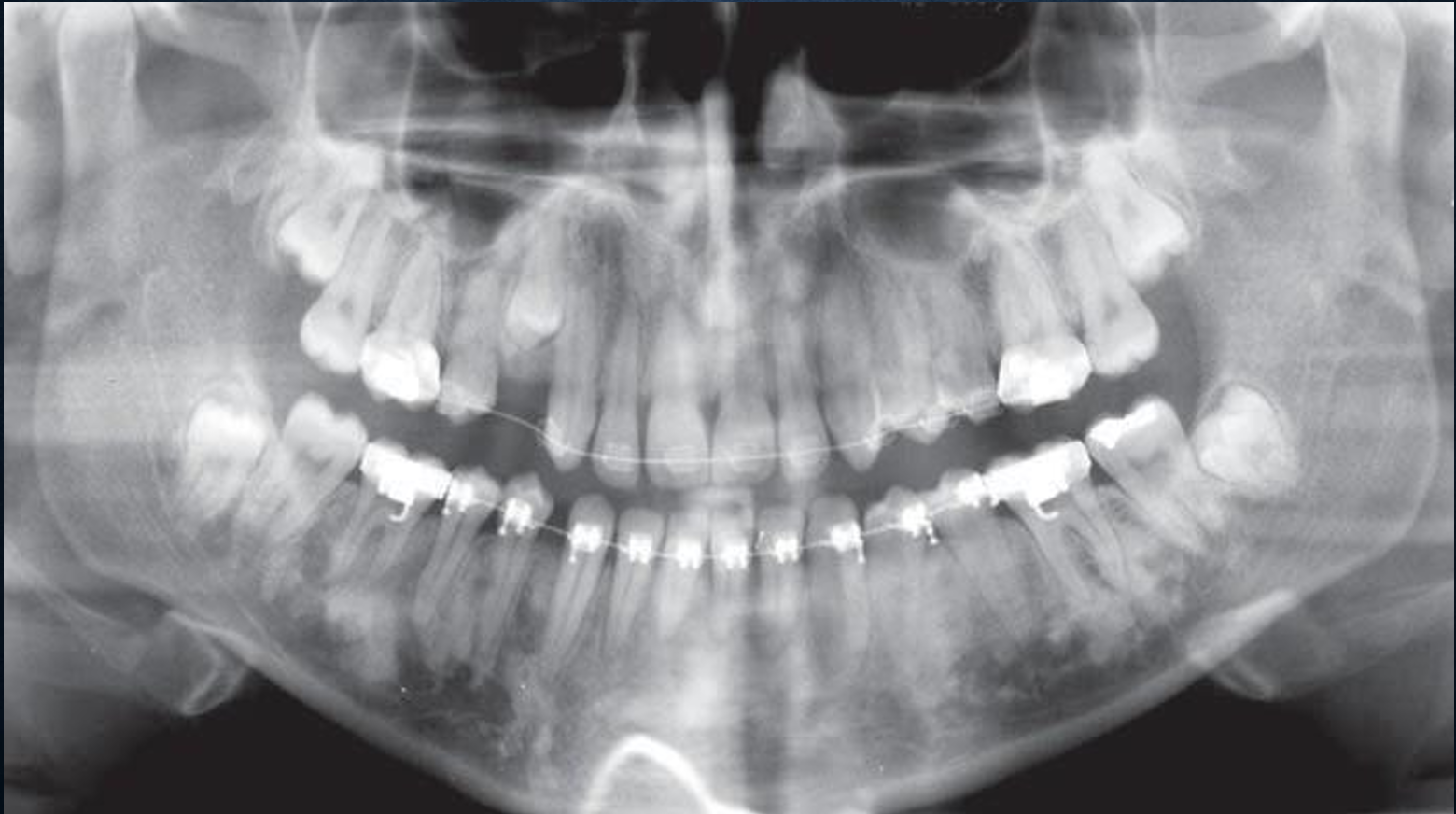
what are the radiographic features of ameloblastoma?
most often in molar ramus region, may extend into symphyseal area, in maxilla is 3rd molar area
well defined, curved, in the maxilla the periphery may be ill defined
internal structure is radiolucent and septated
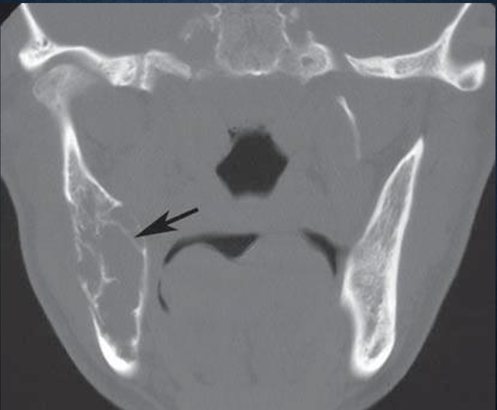
can cause root resoprtion, expansile in mesio-distal and bucco-lingual direction, tooth displacement, thinning of the cortical plates, on CT the cortical plates may show perforation, displacement of IAN inferiorly
what are the differential diagnoses for ameloblastoma?
OKC, odontogenic myxoma, dentigerous cyst
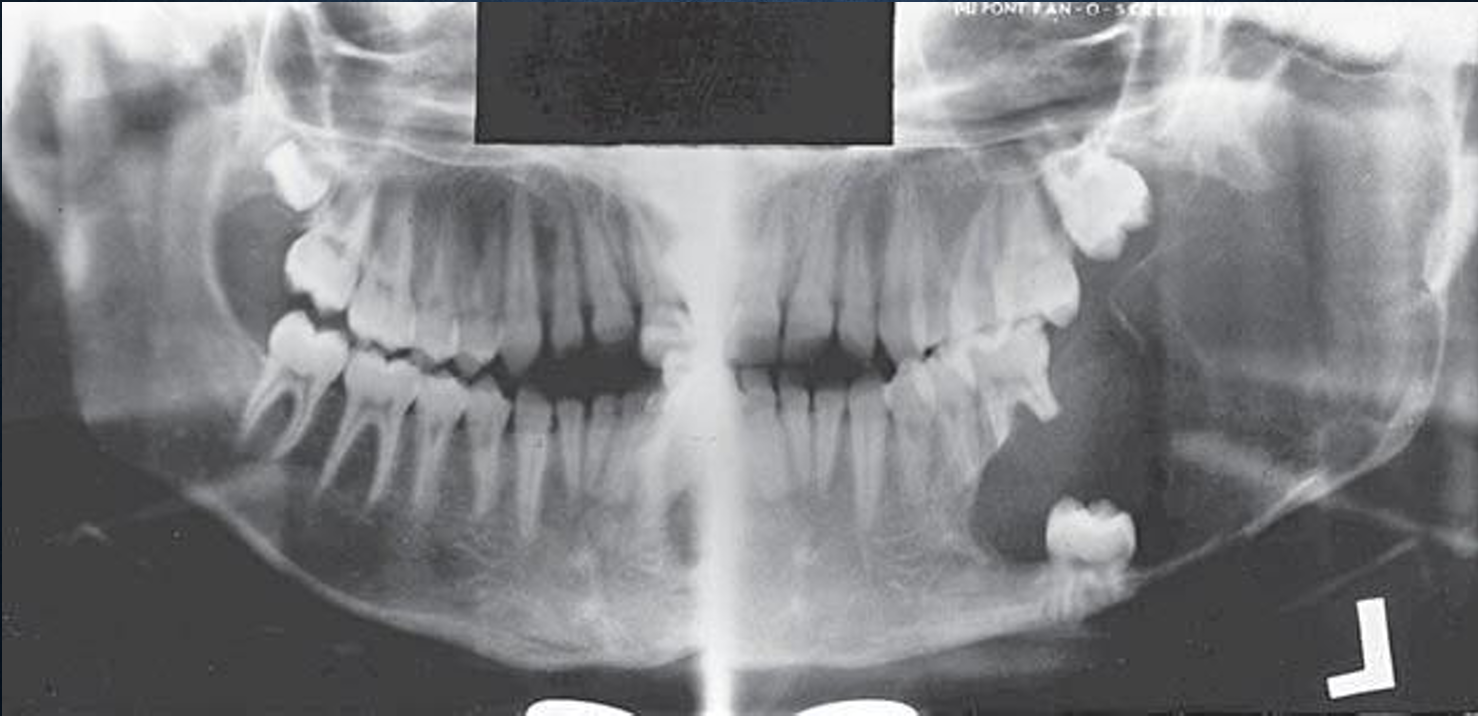
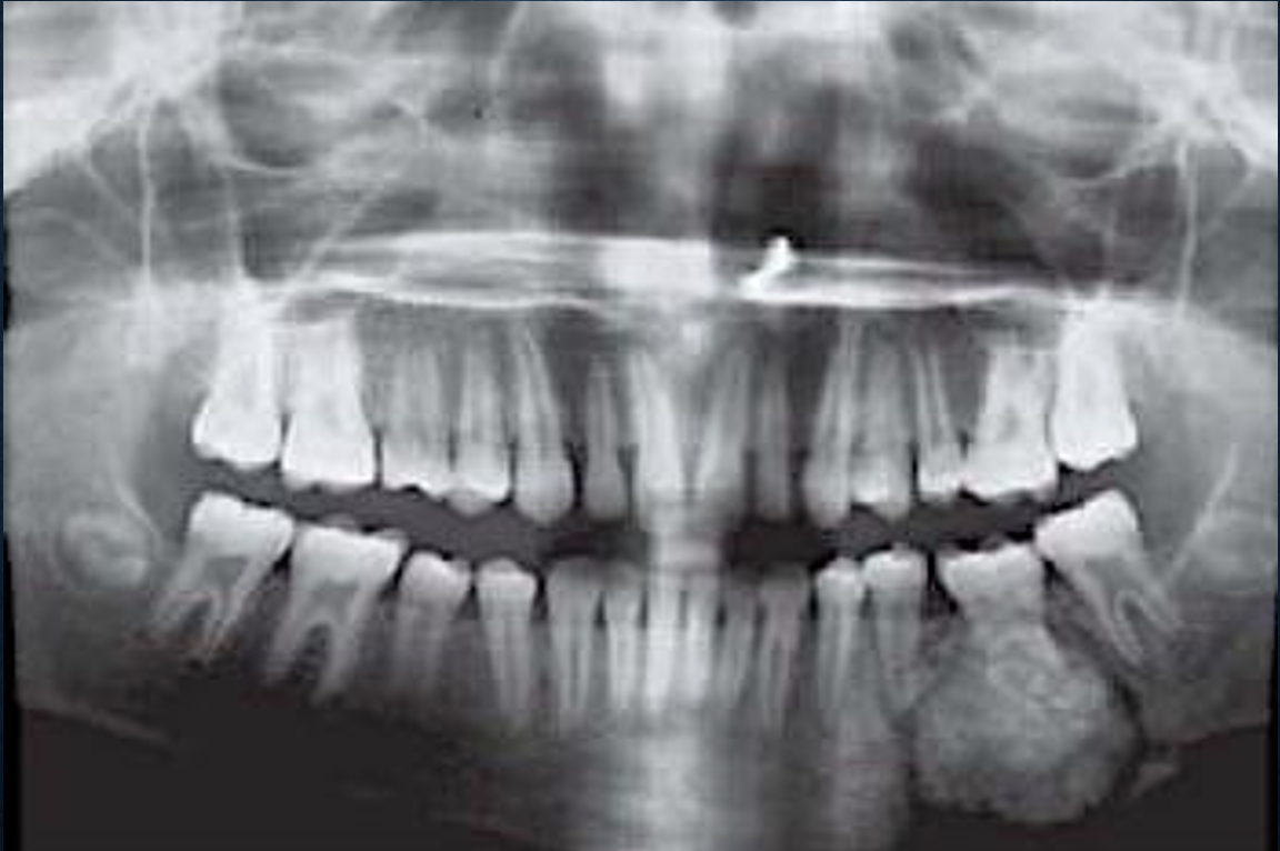
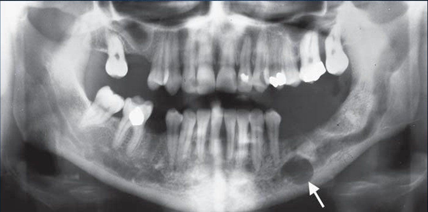
what is this?
ameloblastoma
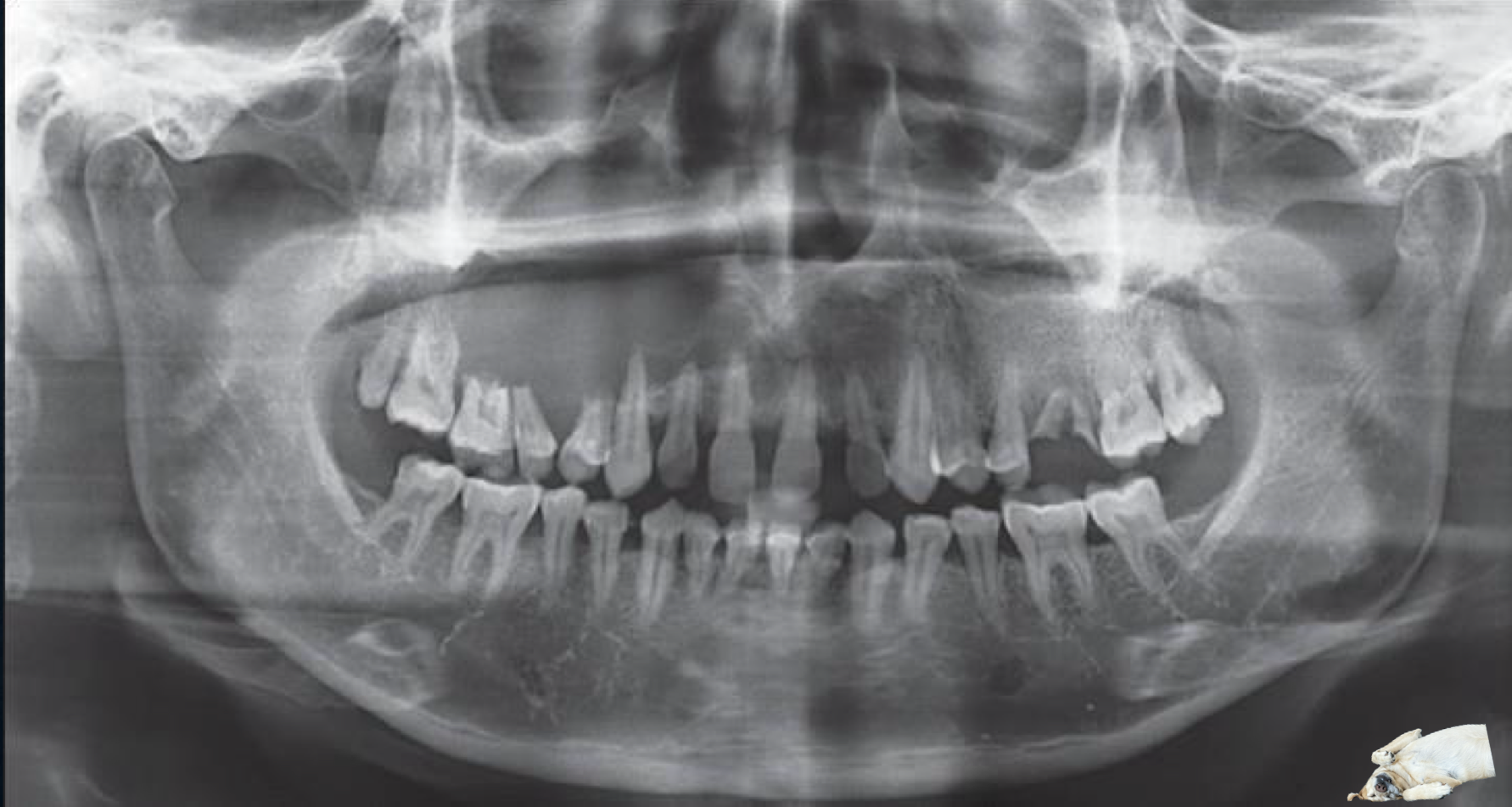
what is this?
ameloblastoma with luna
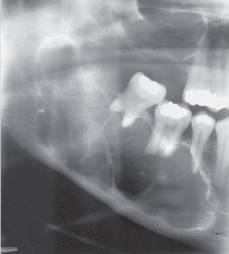
what is this?
ameloblastoma
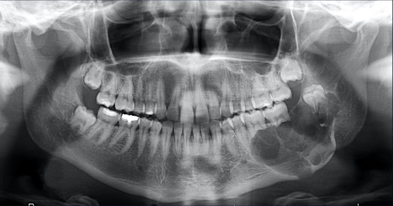
what is this?
ameloblastoma
what is this?
ameloblastoma, but on CBCT
what is this?
luna inside an ameloblastoma
what is this?
ameloblastoma
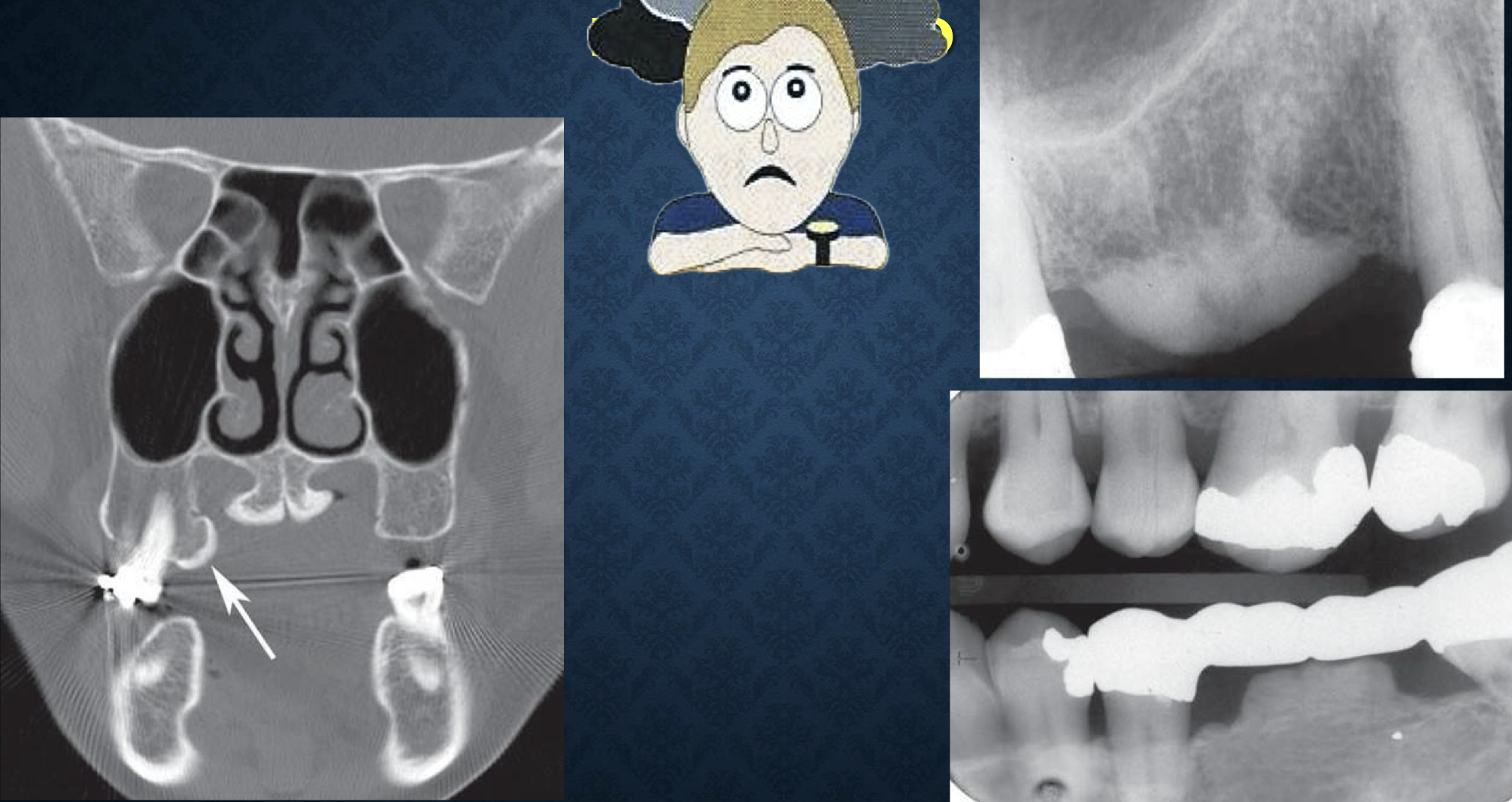
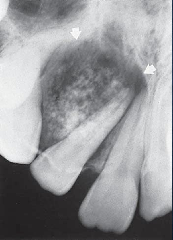
calcifying epithelial odontogenic tumor (CEOT)
aka pindborg tumor
rare, located within bone, produce mineralized substance
men>women, age 8-92, expansile lesion
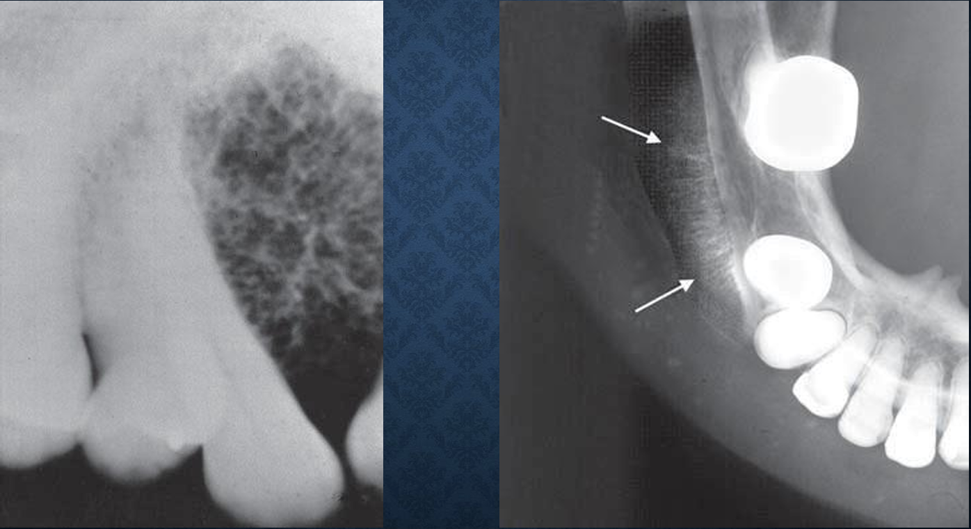
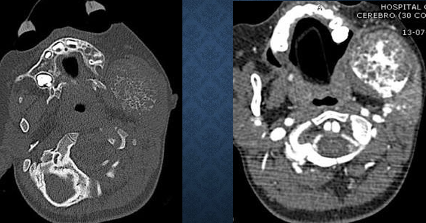
what are the radiographic features of CEOT?
mandible:maxilla 2:1, premolar to molar region, 52% with uninterrupted tooth
well defined cyst like cortex or irregular and ill defined
uni or multilocular, scattered radiopaque foci close to crown
can cause displacement of teeth or prevent eruption, expansile
what are differential diagnoses for CEOT?
adenomatoid odontogenic tumor, ameloblastic fibro-odontoma, dentigerous cyst
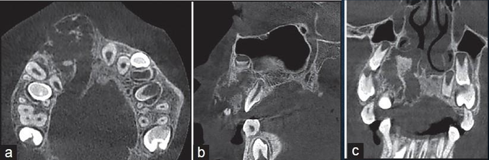
what is this?
an important image of a CEOT
what is this?
CEOT
what is this?
CEOT
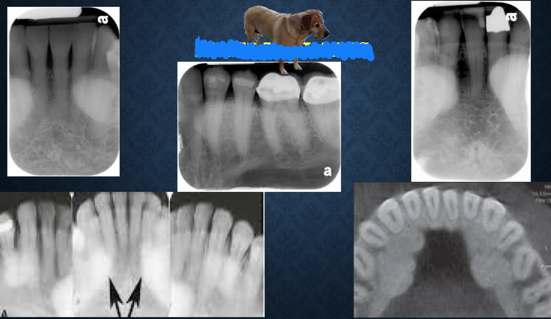
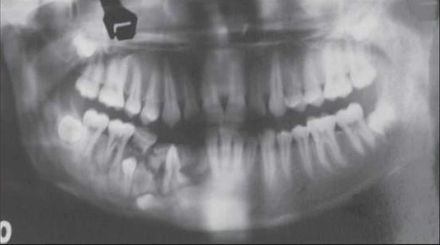
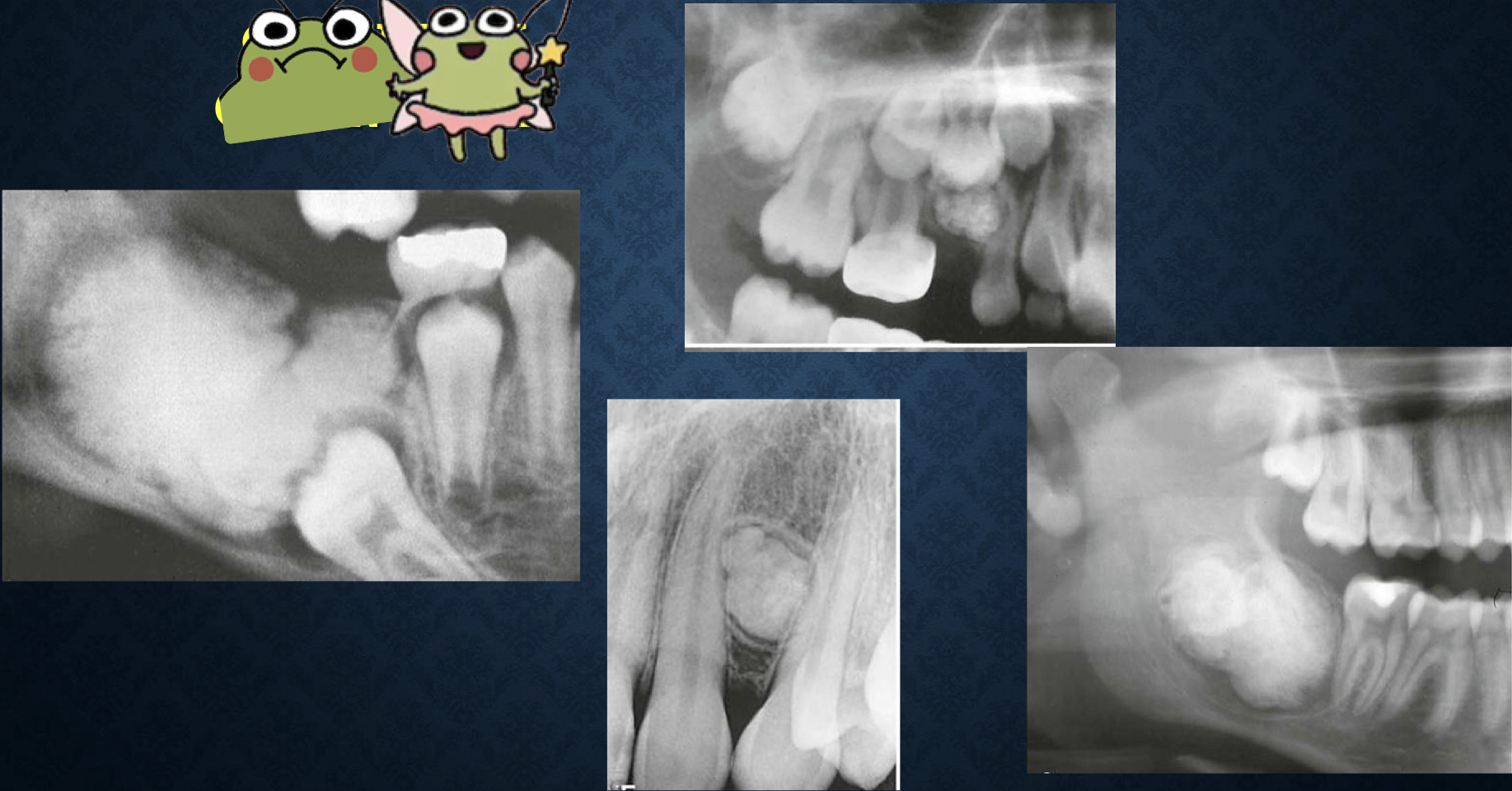
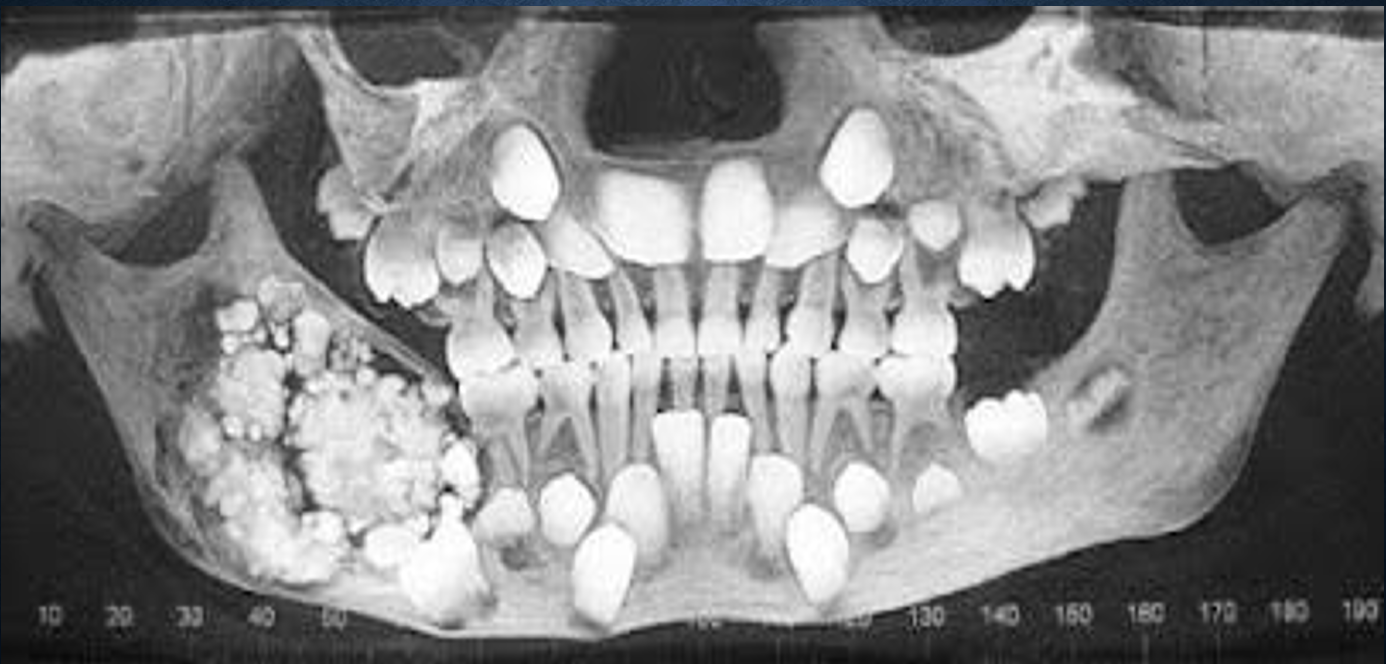
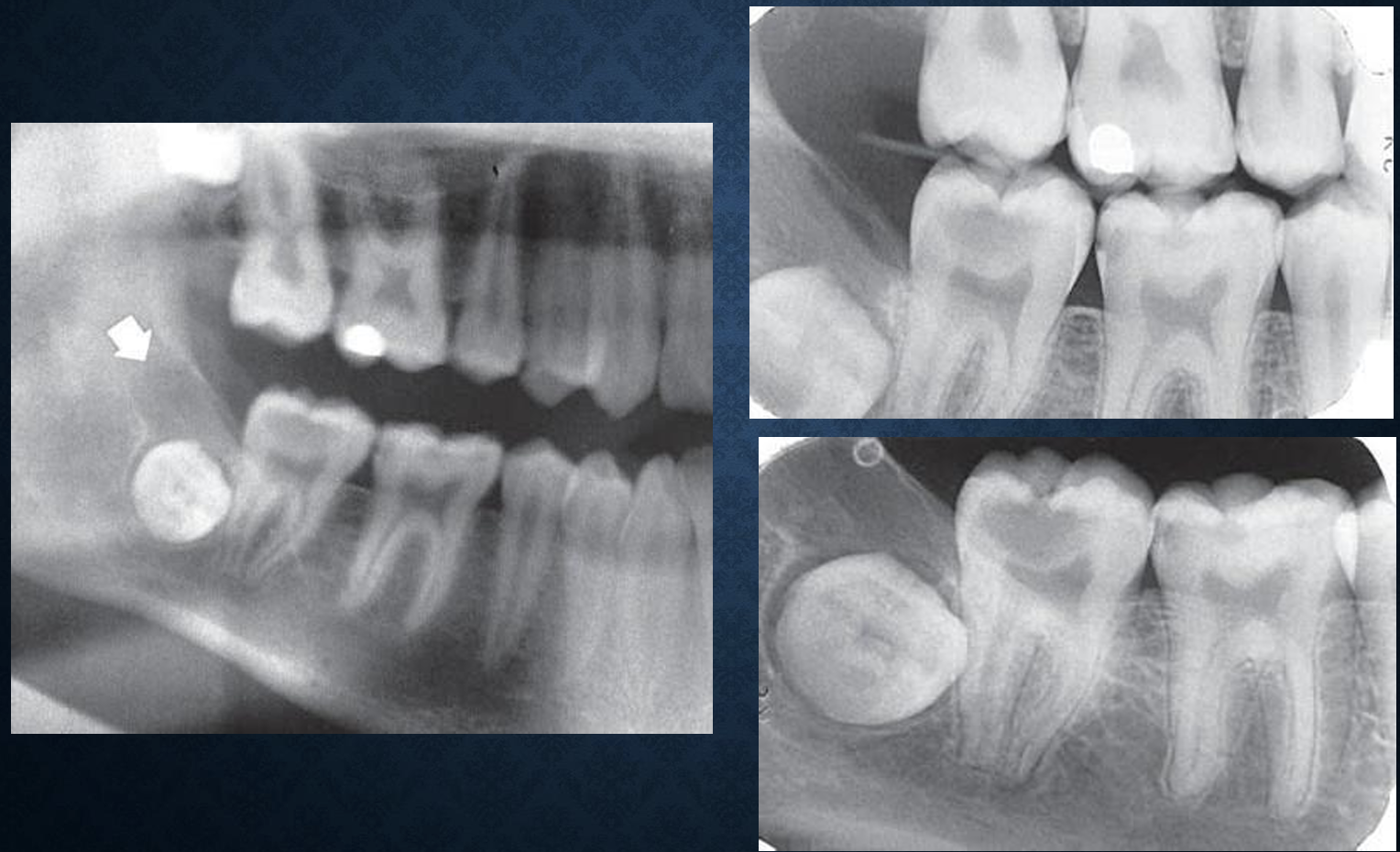
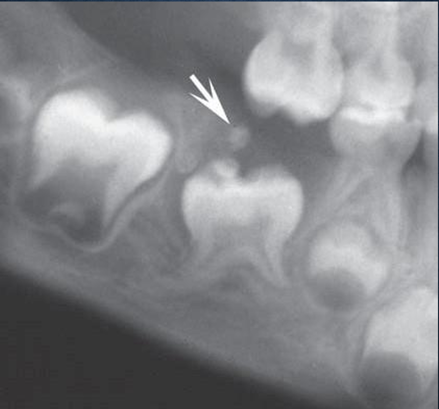
odontoma
aka compound odontoma, complex odontoma, odontogenic hamartoma
radiographically and histologically characterized by production of mature enamel, dentin, cementum, and pulp
compound or complex
interfere with eruption
compound odontoma
multiple, well defined teeth
found mostly in anterior maxilla with crown of unerupted teeth
complex odontoma
nondescript mass of dental tissue
found mostly in mandible, 1st and 2nd molars
what are the radiographic features of odontomas?
well defined, smooth or irregular, corticated border wtih a radiolucent area adjacent
radiopaque, differ based on type
can cause impaction, malpositioning, distema, or malformation
what are differential diagnoses of complex odontomas?
periapical cemental dysplasia
what is this?
complex odontoma
what is this?
odontoma
what is this?
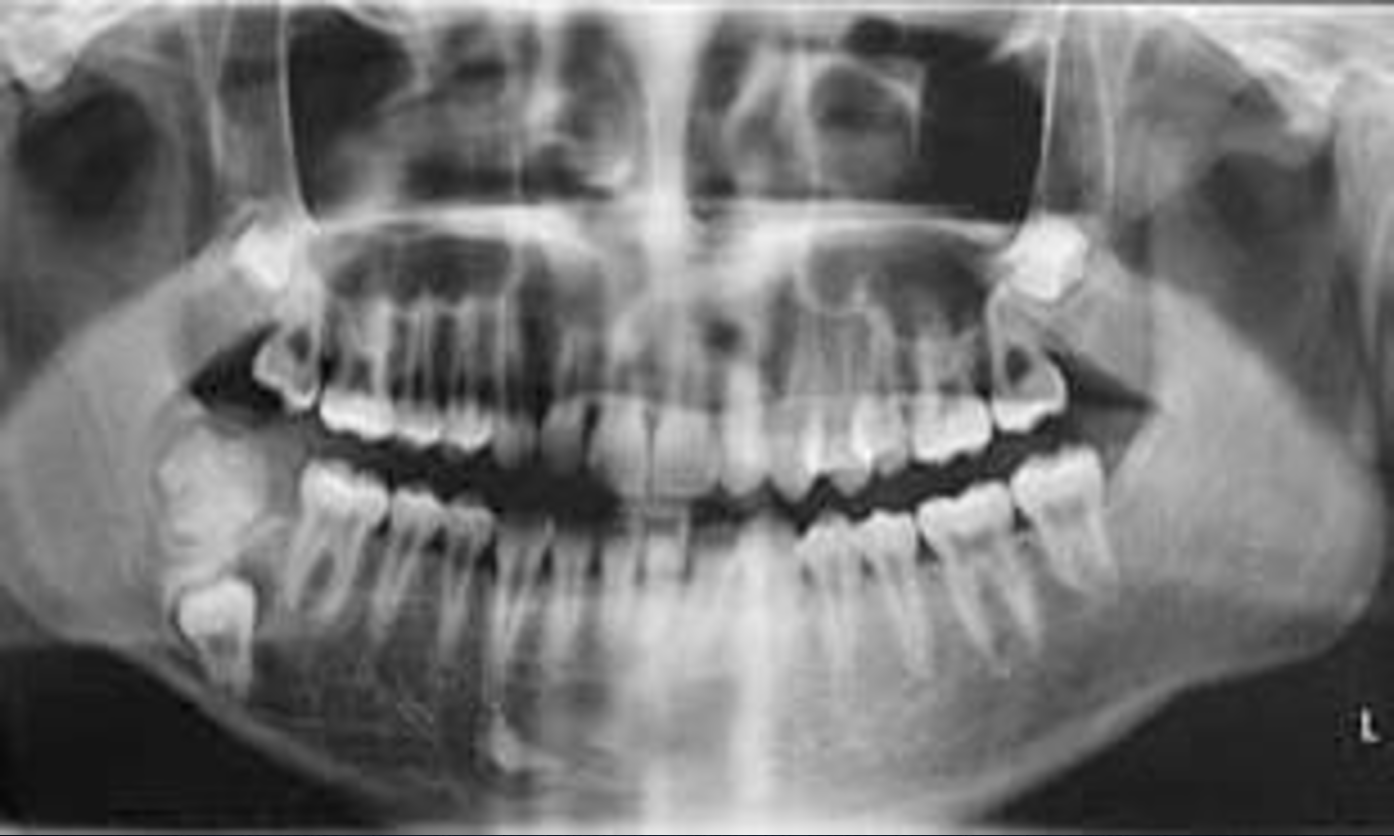
compound odontoma
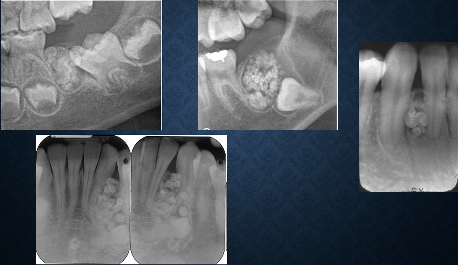
what is this?
compound odontoma
what is this?
compound odontoma
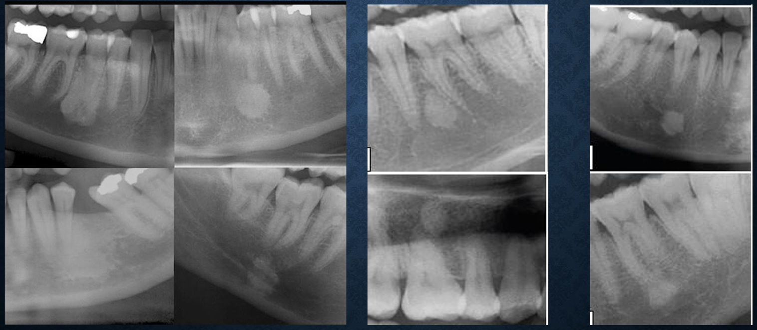
ameloblastic fibroma
aka fibroadamantoblastoma, granular cell ameloblastic fibroma, mixed odontogenic tumor
mixed odontogenic tumor characterized by neoplastic proliferation of the epithelium
age 5-20, missing tooth, painless slow growing
*age is the key
what are the radiographic features of ameloblastic fibroma?
mandible, premolar to molar region, follicular relationship with unerupted tooth
well defined and corticated with cystic manner
unilocular and in rare cases multilocular
in large lesions can expand the cortical plates, prevent the eruption and displace the tooth in apical direction
what are differential diagnoses for ameloblastic fibromas?
dentigerous cyst, hyperplastic follicle, ameloblastoma
what is this?
ameloblastic fibroma
ameloblastic fibro-odontoma
ameloblastic fibroma with enamel and dentin within the lesion
age 5-20, missing tooth
what are the radiographic features of ameloblastic fibro-odontomas?
posterior aspect of the mandible, epicenter is occlusal to the developing tooth
well defined and corticated
mixed lesion, radiolucent with radiopacities, impacted tooth
what are the differential diagnoses for ameloblastic fibro-odontomas?
ameloblastic fibroma, odontoma
what is this?
ameloblastic fibro-odontomas
what is this?
ameloblastic fibro-odontoma
what is this?
ameloblastic fibro-odontoma
adenomatoid odontogenic tumor
adenoameloblastoma and ameloblastic adenomatoid tumor
noaggressive tumors of odontogenic epithelium
age 5-50, 70% at 2nd decade, female > males 2:1, slow growing, painless, associated with missing teeth
what are the radiographic features of adenomatoid odontogenic tumors?
~75% in maxilla, incisor, canine, premolar region
well defined, corticated, or sclerotic border
mixed appearance, calcification seen with well/ill defined borders
can displace teeth, root resportion is rare, may inhibit too eruption
what are differential diagnoses for adenomatoid odontogenic tumors?
OKC, dentigerous cysts
what is this?
adenomatoid odontogenic tumor
what is this?
adenomatoid odontogenic tumor
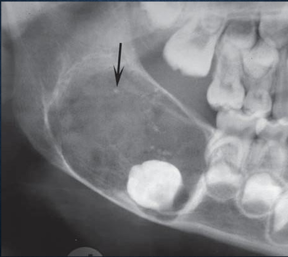
odontogenic myxoma
aka myxoma, myxofibroma, fibromyxoma
not encapsulated and intend to infiltrate the surrounding cancellous bone
slow growing, high recurrance rate
what are the radiographic features of odontogenic myxoma?
mandible:maxilla 3:1, premolar to molar region, rarely in ramus and condyle, maxilla: alveolar process at premolar to molar and zygomatic process
mmost often poorly defined margin
multilocular/unilocular, straight septa
can displace and loosen teeth, expansile
additional imaging with CT, particularly MRI
what are the differential diagnoses for odontogenic myxoma?
ameloblastoma, CGCG, central hemangioma
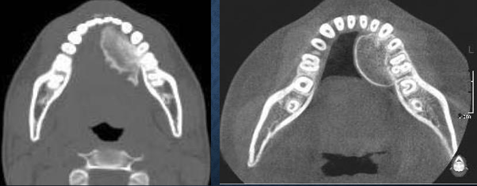
what is this?
odontogenic myxoma
what is this?
odontogenic myxoma
what is this?
odontogenic myxoma
benign cementoblastoma
aka cementoblastoma and true cementoma
slow growing, bulbous growth at the apex of the tooth root
males > females, age 12-65, tooth is vital and painful
what are the radiographic features of benign cementoblastoma?
mandible 78%, premolar and 1st molar ~90%
well defined radiopacity with corticated border
radiopaque internal structure
can cause external root resorption, expansion with intact cortical plates
what are differential diagnoses for benign cementoblastoma?
periapical cemental dysplasia, hypercementosis, periapical sclerosing osteitis
what is this?
benign cementoblastoma
what is this?
benign cementoblastoma
neurilemmoma
aka schwannoma
arising from schwann cell, no potential for malignancy
slow growing, swelling, paresthesia
what are the radiographic features of neurilemmoma?
mandible:maxilla 10:1, expanded IAC
margins are well defined, corticated, fusiform
internal structure is uniformly radiolucent, may have scalloping outline
can cause enlargement of the foramen, may cause root resorption
what are the differential diagnoses of neurilemmoma?
vascular lesions such as hemangioma or AV fistula
what is this?
neurilemmoma
what is this?
neurilemmoma
neuroma
aka amputation neuroma and traumatic neuroma
NOT a neoplasm, over-growth of severed nerve
slow growing reactive hyperplasia, that become larger >1cm, pain
what are the radiographic features of neuroma?
mental foramen > anterior maxilla > posterior mandible
well defined, corticated, varies in shape
internal structure is radiolucent
may cause some expansion of IAC
what are the differential diagnoses of neuroma?
NOT possible to differentiate this lesion from other neural lesions

what is this?
neuroma
*neurofibromatosis
aka von recklinghausen disease
syndrome with cafe au lait spots on the skin, multiple peripheral nerve tumors and other dysplastic abnormalities
most common genetic diseases, 2 major classifications NF-1 (generalized) and NF-2 (central)
*what are the radiographic features of neurofibromatosis?
alteration to the shape of the mandible
enlargement of the coronoid notch
an obtuse angle between body and ramus
deformity of condylar head
lengthening of condylar neck
lateral bowing and thinning of ramus
enlarged IAC, mental, and mandibular foramen
erosive changes to outer cortex
interference with normal eruption
what is this?
neurofibromatosis
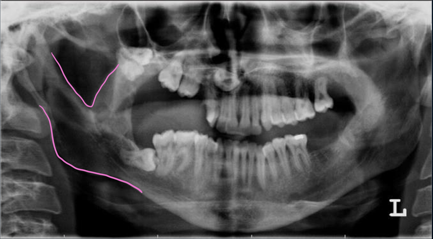
what is this, and what are the pink line demonstrating?
neurofibromatosis
obtuse angle of mandible and enlargement of the coronoid notch
osteoma
may arise from cartilage or embryonic periostium
asymmetry caused by the swelling and painless
cortical type in men, cancellous type in women
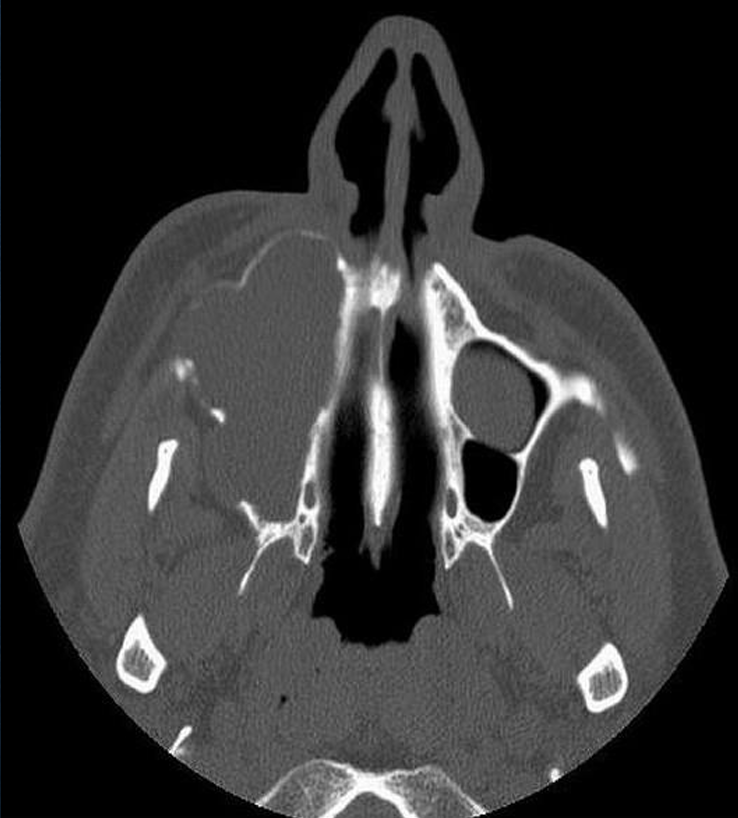
what are the radiographic features of osteoma?
mandible > maxilla, lingual side of the ramus of the mandible or inferior mandibular border apical to molars, condyle and coronoid process, paranasal sinus
well defined
interal structure is uniformly radiopaque
can cause displacement of adjacent structures
what are the differential diagnoses for osteoma of head of condyle and coronoid process?
osteochondroma, osteophytes or condylar hyperplasia
osteochrondroma
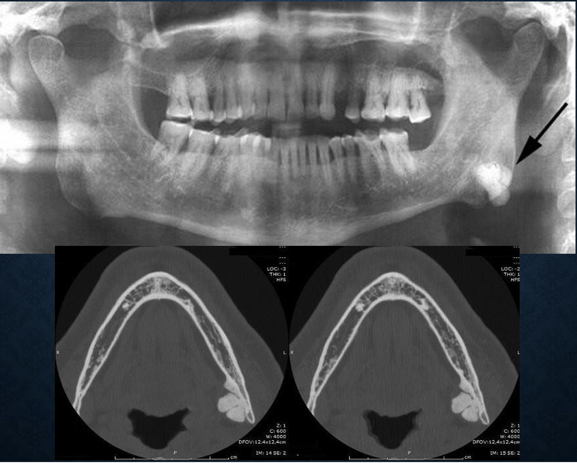
what is this?
osteoma
what is this?
osteoma
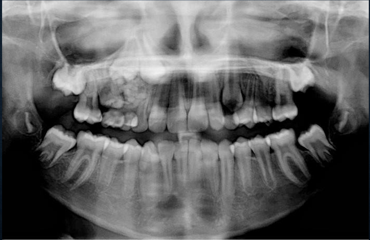
gardner’s syndrome
aka familial multiple polyposis
syndrome, hereditary, multiple osteomas, dense bone islands, epidermoid cysts, subcutaneous desmoid tumors and mutliple polyps of small and large intestine
what are the radiographic features of gardner’s syndrome?
multiple dense bone islands
osteomas most commonly seen in frontal, mandible, maxilla, and sphenoid bones
more than 5 dense bone islands you should consider a syndrome
multiple unerupted teeth permanent and supernumerary
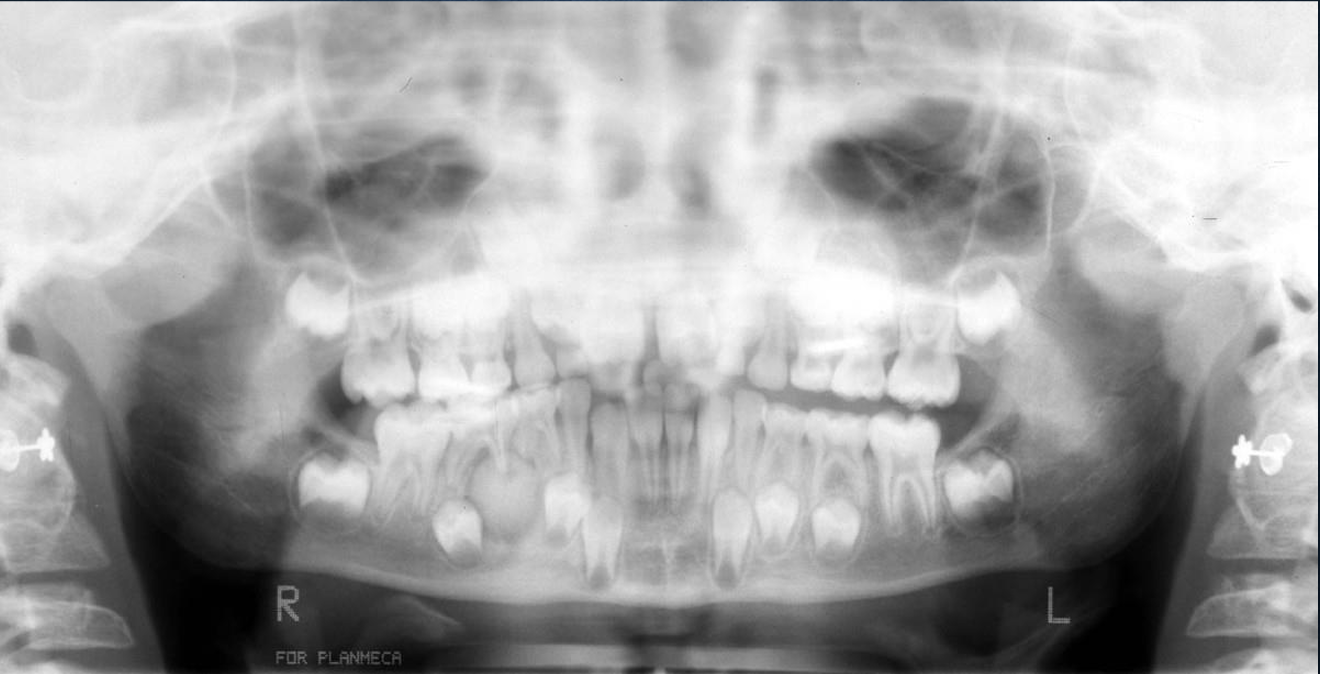
what is this?
gardner’s syndrome
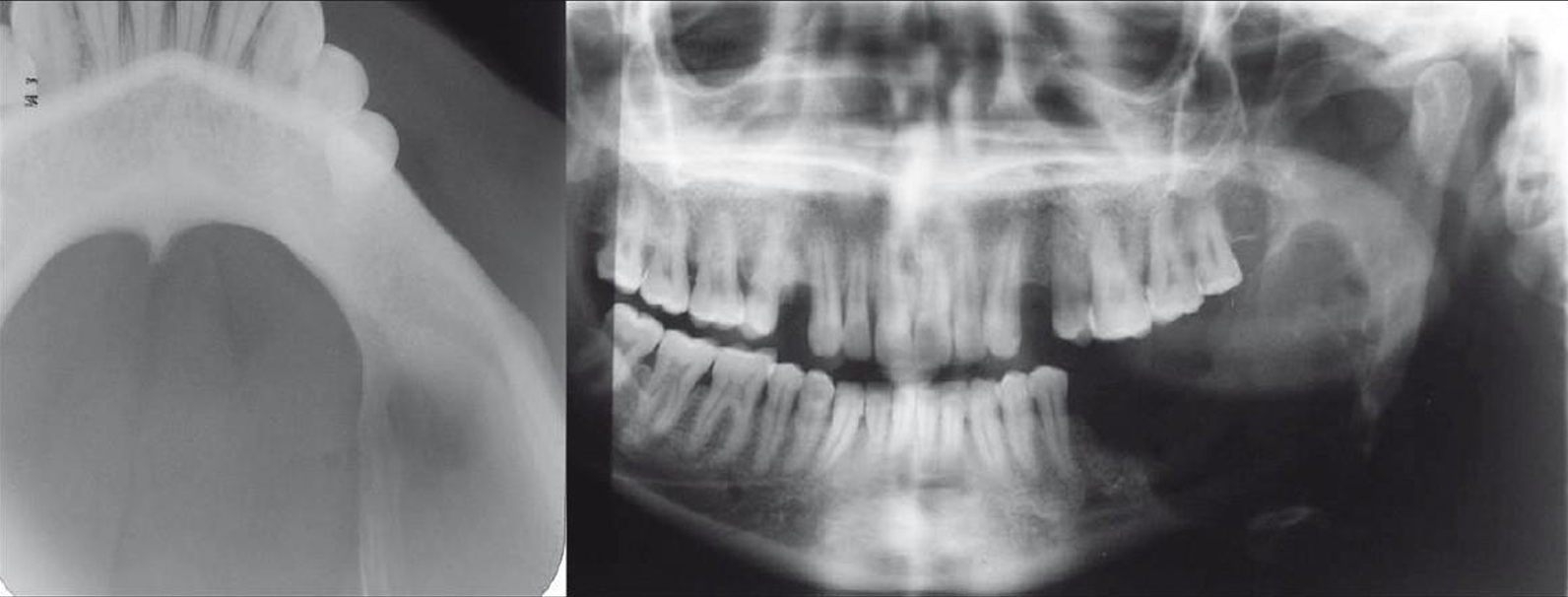
what is this?
gardner’s syndrome
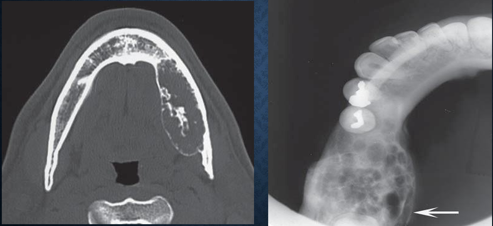
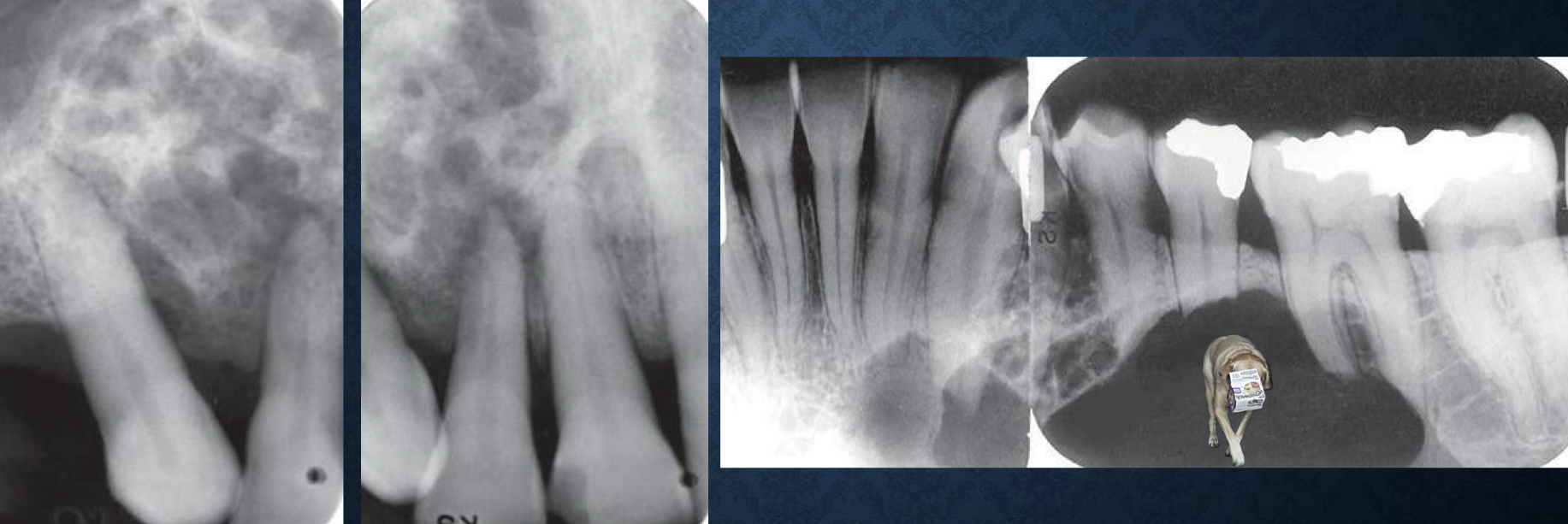
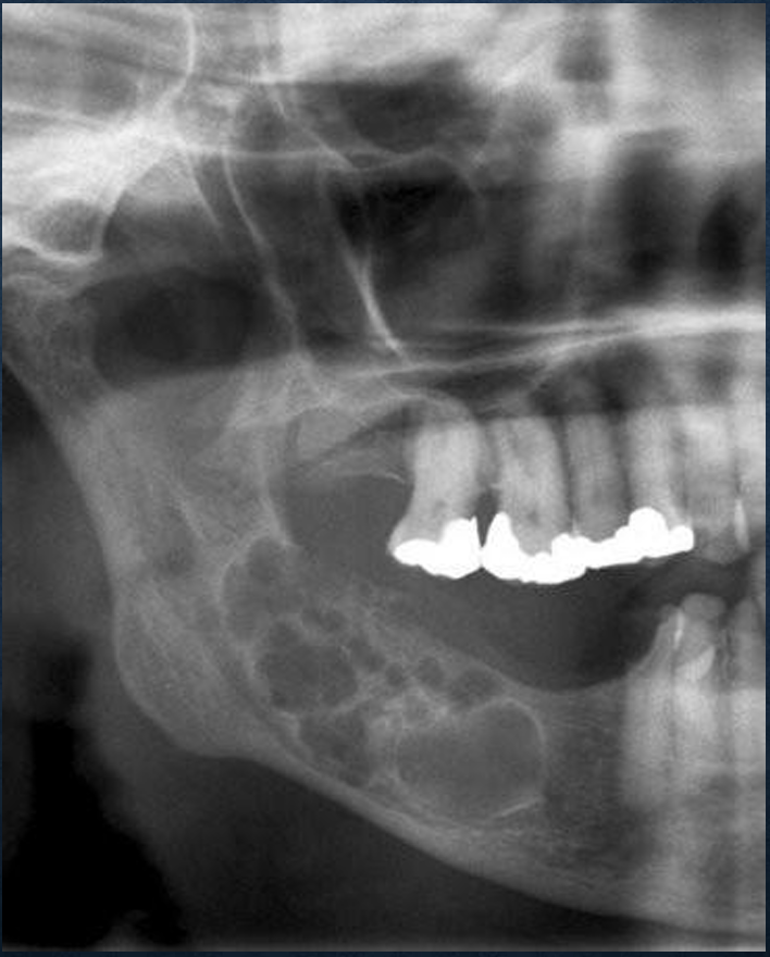
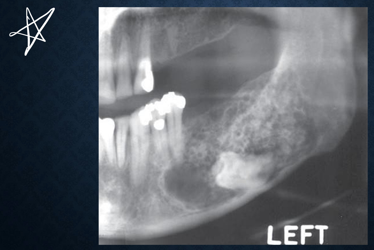
central hemangioma
proliferation of blood vessels creating a mass, resembling a neoplasm, central type is intraosseous
females > males, slow growing, non tender, expansile, bruit detected on auscultation
what are radiographic features of central hemangioma?
mandible > maxilla 2:1, posterior body and ramus within IAC
may be well defined/ill defined, linear spicules of bone emanating with periostium displaced
internal has mixed appearance, coarse internal trabeculae
can resorb or displace teeth, enlargement of IAC and mental foramen, premature eruption
further imaging with MRI, CT with contrast, conventional angiography
what are differential diagnoses of central hemangioma?
myxoma, osteogenic sarcoma
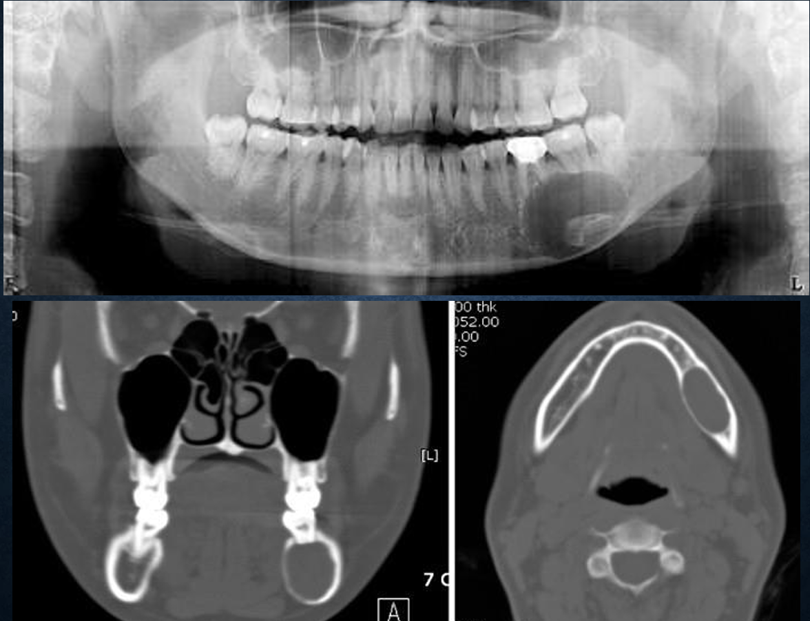
what is this?
central hemangioma
what is this?
central hemangioma
what is this?
central hemangioma
ossifying fibroma
aka cemento-ossifying fibroma, cementifying fibroma, juvenile aggressive ossifying fibroma
indolent to aggressive behavior, usually in young adults, females > males
what are radiographic features of ossifying fibroma?
mandible, premolar to molar area
well defined
internal structure is mixed, radiolucent, or radiopaque
can cause displacement of adjacent structures, root resorption
what are the differential diangoses for ossifying fibroma?
fibrous dysplasia, POD