Eye
1/16
Earn XP
Description and Tags
Parts of the eyeball
Name | Mastery | Learn | Test | Matching | Spaced | Call with Kai |
|---|
No analytics yet
Send a link to your students to track their progress
17 Terms
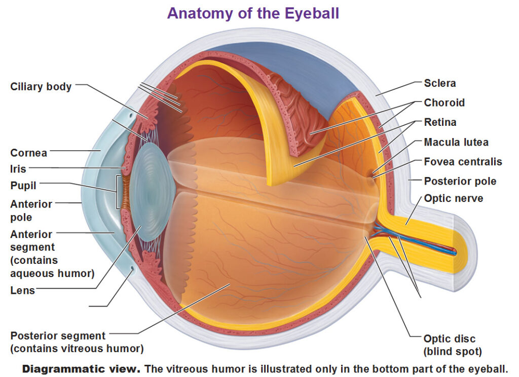
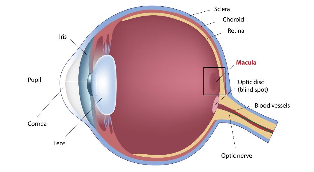
Choroid
A vascular, thin tissue layer located between the retina and the sclera.


Ciliary body
A ring-shaped structure in the eye located behind the iris


Cornea
A transparent, dome-shaped outer layer that acts as a shield

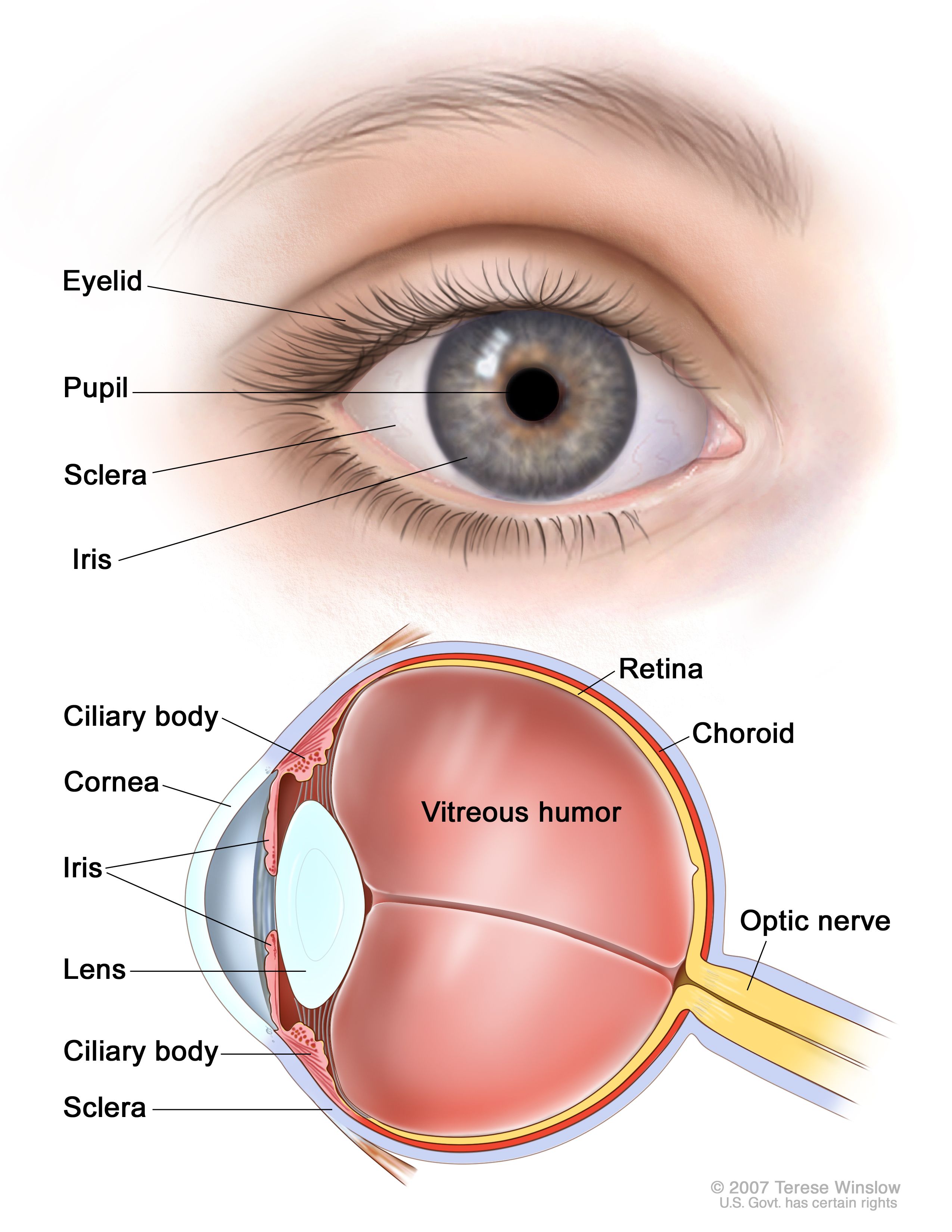
Iris
Colored part of the eye


Lens
A transparent, biconvex, and flexible structure located behind the iris

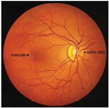
Macula
A small, central area of the retina responsible for sharp, detailed, and color central vision

Fovea Centralis
A tiny, central pit located in the macula of the retina
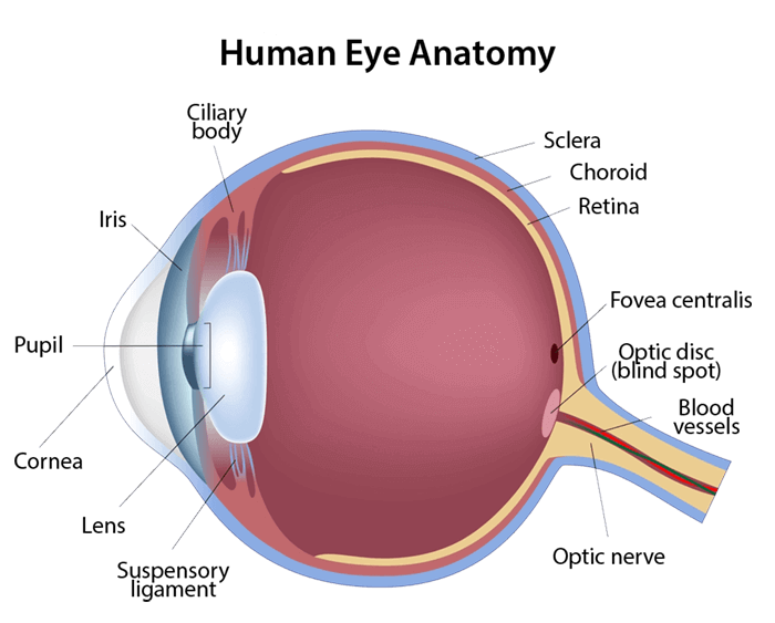

Optic Disk
A raised spot on the retina where nerve fibers meet to form the optic nerve


Optic Nerve
A bundle of nerve fibers that transmits visual information from the retina to the brain


Pupil
Black circular opening in the center of the eye


Retina
A layer of photoreceptors cells and glial cells within the eye


Pigmented epithelium
A single layer of hexagonal, pigmented cells located between the retina and the choroid in the eye
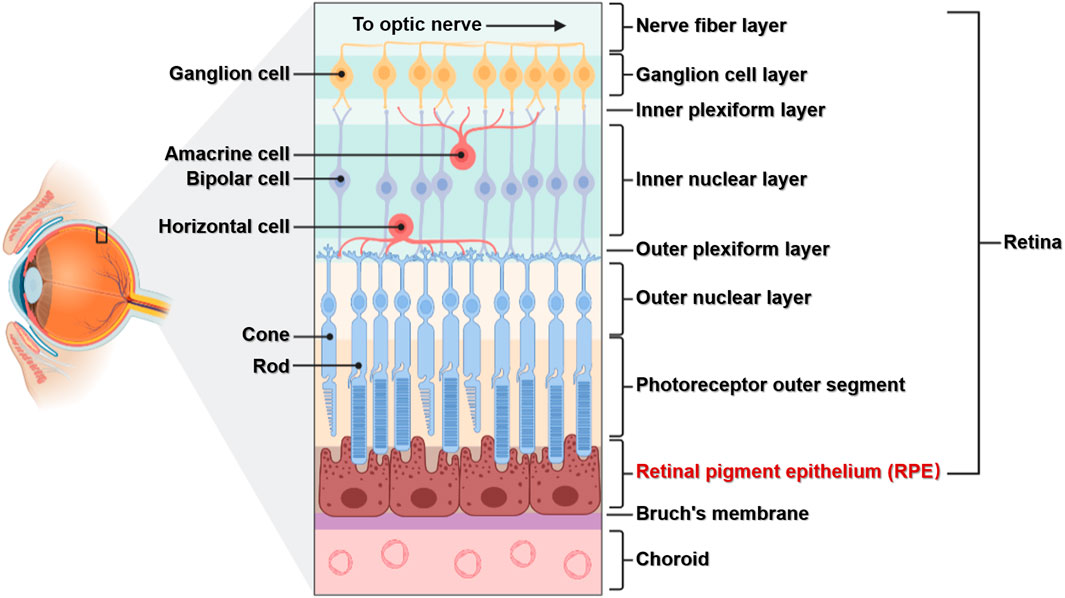

Photoreceptor cell layer
Rods and cones


Bipolar cell layer
Inner nuclear layer (INL) of the retina

Ganglion cell layer
The innermost layer of the retina, containing the cell bodies of retinal ganglion cells (RGCs)


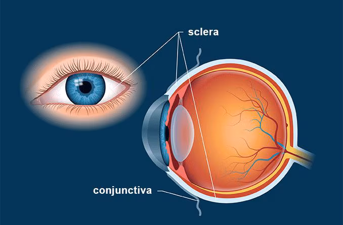
Sclera
The white of the eye
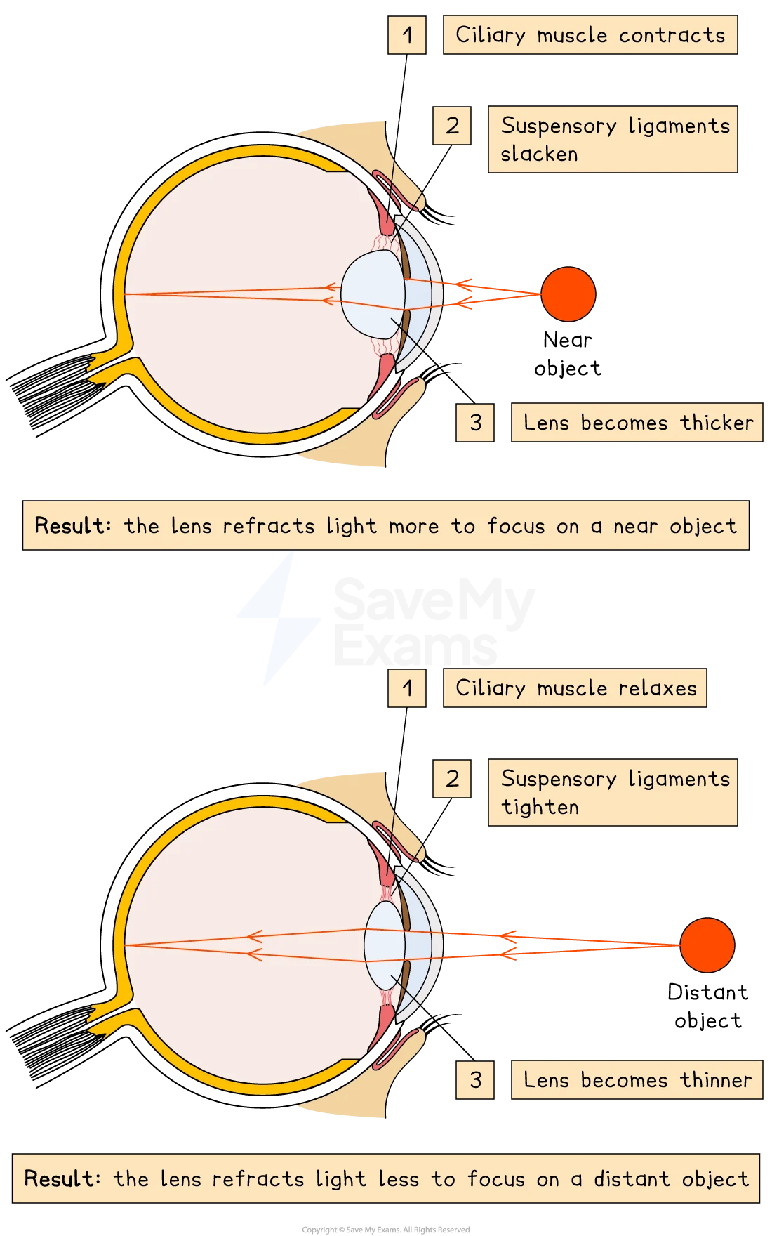
Suspensory Ligaments
Attached below and above the lens that aids in the dilation and contraction phases.
