Unit 4 Senses Combined Terms
1/110
There's no tags or description
Looks like no tags are added yet.
Name | Mastery | Learn | Test | Matching | Spaced | Call with Kai |
|---|
No analytics yet
Send a link to your students to track their progress
111 Terms
Peripheral nervous system
the sensory and motor neurons that connect the CNS to the rest of the body
Stimulus
change that is detectable by the body
Response
a reaction to a stimulus
Sensory Receptor
neuron that reacts to a specific stimulus by sending impulses to other neurons and eventually to the central nervous system
Photoreceptors
respond to light
Mechanoreceptors
respond to touch, pressure, vibration, and stretch
Thermoreceptors
detect changes in temperature
osmoreceptors
detect the osmotic pressure of body fluids
chemoreceptors
respond to chemicals
Nocireceptors
pain receptors, sense tissue damage or intense stimulation of any receptor
special senses
vision, hearing, taste, smell, equilibrium
general senses
Somatic and visceral- temperature, pain, touch, pressure, vibration, proprioception
somatic senses
touch, temperature, pain, proprioception
visceral senses
information about conditions within internal organs
perception
The act of becoming aware through the senses
adaptation
decreased receptor response during prolonged stimulation
free nerve endings
pain and temperature, itch
encapsulated nerve endings
dendrites enclosed in connective tissue capsule for pressure, vibration, and some touch sensations
separate specialized cells
hair cells in inner ear, photoreceptors in retina of eye
touch receptors (examples)
meissner corpuscles, hair root plexus, merkel disc or tactile discs, ruffini corpuscles
pressure receptors (examples)
Pacinian corpuscles
Vibration receptors (receptors)
Meissner and Pacinian
Gustation
sense of taste
5 primary tastes
sweet, salty, bitter, sour, umami
Location of taste buds (larger organs)
tongue, pharynx and epiglottis
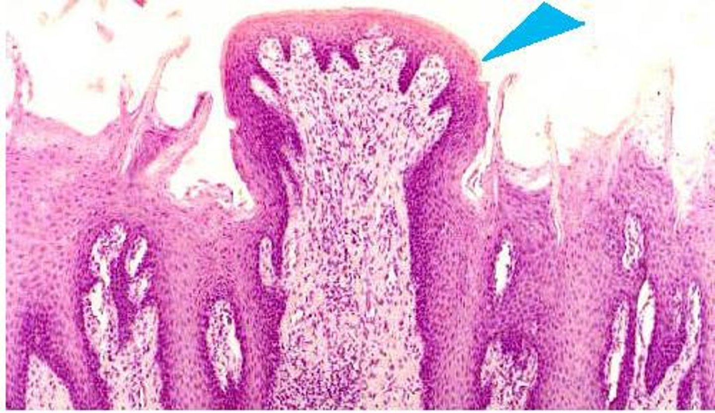
Papillae
Structures on the tongue that contain taste buds
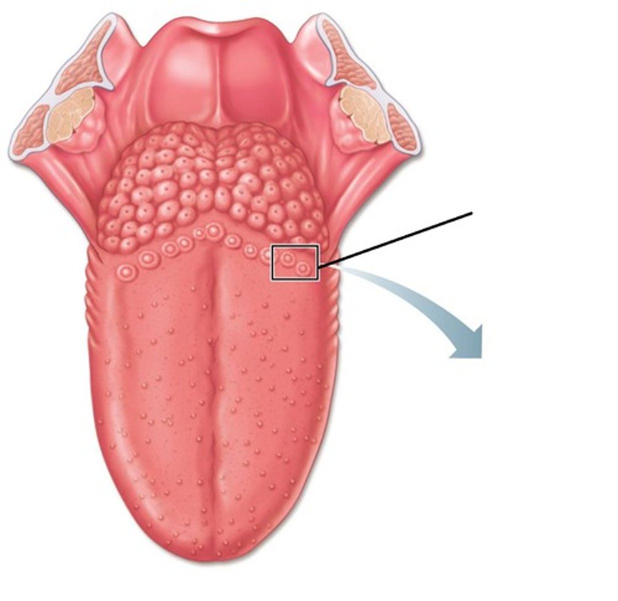
Vallate papillae
Posterior, arranged in the form of a V
Fungiform papilla
located allover tongue, rounded
filiform papillae
Only touch receptors
gustatory receptor cells
sensory cells in the taste bud that transduce the chemical stimuli of gustation
Gustatory hair
Microvilli, project through taste pore

Basal cells
stem cells that replace taste cells every 7 to 10 days
Saliva
digestive juice produced by salivary glands, dissolved tastant
Sequence of stimulation of taste receptors
Tastant dissolves in saliva →
Enters taste pore → contacts gustatory hair→
Electrical signal produced →
Causes gustatory cell to release neurotransmitter
That activates dendrites of first-order neurons
Gustatory pathway
Cranial nerves transmit impulses
Facial (CN VII) from anterior of tongue
Glossopharyngeal (CN IX) from posterior
Vagus (CN X) from pharynx, epiglottis
To medulla oblongata
→ Thalamus → primary gustatory area of cerebral cortex
→Limbic system or hypothalamus
Cranial Nerves involved in taste
facial, glossopharyngeal, vagus
Limbic system
neural system located below the cerebral hemispheres; associated with emotions and drives
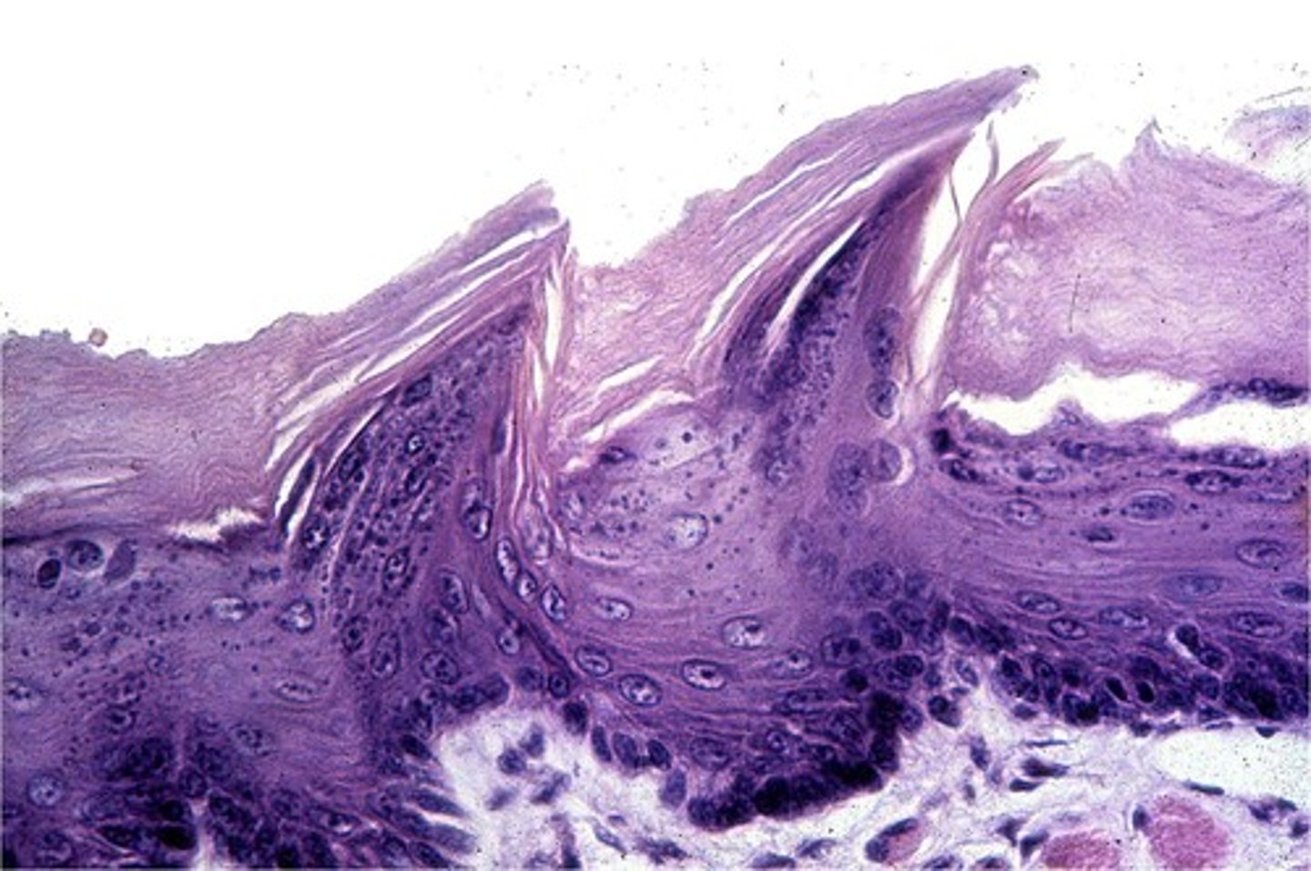
Olfaction
sense of smell
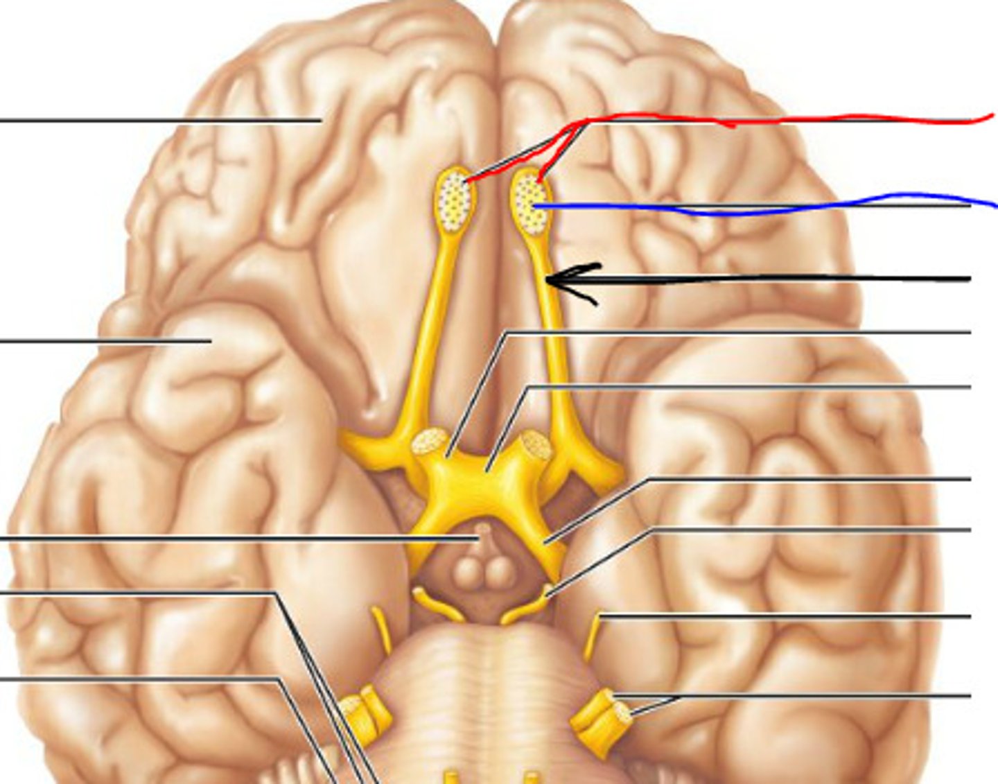
Parts of the brain involved in olfaction
temporal lobe of the cerebral cortex, hypothalamus, limbic system
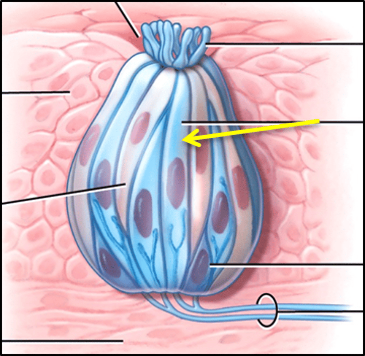
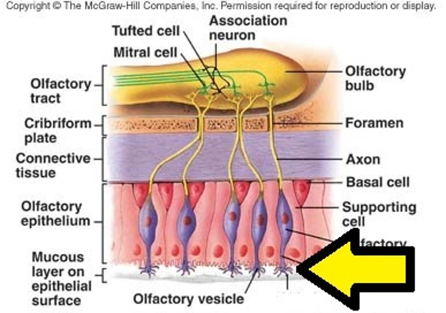
Olfactory receptors
nerve endings that act as the receptors for the sense of smell
Supporting cells
cells that insulate, support and protect neurons
Basal Cells
stem cells that produce new neurons (rare)
nasal mucosa
the lining of the nose, contains olfactory receptors
Olfactory bulb
a brain structure located above the nasal cavity beneath the frontal lobes
In order to be smell substances must be
Volatile
Water soluble
Lipid soluble
Volatile
able to evaporate easily
water soluble
able to be dissolved in water
lipid soluble
able to be dissolved in fats
odorant molecules
volatile chemicals that bind to receptor proteins in olfactory neurons to stimulate the sense of smell
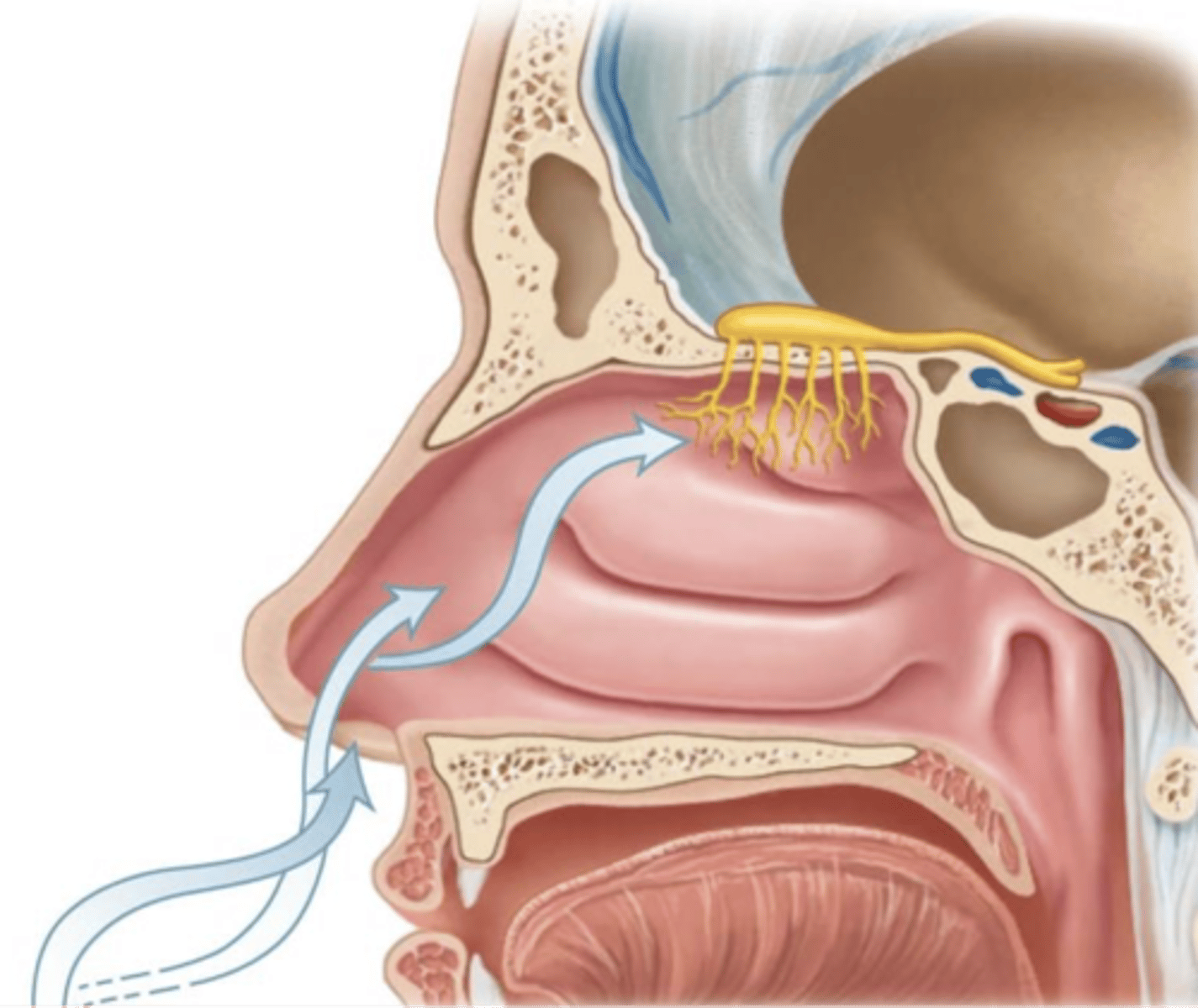
Pathway of smell
Olfactory cells convey nerve impulses to the olfactory nerves, olfactory bulbs, olfactory tracts, cerebral cortex, limbic system, and the hypothalamus
Olfactory hairs
cilia that extend from the olfactory receptor cells into the nasal cavity
chemoreceptors
respond to chemicals
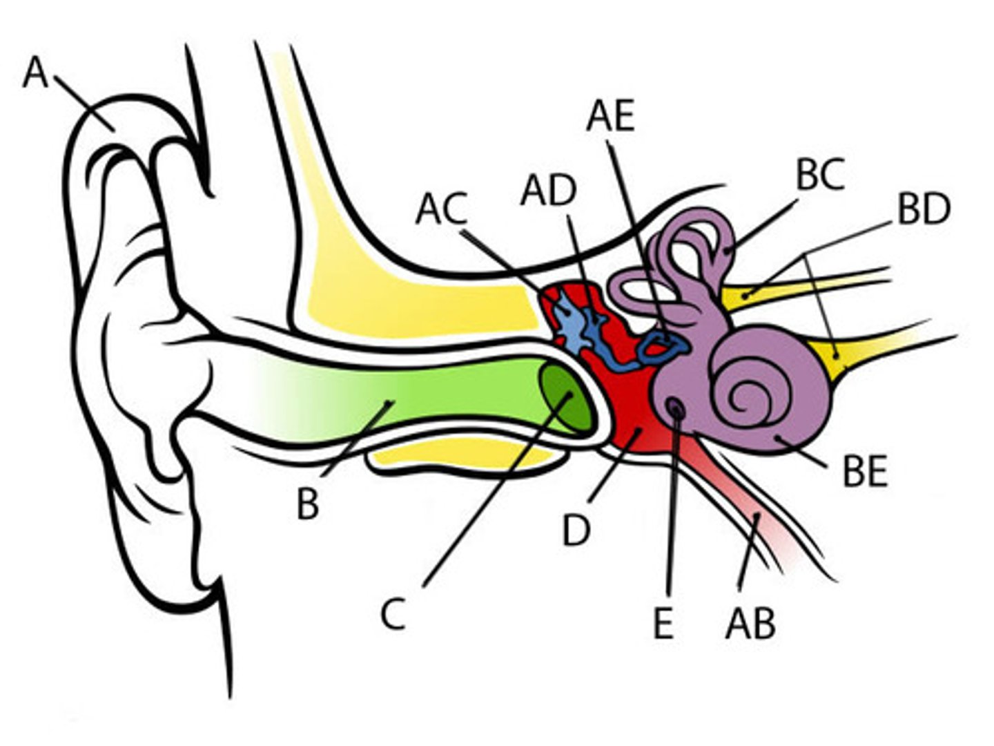
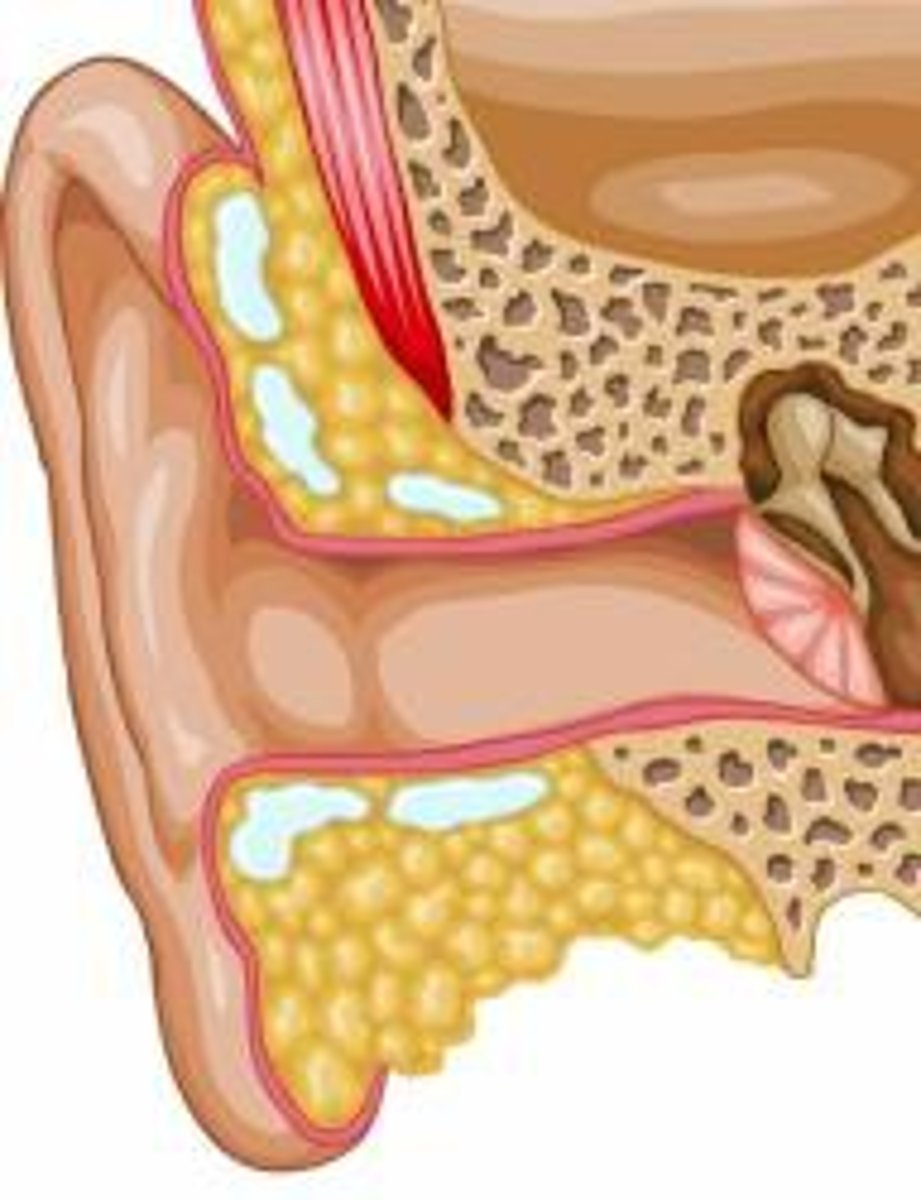
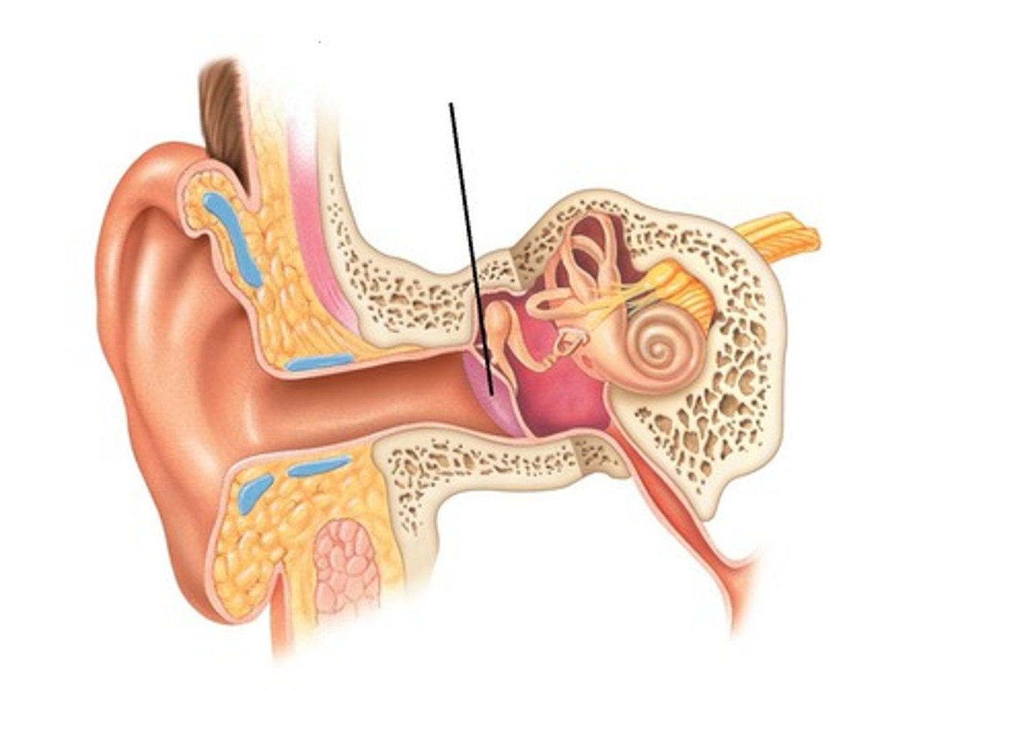
auricle (pinna)
outer ear
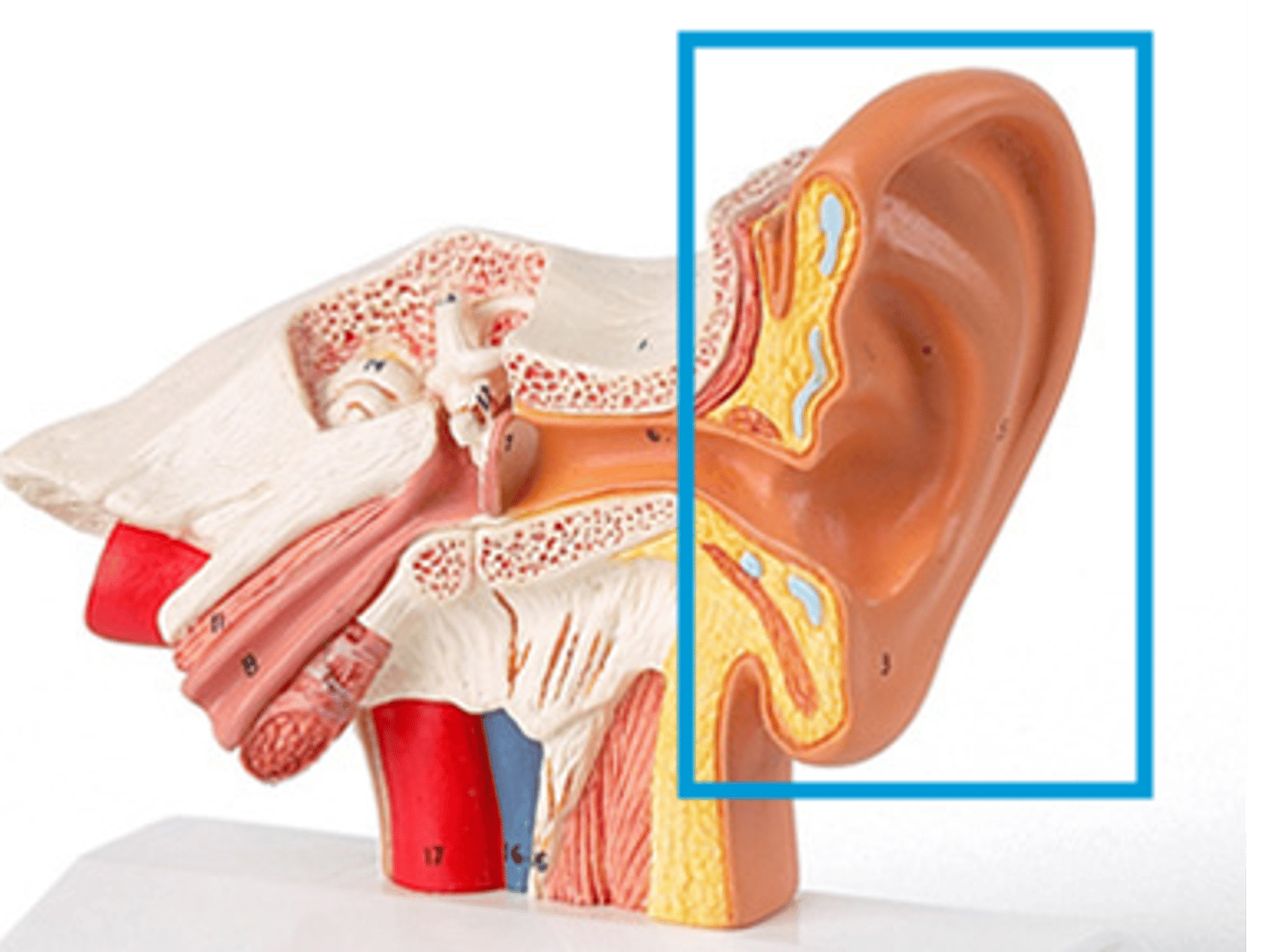
External auditory canal (meatus)
channel that leads from the pinna to the eardrum
tympanic membrane
The eardrum. separates the outer ear from the middle ear and vibrates in response to sound waves
Semicircular canals
three fluid-filled canals in the inner ear responsible for our sense of balance
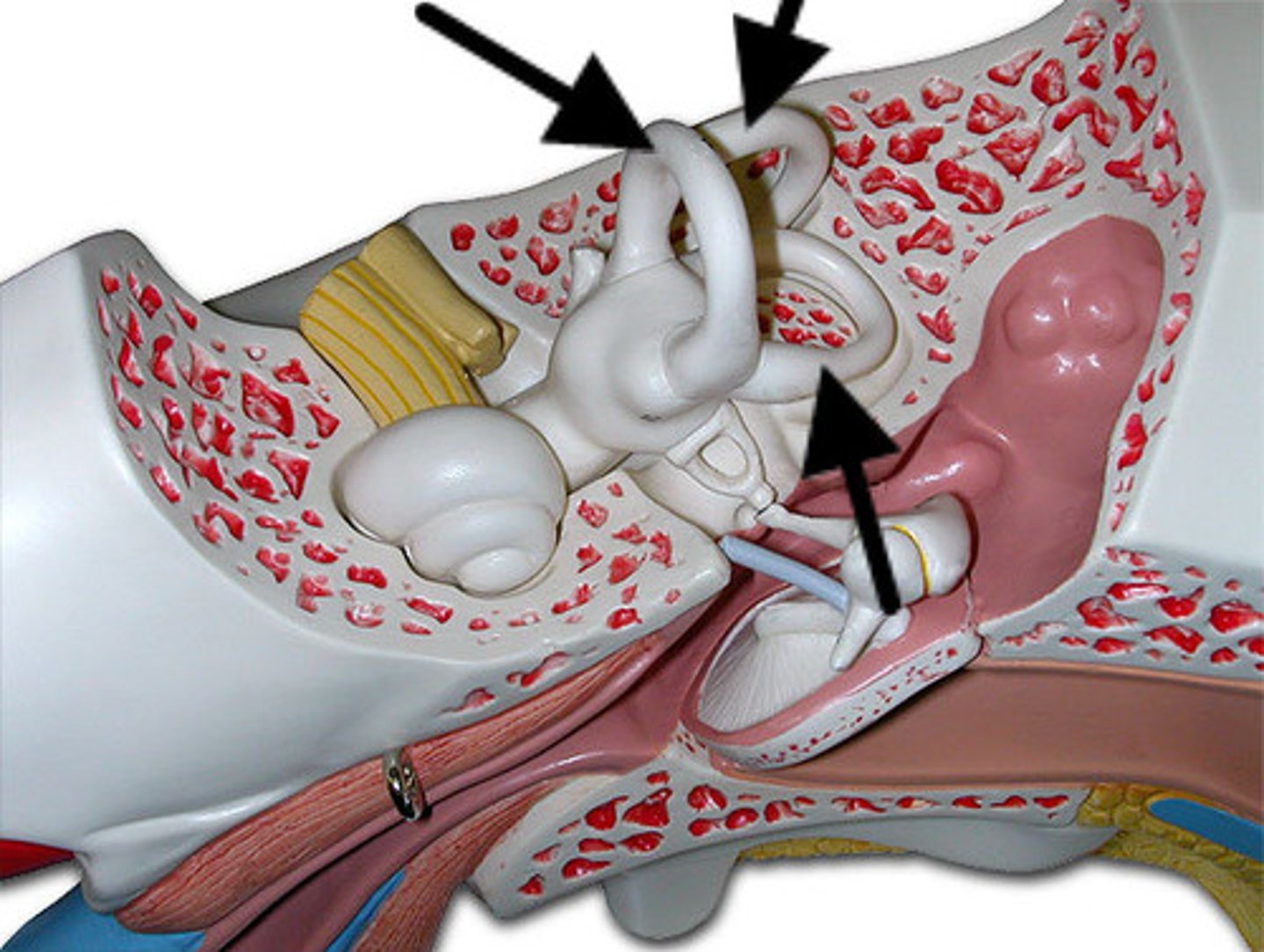
Ossicles
malleus, incus, stapes; three tiny bones in the middle ear
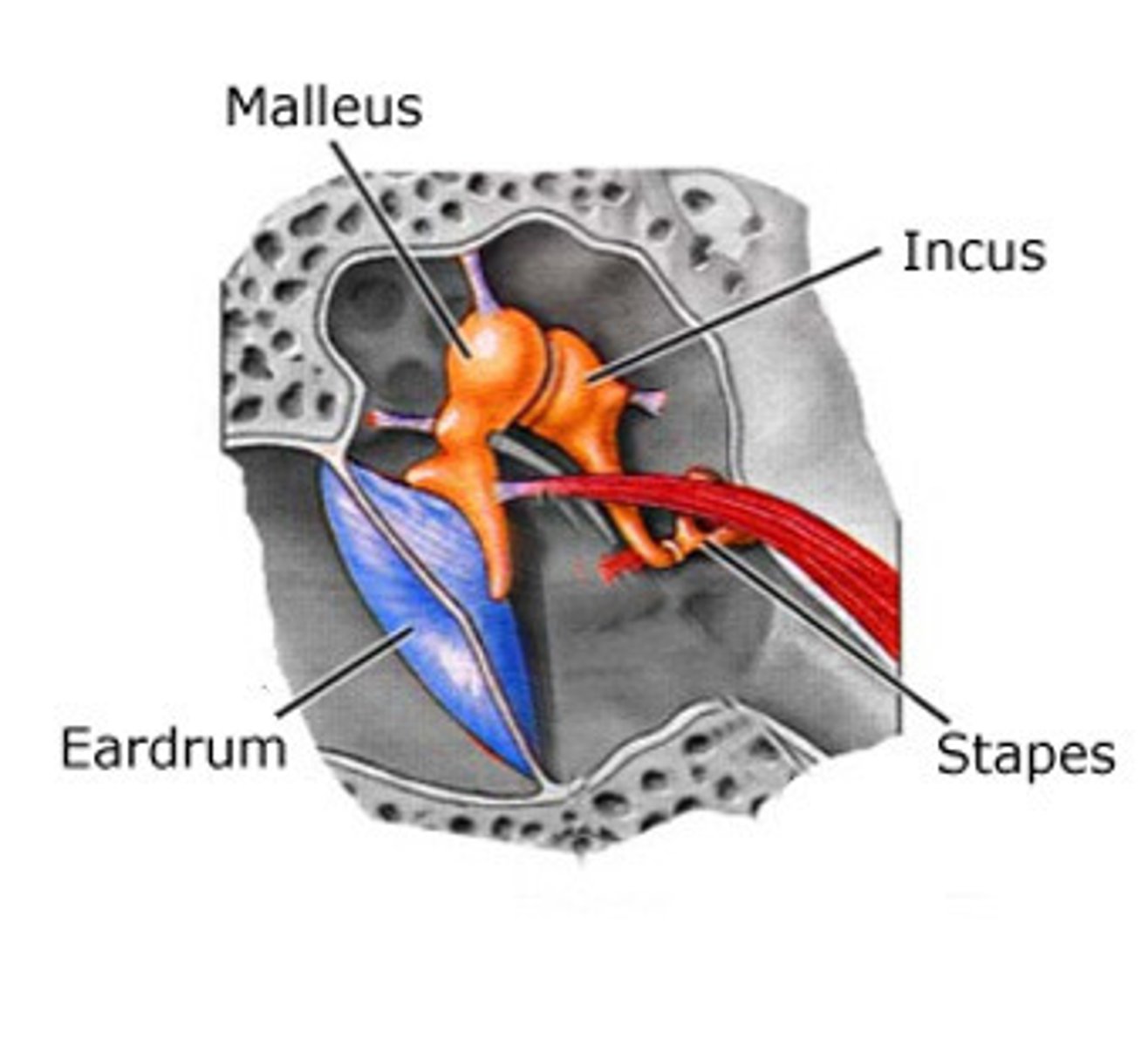
malleus
hammer; first of the three auditory ossicles of the middle ear
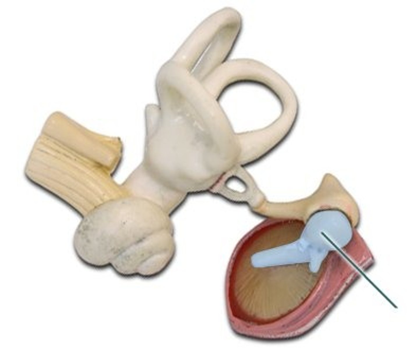
incus
anvil; middle of the three auditory ossicles of the middle ear
Stapes
stirrup; last of the three auditory ossicles of the middle ear (Smallest bone in the body)
Cochlea
a coiled, bony, fluid-filled tube in the inner ear through which sound waves trigger nerve impulses
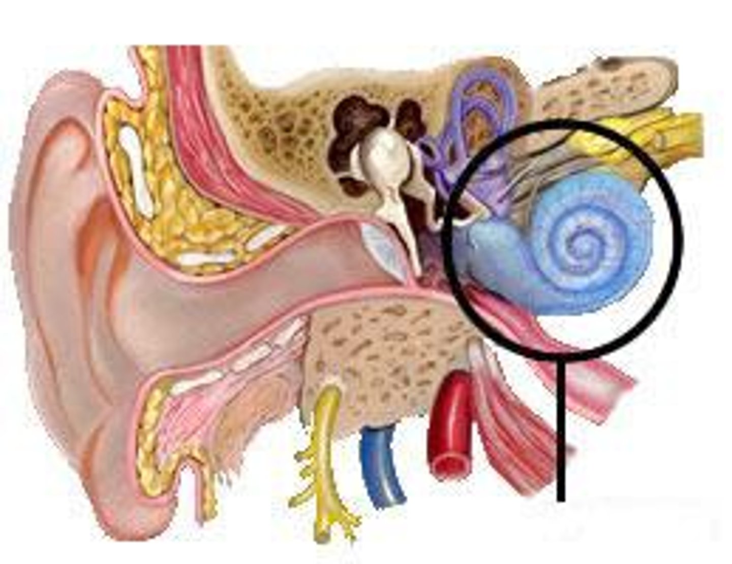
Vestibular cochlear nerve
transmits signals from the cochlea to the brain
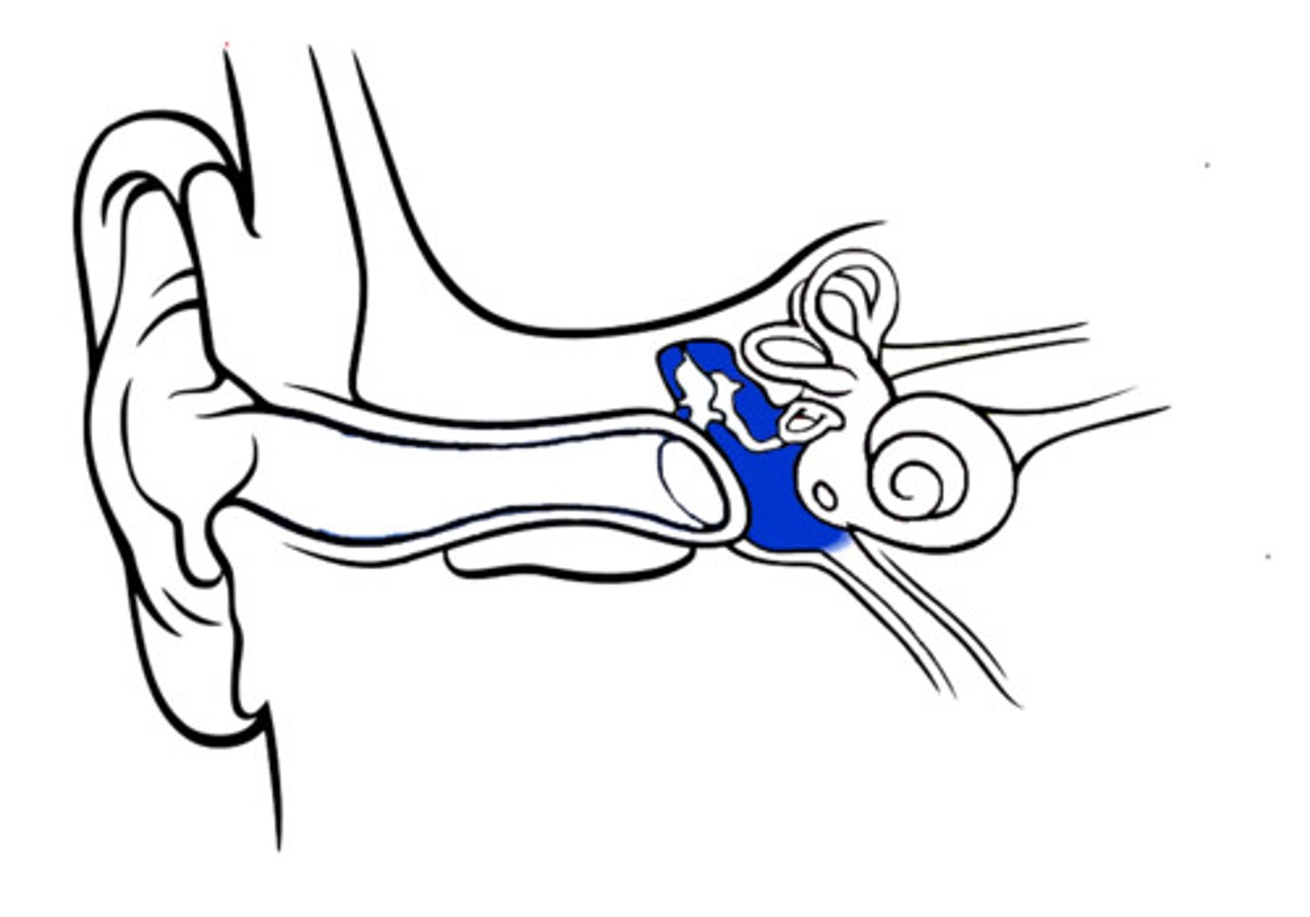
Tympanic cavity
opening of middle ear
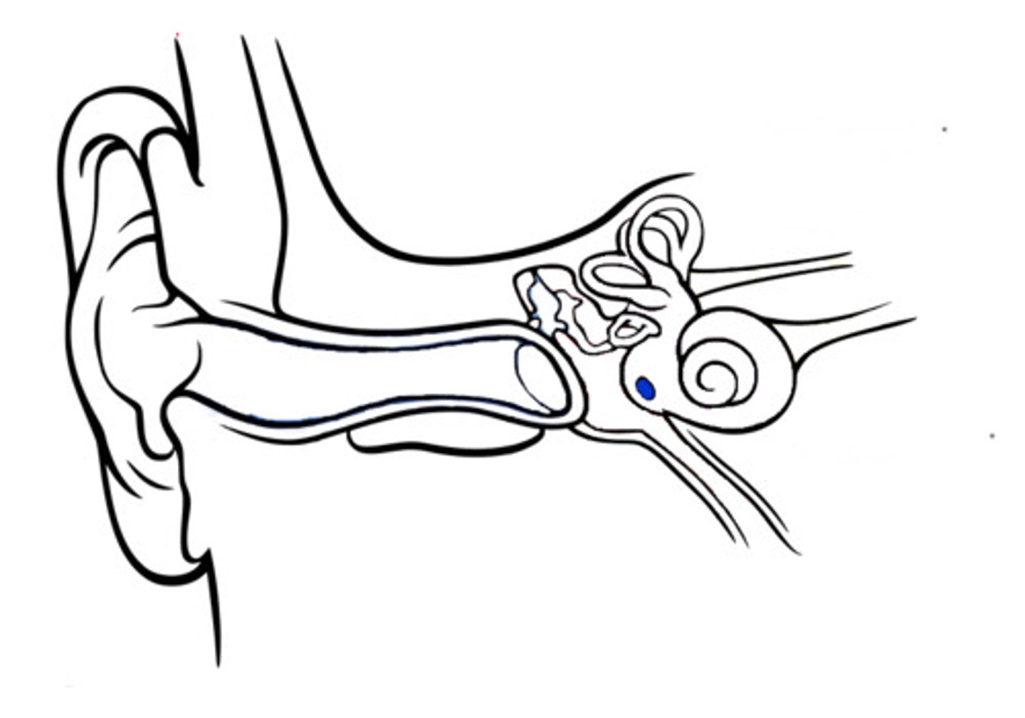
Round window
The membrane that relieves pressure from the vibrating waves in the cochlear fluid.
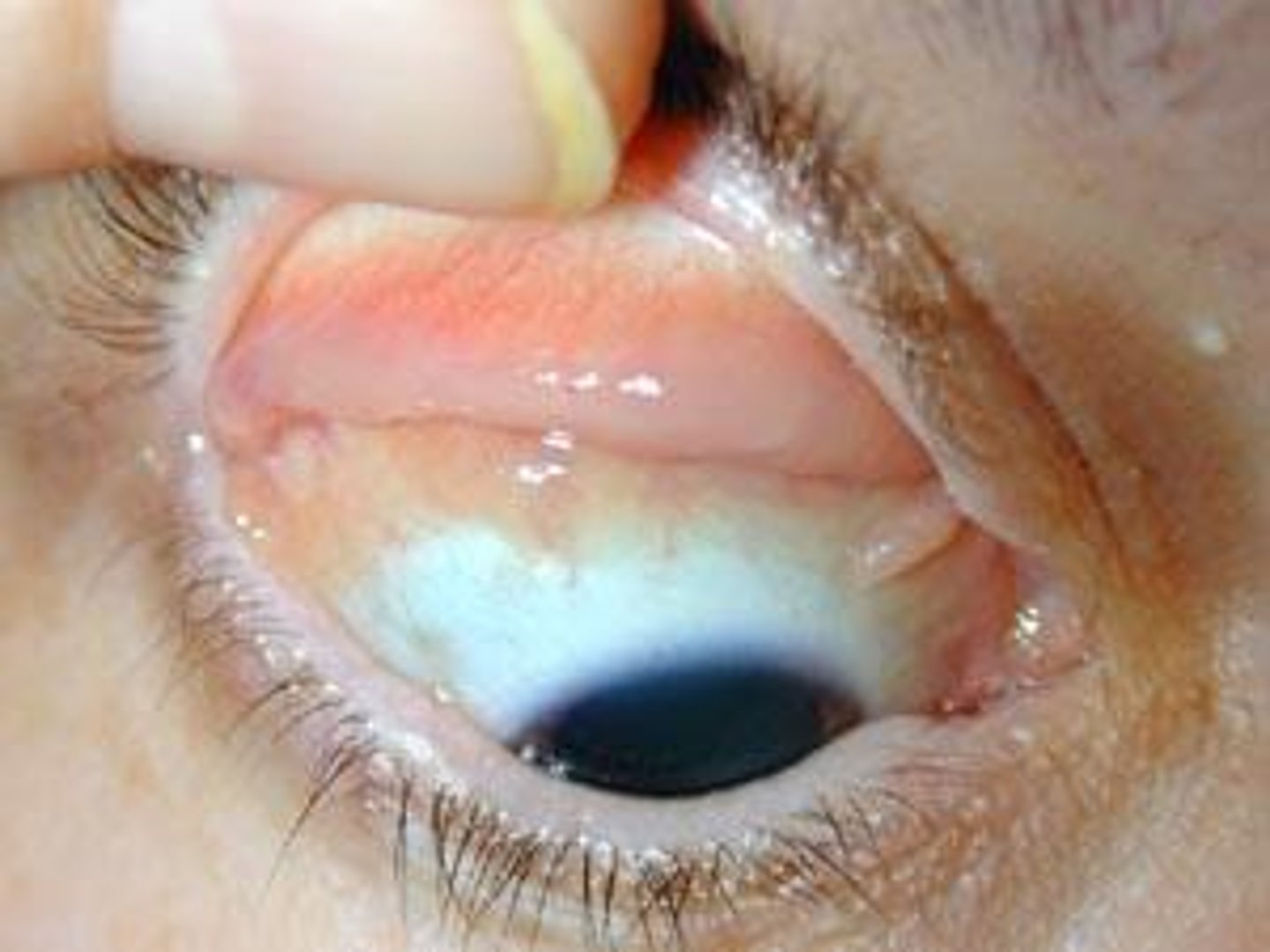
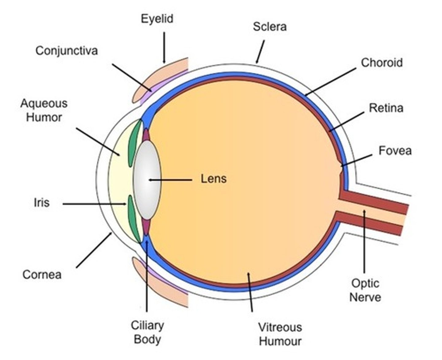
conjuctiva
Thin outer lining of the eye and eyelid
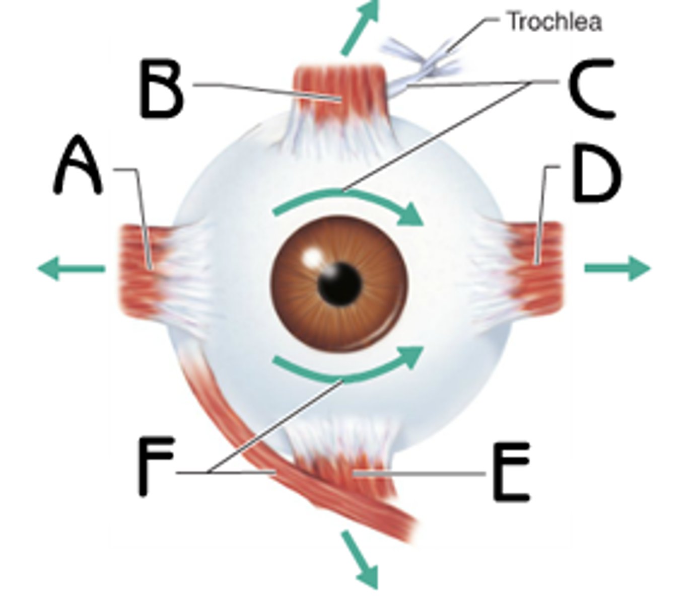
Extrinsic Eye Muscles
move the eyeball
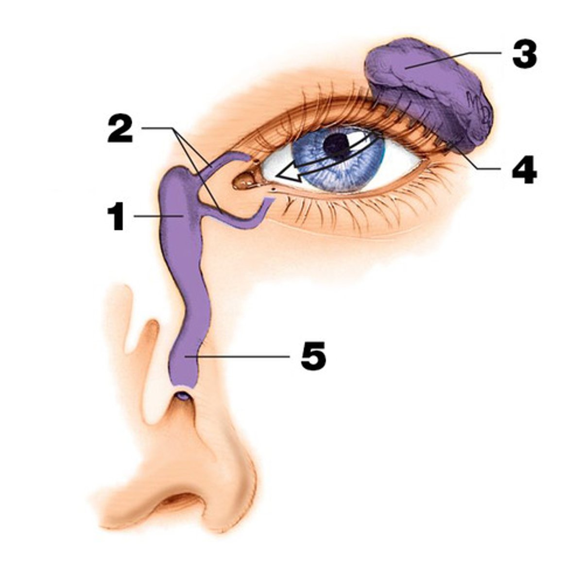
Lacrimal Glands
glands that secrete tears, drain into nasal cavity
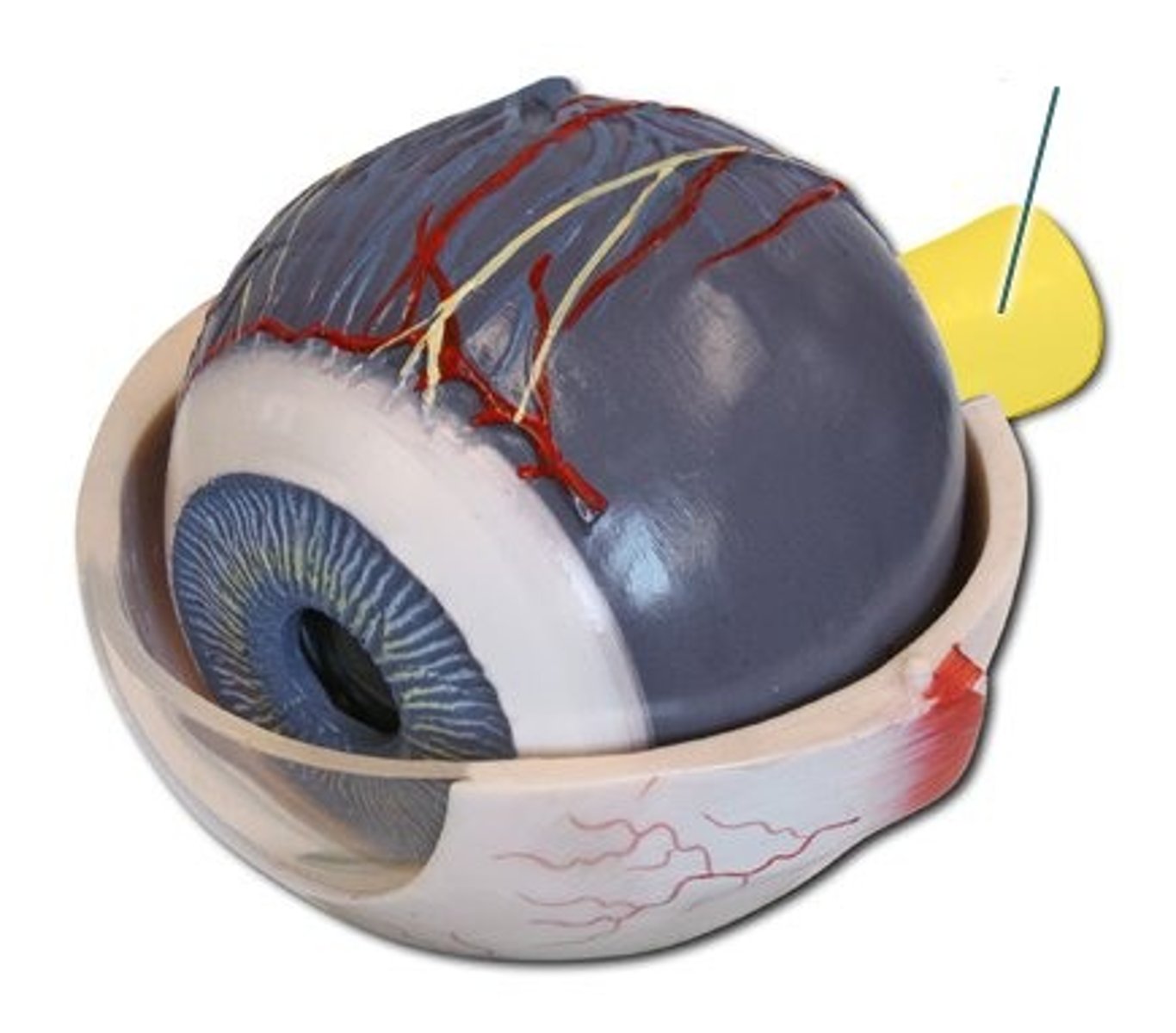
Outer Tunic of the Eye
cornea, sclera, optic nerve
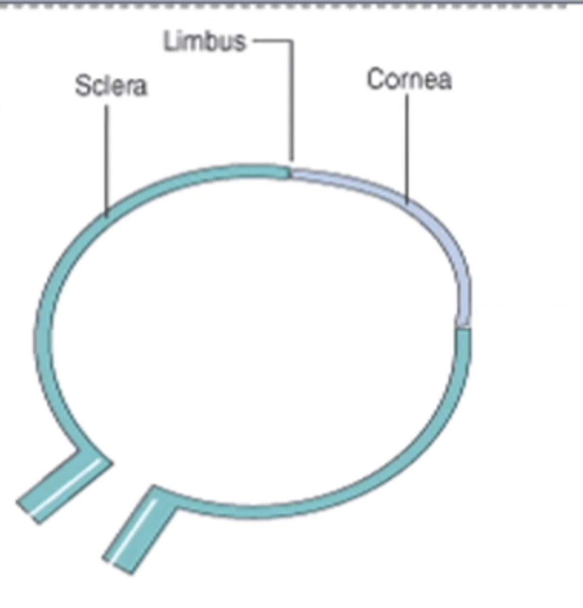
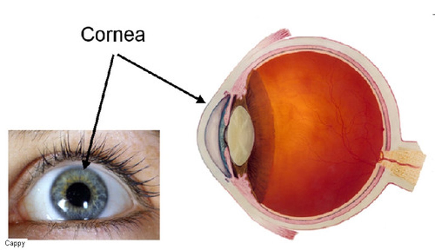
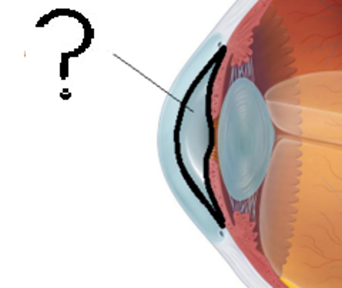
Cornea
the transparent layer forming the front of the eye, focuses the light
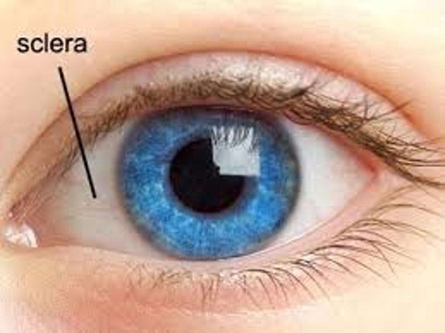
sclera
white part of the eye
optic nerve
the nerve that carries neural impulses from the eye to the brain
Middle Tunic of eye
choroid, ciliary body, iris, and lens, pupil, aqueous humor
Choroid
middle, vascular (blood vessels) layer of the eye, between the retina and the sclera
Ciliary body
Composed of ciliary muscle and ciliary processes,
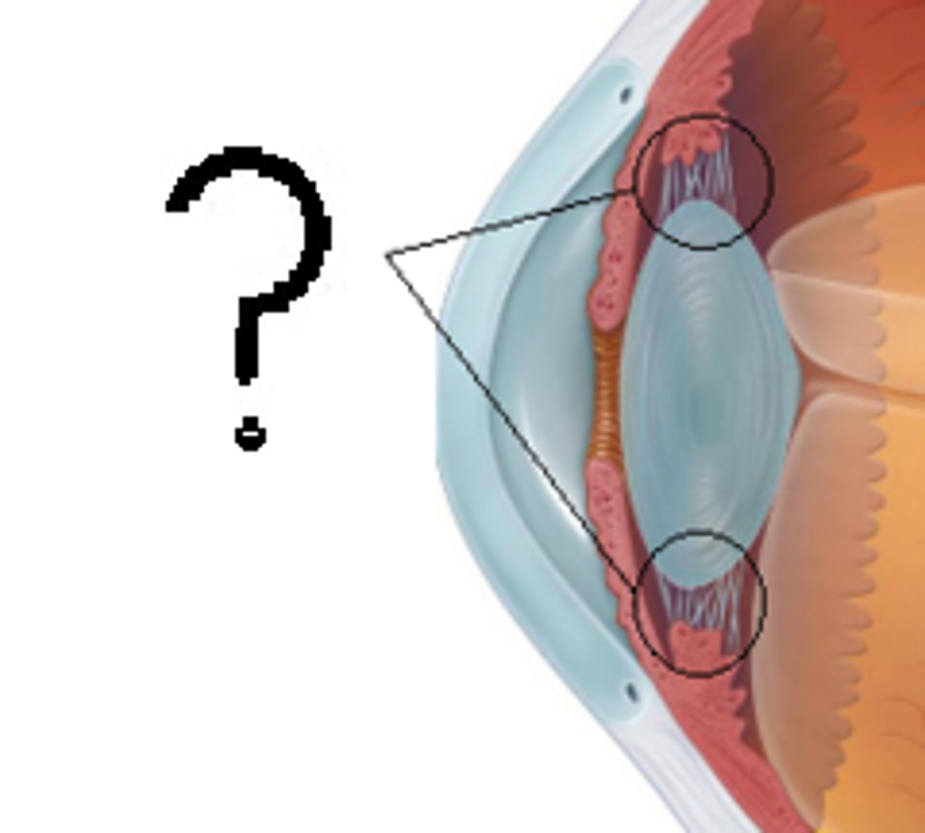
Lens
Focuses light onto retina
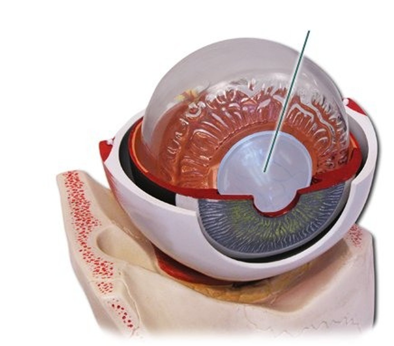
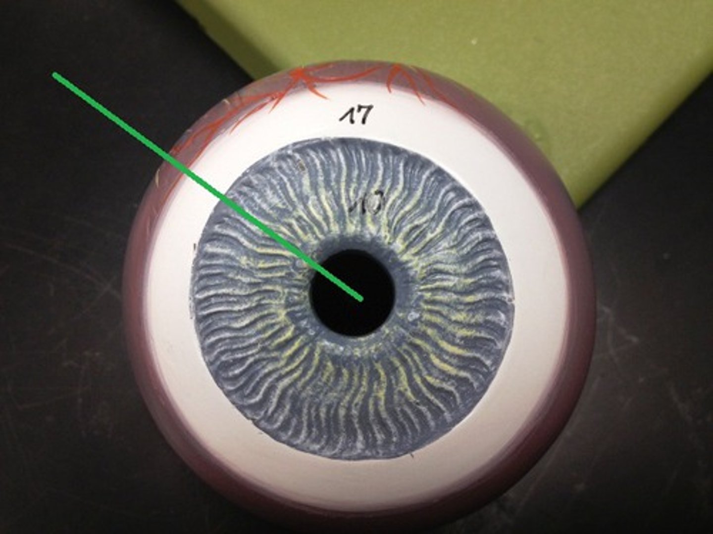
iris
Colored part of the eye
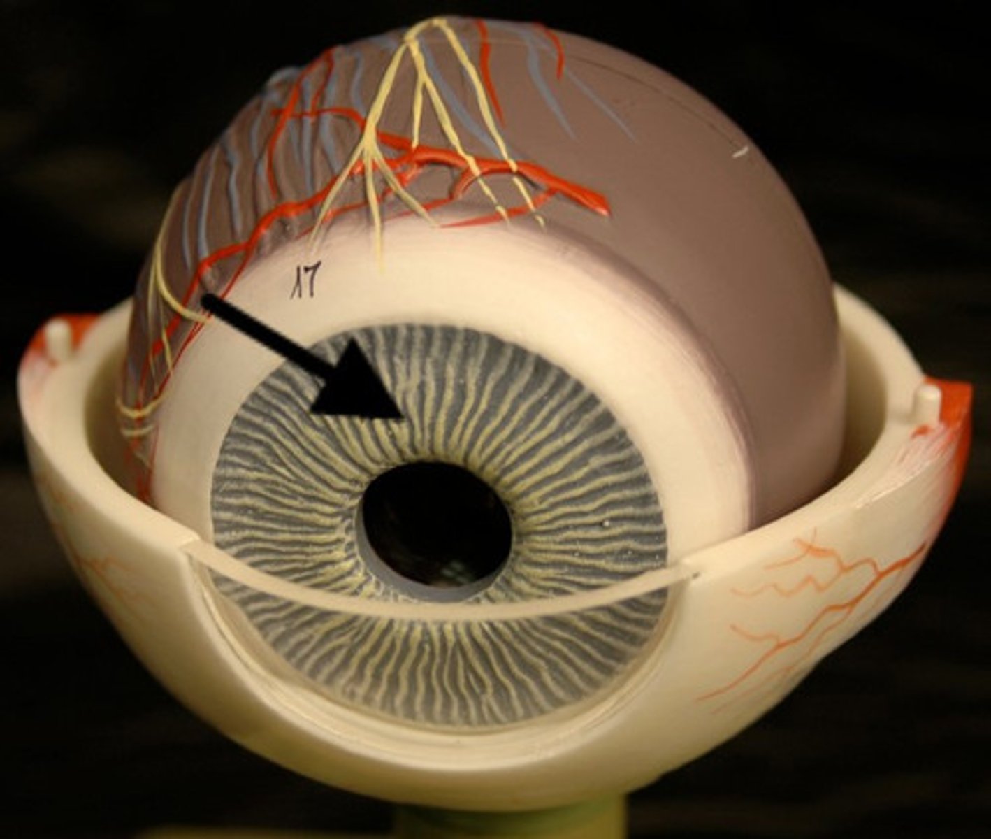
pupil
The opening through which light enters the eye
aqueous humor
fluid in the eye, found between the cornea and the lens
inner tunic
retina, fovea centralis, optic disc, vitreous humor
retina
the light-sensitive inner surface of the eye, containing the receptor rods and cones plus layers of neurons that begin the processing of visual information
fovea centralis
area consisting of a small depression in the retina containing cones and where vision is most acute
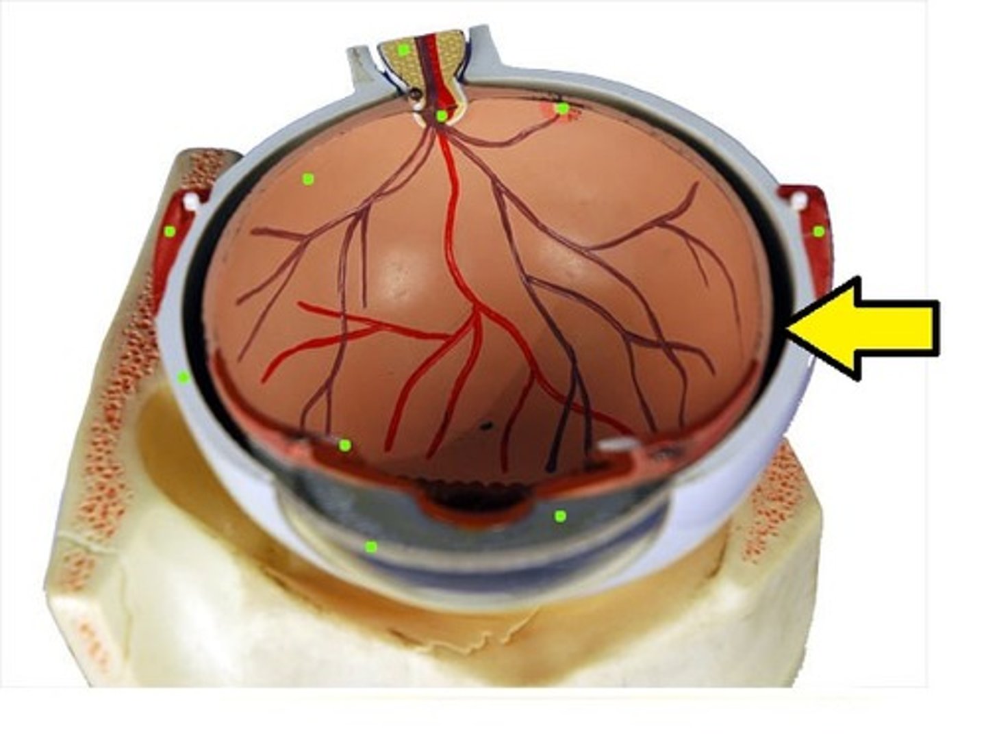
optic disc
Blind Spot- Region at the back of the eye where the optic nerve meets the retina
vitreous humor
the transparent jellylike tissue filling the eyeball behind the lens.
Describe the different types of receptors.
-Thermoreceptors :Respond to heat energy
-Chemoreceptors: Respond to chemical energy
-Photoreceptors: Respond to light energy (i.e., rods and cones)
-Mechanoreceptors: Respond to mechanical energy (e.g., blood pressure)
Distinguish between the Meissner's corpuscles and the Pacinian corpuscles.
Describe the acute pain, chronic pain, viscerial pain and referred pain.
-acute pain: short lived
warning of injury
presents symptoms at the tissue level
resolves when healed
body emits a reflex response through the spinal cord
-Chronic Pain: pain lasting for more than 6 months of expected healing time
symptoms may be psychological and can interfere with daily activities
concern when treating: narcotic dependence
-Referred Pain: Pain is remote from site of injury
What is the difference between a somatic sense and special sense?
Somatic Senses: Receptors associated with the skin, muscles, joints, and viscera make up the somatic senses.
Special Senses: These include the senses of smell, taste, hearing, static equilibrium, dynamic equilibrium, and sight.
Define and give an example of sensory adaption.
The ability of the nervous system to become less responsive to a maintained stimulus
Ex: Getting used to the temperature of the ocean
What are olfactory receptors and where are they located?
Smell Receptors, in the Mucosa
List the four different types of taste sensations. What is umami?
Sweet, sour, salty, bitter, umami (Savory)
Describe the path sound waves take within the ear and are eventually processed by the brain.
Steps of Hearing:
1.Sound waves enter external auditory meatus eardrum vibrates
2.Auditory ossicles (malleus, incus, stapes) amplify vibrations
3.stapes it oval window and transmits vibrations to cochlea
4.coati contain receptor cells (hair cells) Thant deform from vibrations
5.impulses sent to the vestibule cochlear nerve
Compare static equilibrium; what part of the ear maintains your equilibrium?
-Static equilibrium: Sense the position of the head, maintain stability and posture
-Dynamic equilibrium (semicircle canals): Balance the head during sudden movement
What is the difference between rods and cones?
rods detect light; cones detect color
Describe how we see images and the part of the brain that interprets those images.
-Light refraction: The bending of light around an object images viewed by the eye are upside-down, our brains interpret in properly
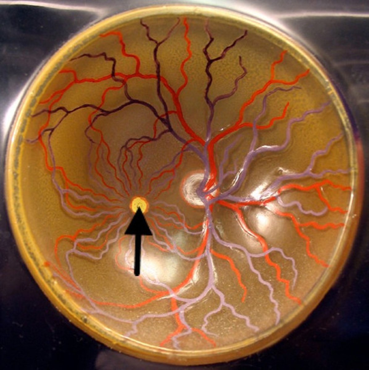
Know what the blind spot is, where it's located and why it exists.
On the back of our eye, the retina is the stuff that detects the light. All the information that the retina picks up is sent to the brain through the optic nerve. The only problem is that the optic nerve needs a way to get out of the eye. The place where it leaves is where we have our blind spot.
Define refraction and discuss how it affects your vision. What is the fovea centralis?
Refraction is when light bends around object; we see everything upside down.
-Fovea Centralis: region of sharpest vision.
What is myopia, hyperopia and astigmatism? What is a cataract?
-Myopia (Nearsightedness): Eyes that're too long. Can see up close, but not far away.
-Hyperopia (far-sighted): Eyes are too short. Can see far away, but not up close.
-Cataracts: Clouding of the lens that leads
What are the muscles that move the eye called?
-superior rectus
-trochlea
-superior oblique
-medial rectus
-inferior rectus
-inferior oblique
-lateral rectus
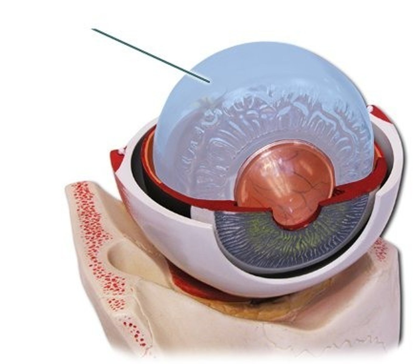
Label the parts of the eye on a diagram and know the functions.
Eye Diagram
auricle
A