Disorders of Cornea , iris + cataracts
 Call Kai
 Learn
Learn Practice Test
 Spaced Repetition
 Match
Match Flashcards
 Knowt Play
1/50

There's no tags or description
Looks like no tags are added yet.
Name | Mastery | Learn | Test | Matching | Spaced | Call with Kai |
|---|
No analytics yet
Send a link to your students to track their progress
51 Terms
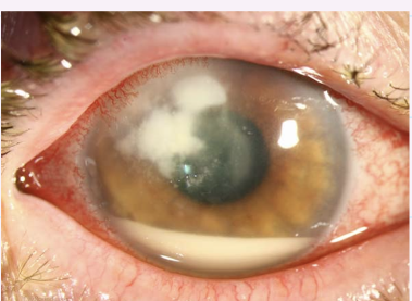
What is this condition and describe?
Bacterial/Fungal Keratitis
conjunctival hyperaemia
epiphora
corneal lesion - mid-peripheral and inferior
stromal infiltration
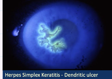
Describe this image and give diagnosis?
Herpes Simplex Keratitis
dendritic ulcer
stained using fluorescein
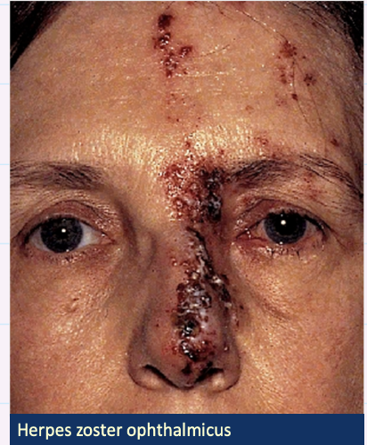
Describe this image and give diagnosis?
Herpes Zoster opthalmicus
vesicular rash affecting forehead, upper eyelid and nose - hutchingsons sign
redness
uni-lateral
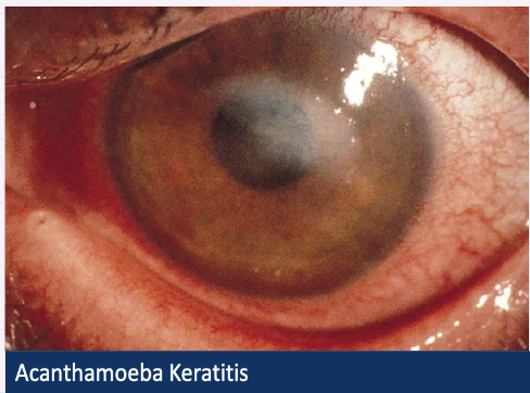
Describe image + give diagnosis?
Acanthamoeba Keratitis
white corneal ulcer
conjunctival hyperaemia
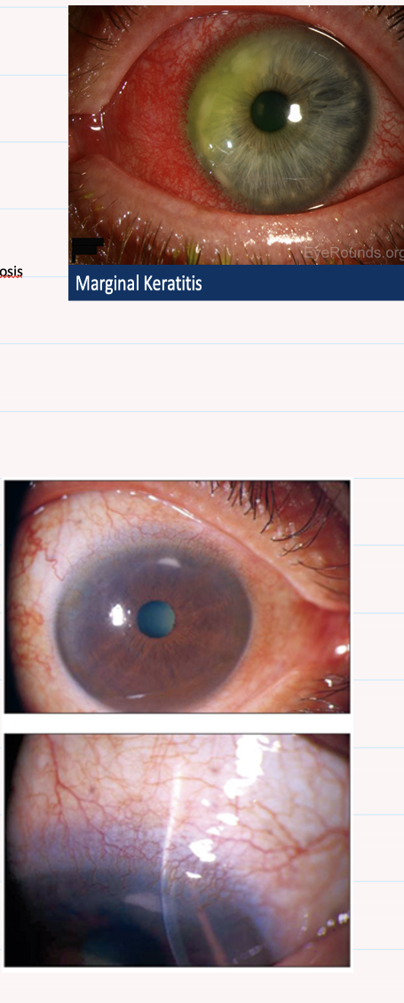
Describe this image and give diagnosis?
Marginal Keratitis
small lesions in peripheral cornea
conjunctival hyperaemia
arcuate shaped ulcer
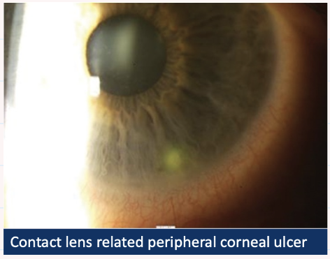
Describe + give diagnosis?
CL associated peripheral corneal ulcer / CL associated Keratitis
stained peripheral corneal ulcer
mild conjunctival hyperaemia
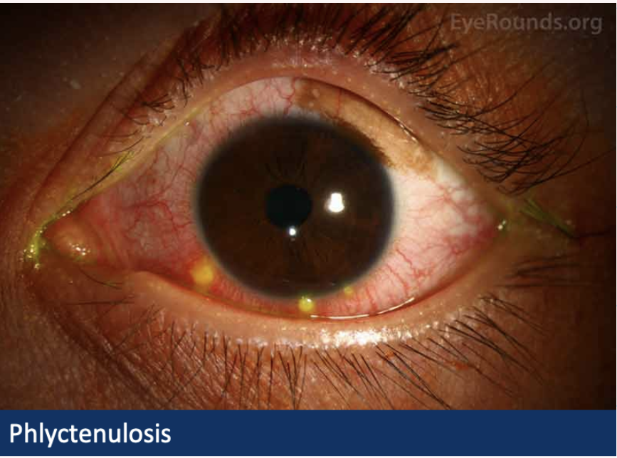
Describe and give diagnosis - good to know
Phlyctenulosis
sub epithelial nodules (phylcten) located in conjunctiva
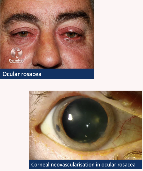
Describe and give diagnosis
Ocular Rosacea
hyperaemic thickened lids
telangiectasia of lid margins
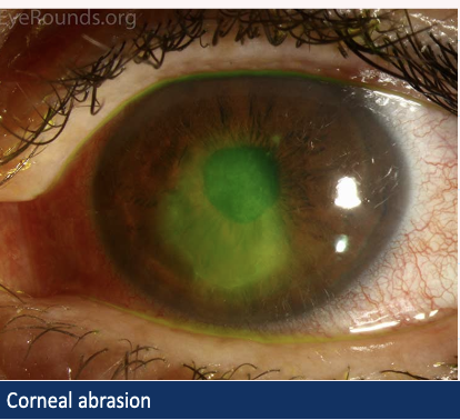
Describe and give diagnosis
Corneal abrasion
epithelial defect which stains with fluorescein
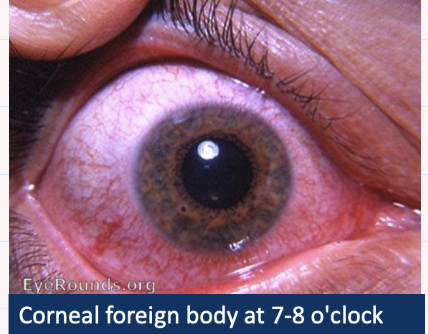
Describe and give diagnosis
corneal foreign body
at 7-8 o'clock small and round mid-periphery
rust ring
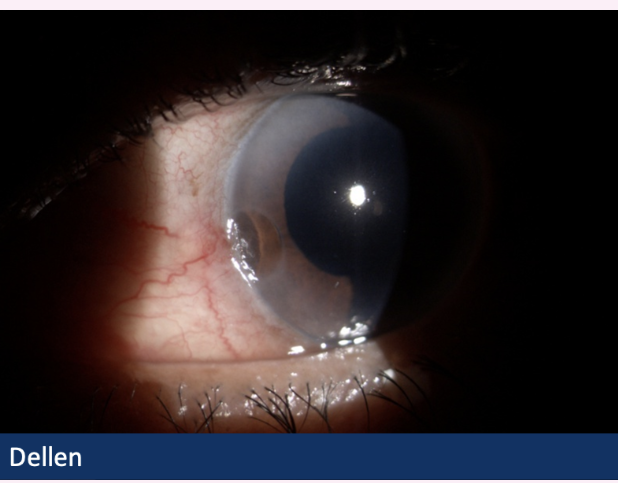
Describe and what’s the diagnosis?
Dellen
localised depression (stromal thinning and compaction)
at 9’o’clock in peripheral cornea
conjunctival hyperaemia adjacent to thinning
adjacent elevation - pinguecula
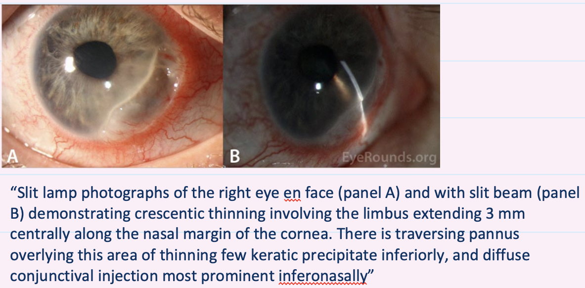
Describe and give diagnosis
Mooren’s ulcer
crescent shaped , peripheral cornel thinning from 3 to 6’o’clock
conjunctiva injection inferonasally
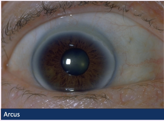
Describe and give diagnosis
Arcus
circumferential opacity - white ring in peripheral cornea
clear zone of 1mm between edge of opacity and limbus
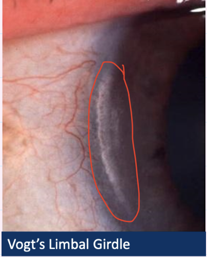
Describe and give diagnosis
Vogt Limbal Girdle
chalky white crescent near limbus , 3 and 9 o’clock
clear zone between crescent and limbus
mild conjunctiva hyperaemia
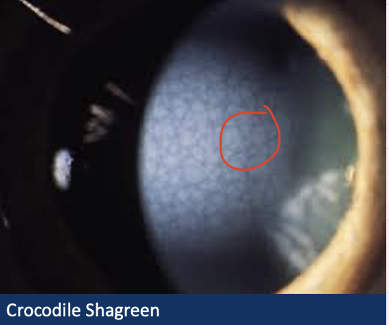
Describe and give diagnosis
Crocodile Shagreen
Faint reticular polygonal network of stromal opacities
white/grayish opacities
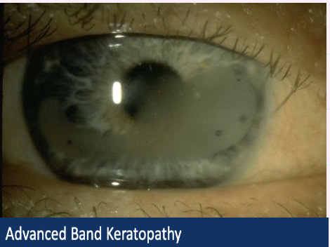
Describe and give diagnosis
Band Keratopathy
White opacification at 3 o'clock + 9 o’clock
in periphery of cornea, extends to centre of cornea
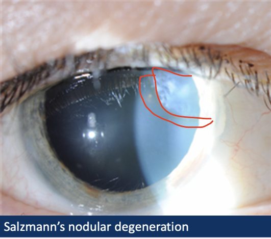
Describe and give Diagnosis
Salzmann’s nodular degeneration
superficial blue-white nodules
slightly elevated in cornea
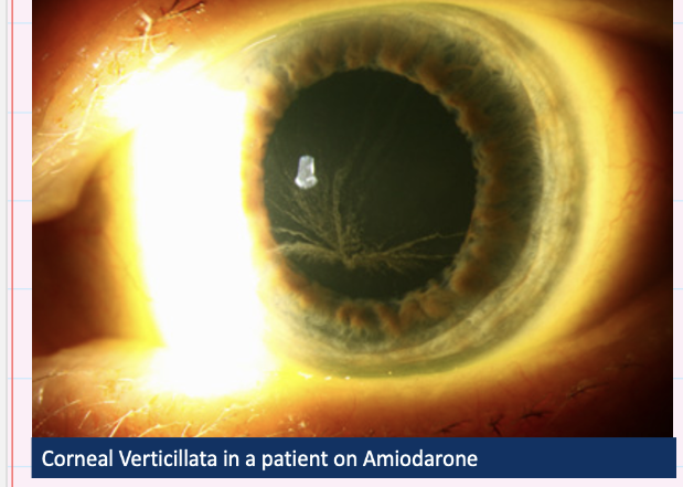
Describe and give diagnosis
Corneal Verticilata
white , whirl shaped opacities
inferior cornea branches out
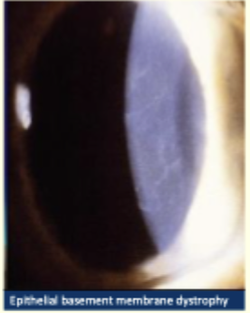
Describe and give diagnosis
Epithelial basement membrane dystrophy
fine refractive lines - fingerprints
diffuse grey patches - maps
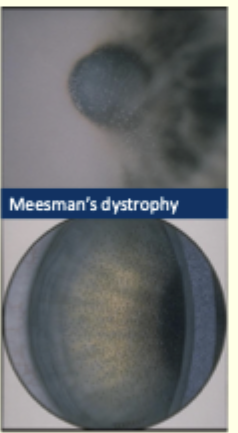
Describe the image and give diagnosis
Meesman’s dystrophy
multiple, tiny uniform epithelial microcysts
maximum in centre , less in periphery and spare the limbus
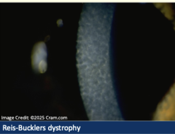
Describe and give diagnosis
Reis Bucklers dystrophy
grey/white subepithelial opacities
reticular/honeycomb pattern
most dense centrally
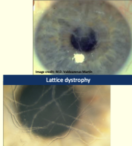
Describe and give diagnosis
Lattice dystrophy
branching criss-crossed lines of amyloid
on central cornea
covering pupil
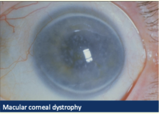
Describe and give diagnosis
Macular corneal dystrophy
greyish opacities
stromal haze
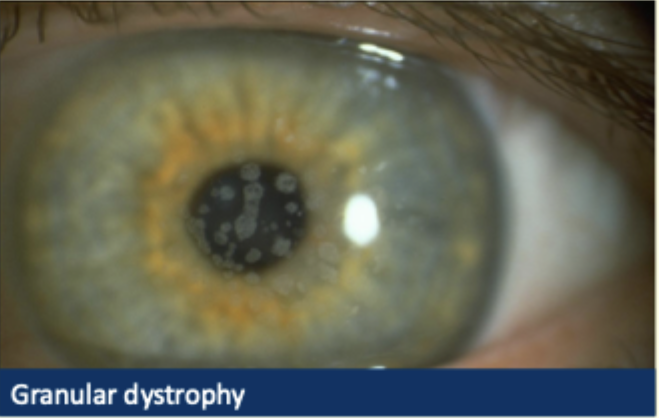
Describe + give diagnosis
Granular dystrophy
white crumb like opacities
in central cornea
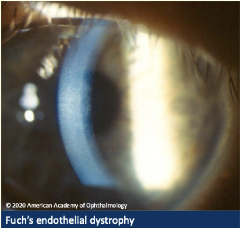
Describe + give diagnosis
Fuchs endothelial dystrophy
corneal oedema
haze of stroma
Corneal guttata —> beaten metal appearance
brown pigment spots
lines are fold in descemets membrane
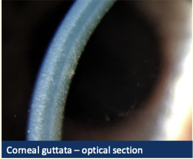
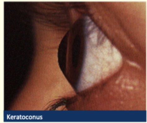
Describe and give diagnosis
Keratoconus
conical distortion of cornea
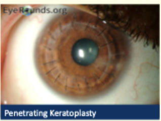
describe and diagnosis
Penetrating Keratoplasty
sutures around peripheral cornea circumferentially
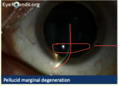
Describe and diagnosis
pellucid marginal degeneration
inferior crescent shaped band at 8 o'clock
peripheral cornea
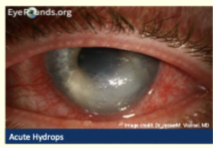
describe and give diagnosis
Acute hydrops
corneal oedema
Grade 3 conjunctival hyperaemia
milky white opacification involving central and inferior cornea
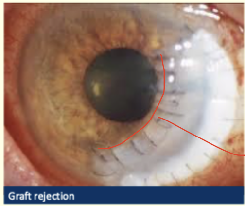
Describe and diagnosis
Graft rejection
Khodadoust line extending from 1 to 7 o’clock
transplant oedema
conjunctiva hyperaemia around limbus
anterior ciliary injection
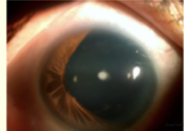
Describe and give diagnosis
Aniridia
large section of Iris missing
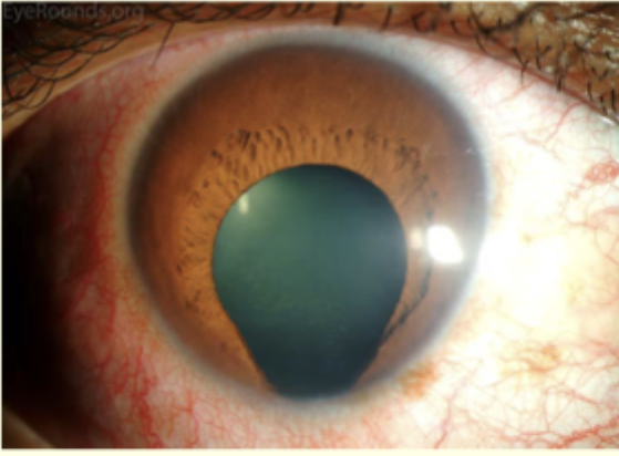
Describe and give diagnosis
Coloboma
key-hole shaped pupil inferiorly - notch in iris
diffuse conjunctival hyperaemia
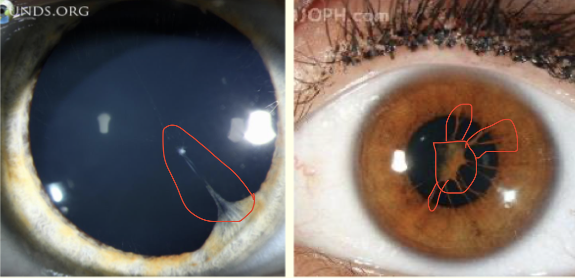
Describe this image and give a diagnosis
Persistent Pupillary Membrane
iris strands across pupil from iris to lens
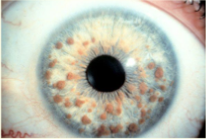
Describe and give diagnosis
Lish Nodules
Brown nodules over iris
round elevations varying in size
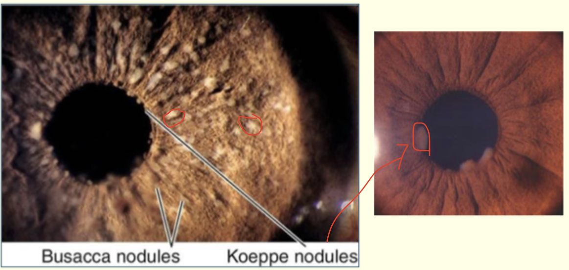
Describe this image and give diagnosis
Busacca and Koeppe nodules
Busacca nodules —> deeper into iris
Koeppe nodules —> margin of iris
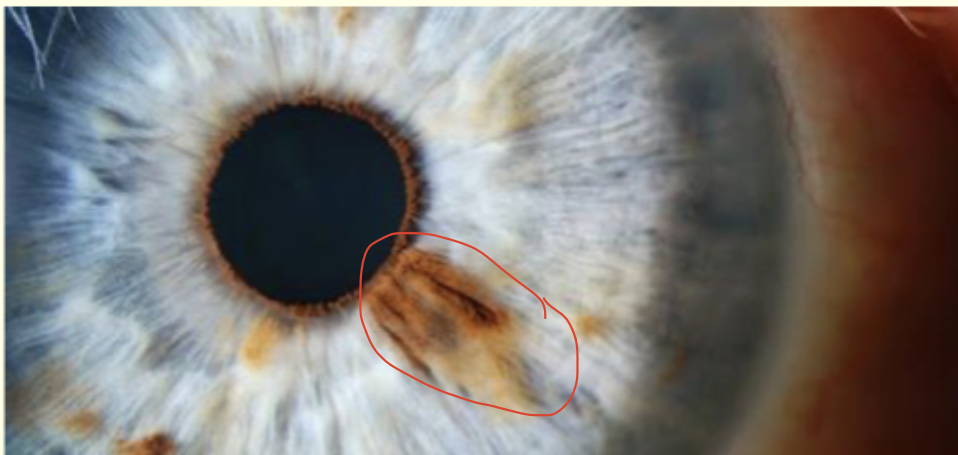
Describe and give diagnosis
Iris Naevus
Flat, brown pigmented lesion
at 5 o'clock extends from pupillary margin to mid-periphery of cornea
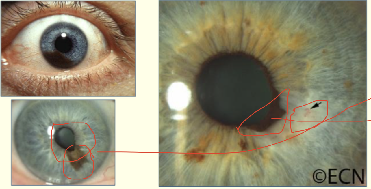
Describe this image and give diagnosis
Iris Melanoma
inferior half of iris
pupil distortion
surface blood vessels
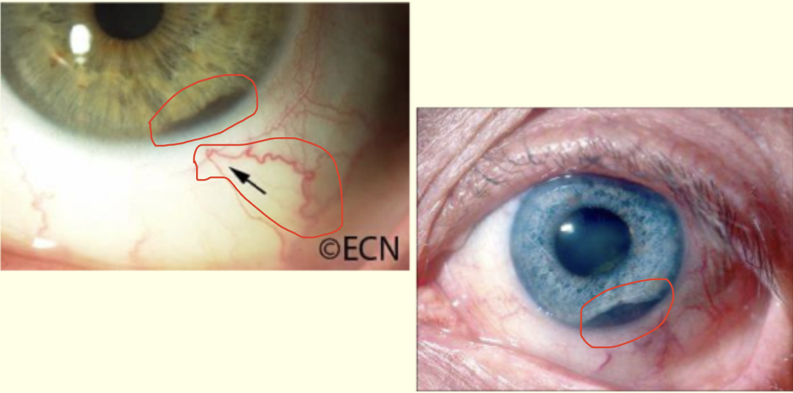
Describe and give diagnosis
Ciliary body tumours
brown pigment at edge of iris
dilated episcleral vessels
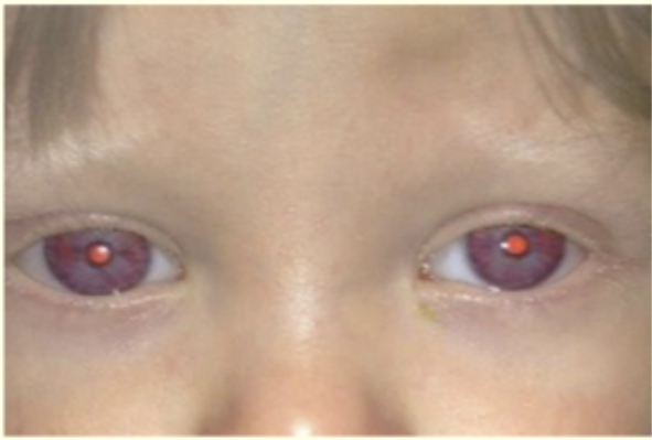
Describe and give diagnosis
Albinism
Iris transillumination
pale blue eyes, pale skin
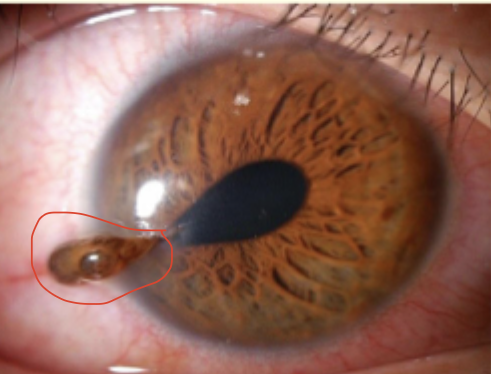
Describe and give diagnosis
Iris Prolapse
Tear drop pupil shape
lesion through cornea
some iris protruding through lesion and cornea
conjunctival hyperaemia
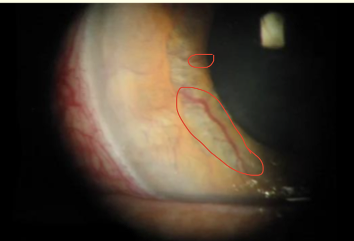
describe + diagnose
Rubeosis
new blood vessels on iris surface
near pupil margin
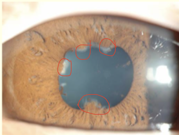
Describe and give a diagnosis
Posterior synechiae
adhesions of iris to lens - 1 inferior at 6' o clock , 1 at 10 o'clock and 2 superior
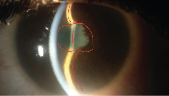
describe and give diagnosis
Iris Bombe
iris pushed forward like doughnut
irregular pupil
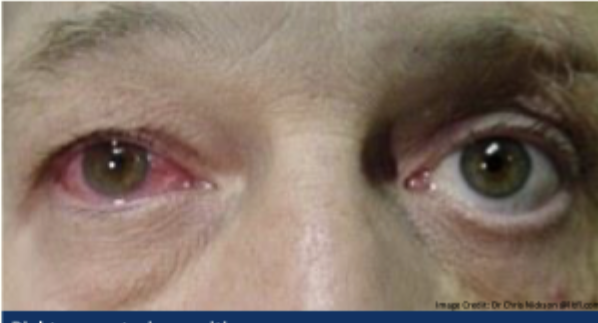
Describe and give diagnosis ?
RE anterior uveitis
conjunctival injection
epiphora
lid oedema
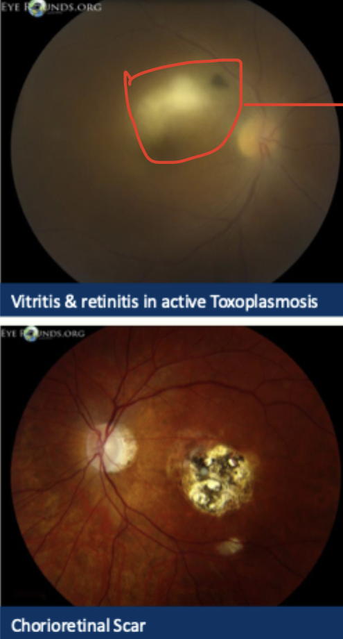
Describe and give diagnosis
yellowish lesion with fluffy edges
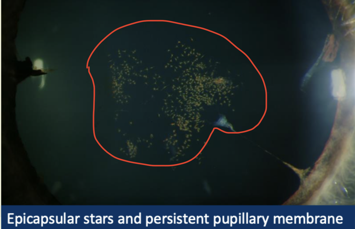
describe and diagnose
Eupicapsular stars and PPM
little stellate pigment spots on anterior surface of lens
little strand at pupil margin
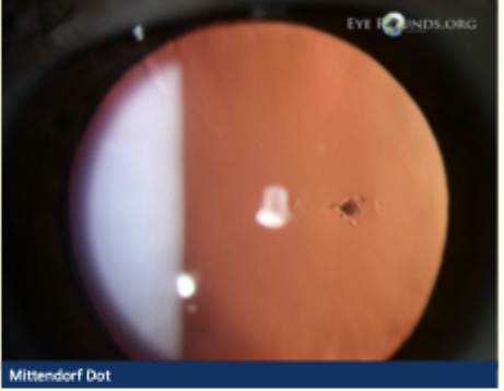
Describe and give diagnosis
Mittendorf Dot
small, round, circular opacity on posterior lens capsule
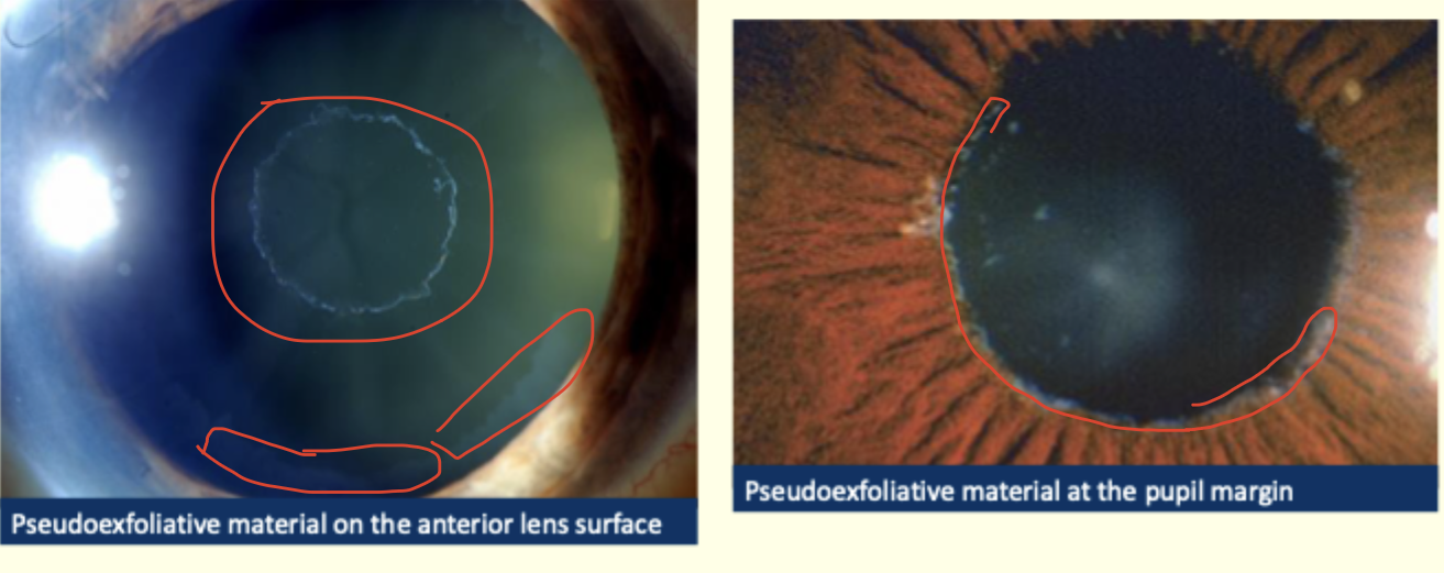
Describe and give diagnosis
Pseudoexfoliation
little, white flaky material - dandruff like
describe and give diagnosis
Ectopia Lentis
dislocation of lens
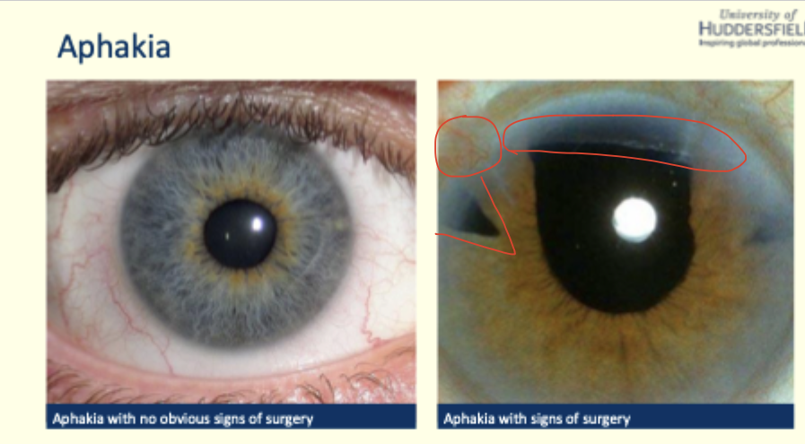
describe and give diagnosis
Aphakia
neovascularisation at top
triangle shape - missing iris , trabeculectomy
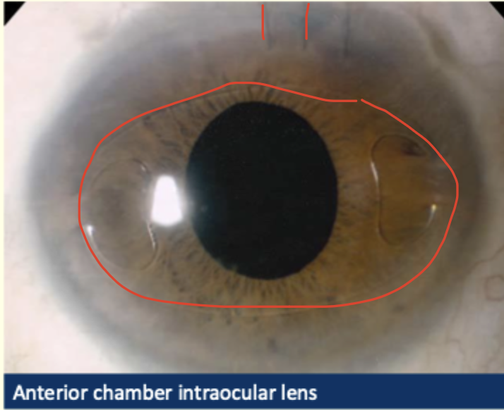
describe and diagnose
AC intraocular lens
stitches at top