Lecture 8 -- Equine Distal Hindlimb and Stay Apparatus
1/15
There's no tags or description
Looks like no tags are added yet.
Name | Mastery | Learn | Test | Matching | Spaced | Call with Kai |
|---|
No analytics yet
Send a link to your students to track their progress
16 Terms
Which joints have little movement in the tarsus?
Proximal inter-tarsal, distal inter-tarsal, and tarso-metatarsal joints.
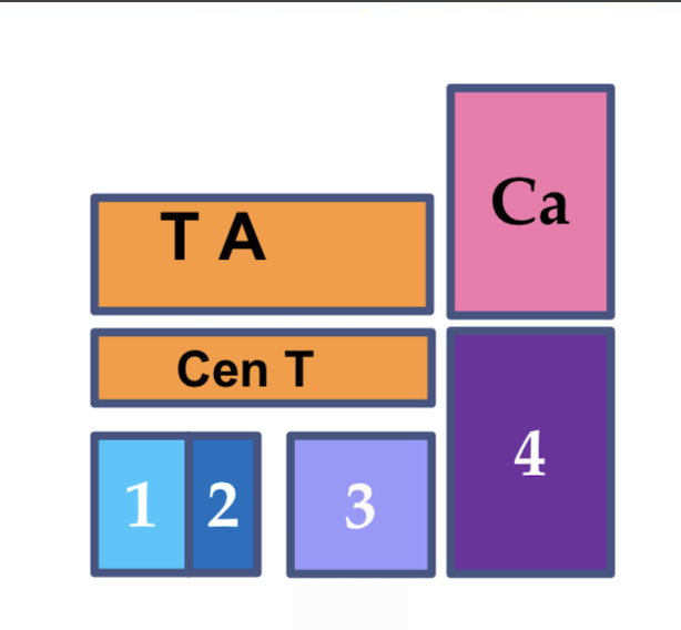
What are the key features of tarsus of horses?
1st and 2nd tarsal bone are fused together
3rd tarsal bone are as large as central tarsal bone
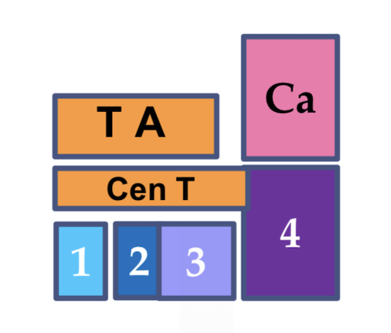
What are the key features of ruminant’s tarsus?
Central and 4th tarsal bone are fused
2nd and 3rd tarsal bone are fused
The trochlea of talus is straight → Trochlear ridges one at either end → Rotation possible at proximal inter-tarsal joint → Lateral “cow” kick
What are the key features of pigs’ tarsus?
All tarsal bones present

The tendon of cranial tibial muscle slips into two parts in horses. What is the medial part named?
Cunean tendon
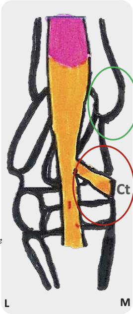
What are the origin and insertions of the cranial tibial muscle in horses?
O: Lateral condyle and tibial tuberosity of tibia
I: 3rd MT and medial aspect hock (T1&2) → Cunean tendon
What are the origin and insertions of the peroneus tertius in horses?
O: Extensor fossa of femur (With long digital extensor)
I: 3rd MT (with cranial tibial) and lateral aspect of tarsus
What are the components of the common calcanean tendon in horses?
Biceps femoris
Semitendinosus
Gracilis
Gastrocnemius
Soleus
Superficial digital flexor
Where are the calcanean bursae located?
Calcaneus & calcanean tendon
Calcanean tendon & SDFT
What structures make up the reciprocal apparatus?
Peroneus tertius
Superficial digital flexor
Describe the mechanism of reciprocal apparatus.
The reciprocal apparatus is a mechanism that links movement of the stifle and hock joints so they flex and extend together
When the superficial digital flexor contracts, stifle flexes → At the same time, the peroneus tertius pulls the metatarsal bones proximally, causing the hock to flex
What prevent collision between forelimb and hindlimb?
Trochlea on talus is not vertical → When tibia moves over talus during flexion, pes rotates lateral to forelimbs → Prevent over-reaching injuries
What are the differences between the pes region from manus in horses?
SDFT:
Attached not only to middle phalanx, but also to calcaneus
No superior check ligament
DDFT:
Poorly developed inferior check ligament
What are the key components of stay apparatus?
Stifle:
Patella & patellar ligaments → Patellar locking mechanism
Hock:
Reciprocal apparatus → Prevention of collapse into flexion
MTP, PIP and DIP joint (Prevention of hyperextension):
Suspensory ligament
Distal sesamoidean ligaments (Short, cruciate, oblique and straight)
SDFT, DDFT and check ligament
Annular ligaments (PAL, PPAL, DAL)
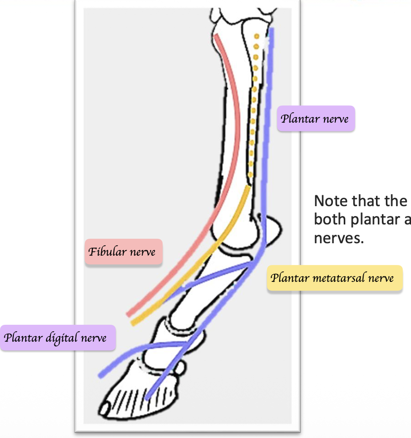
Describe how the tibia and fibular nerve branches at the distal limb.
Tibial nerve:
Plantar nerves → Plantar digital nerve
Plantar metatarsal nerve
Fibular nerve:
Medial and lateral branches of fibular nerve
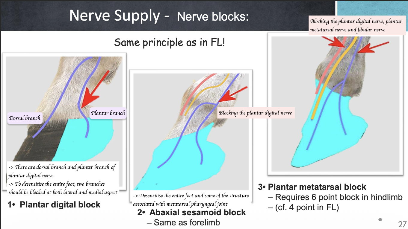
What are the three types of nerve blocks for hindlimb?
Plantar digital block
Block the lateral and medial aspect of plantar branch of plantar digital nerve
Abaxial sesamoid block
Block the plantar nerve (Before it branches off to plantar digital nerve)
Plantar metatarsal block
Requires 6 point block in hindlimb x To block plantar digital nerve, plantar metatarsal nerve and fibular nerve