EXAM 2 - General Sensory Physiology
1/152
There's no tags or description
Looks like no tags are added yet.
Name | Mastery | Learn | Test | Matching | Spaced | Call with Kai |
|---|
No analytics yet
Send a link to your students to track their progress
153 Terms
types of sensory receptors
mechanoreceptors, chemoreceptors, thermoreceptors, nociceptors, photoreceptors, propioreceptors
Mechanoreceptors detect
Sensory receptors responsible for sensing deformation in body tissues
Tactile Mechanoreceptors
touch, pressure, vibration, tickle and itch
thermoreceptors detect
detect heat and cold (temperature)
Nociceptors detect
detect tissue damage with pain receptor
what is pain
Tissue damage
photoreceptors detect
photon of light
chemoreceptors detect
chemicals
taste, smell, CO2, O2
proprioceptor
sense postition
Modalities of sensation
touch, pain, sigh, and sound, etc
each receptor is responsive to
one type of energy stimulus
How the sensation is perceived is determined by what?
1. the characteristics of the receptor
2. the central connections of the axon connected to the receptor
Receptor Excitation - Mechanical deformation
stretch of the membrane - causes ion channels to open- might lead to EPSP
Receptor Excitation - Application of chemicals
opens the ion channels - might lead to EPSP
Receptor Excitation - Change in temperature
alter permeability of membrane - might lead to EPSP
Receptor Excitation - Electromagnetic radiation
changes properties/characteristics of membrane - might lead to EPSP
Receptor potential
The membrane potential of the receptor
A local graded electrical potential produced by a receptor cell in response to a physical stimulus
Each receptor is responsive to
One type of stimulus energy: specificity is key
receptors are specific to one type of energy
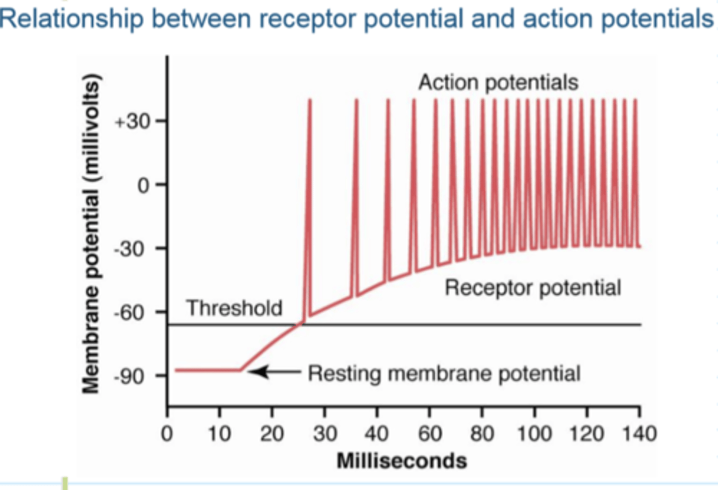
Receptor potential rises above the threshold
makes action potential
The greater the intensity of the stimulus
the greater the receptor potential
the greater the rate that AP are created
larger stimulus=larger receptor potential=faster the AP created
Relationship Between Receptor Potential and Action Potentials
increase intensity: Increases the rate (of AP) and response
sensory system receptor potential threshold
WONKY
you only get an AP 50% of the time
Summary of Sensory Transduction
Stimulus opens ion channels on sensory neuron or sensory cell, which produces a graded potential called a: receptor potential
- Reach threshold and Leads to: Action potential 50% of time
Adaptation of Receptors
When a continuous stimulus is applied, receptors respond rapidly at first, but response declines until all receptors stop firing
Muscle spindle: Adaptation of Receptors
slow adaptation
Adaptation of Receptors: pacinian corpuscle
rapid adaptation
Adaptation
◻ Rate of adaptation varies with type of receptor
◻ Receptors respond when a change is taking place (i.e., think of the feel of clothing on your skin)
Slowly Adapting (Tonic) Receptors - definition
def: fire throughout stimulation (as long as stimulus is present then it is firing/transmitting signal)
Slowly Adapting (Tonic) Receptors function
gives information on DURATION of the stimulus
- keep brain aware of stimulus presence (how long is stimulus present)
Slowly Adapting (Tonic) Receptors - adaptation
Will adapt to extinction as long as the stimulus is
present; however, this may take hours or days
Slowly Adapting (Tonic) Receptors - examples
muscle spindles, GTOs, ruffini endings, chemoreceptors, barro receptors
Rapidly Adapting (Phasic) Receptors - definition
only fire/respond when a change is taking place
onset and offset of a stimulus
Rapidly Adapting (Phasic) Receptors are Important for what
predicting the future position or condition of the body
1. movement
2. balance
Rapidly Adapting (Phasic) Receptors - examples
semicircular canals and picinian corpuscles
Transmission of Receptor Information to the Brain - Velocity
• The larger the nerve fiber diameter the faster the
rate of transmission of the signal
• Velocity of transmission can be as fast as 120
m/sec or as slow as 0.5 m/sec
Nerve Fiber Classification
Type A or C
Type A nerve fiber
myelinated fibers of varying sizes, generally
fast transmission speed.
• subdivided into a, b, d, g (alpha, beta, delta, gamma)
Type C nerve fiber
unmyelinated fibers, small with slow
transmission speed.
Importance of Signal Intensity
Signal intensity is critical for interpretation of the
signal by the brain - best example: PAIN
Gradations in signal intensity can be achieved by
1) increasing the number of fibers stimulated - spatial summation
2) increasing the rate of firing in a limited number of
fibers - temporal summation
spatial summation
increasing the number of fibers stimulated
temporal summation
increasing the rate of firing in a limited number of
fibers
Sensory Modulation - filtering
used as a mechanism to increase sensitivity
EX: at a party trying to listen to one person, need to filter out background noise. The person will enter the CNS regular intervals. the background noise will enter through random intervals
Sensory Modulation - lateral inhibition
neuron influence input by nearby receptor cells
LOCALIZING THE AREA OF THE STIMULUS
Lateral Inhibition
The pattern of interaction among neurons in the visual system in which activity in one neuron inhibits adjacent neurons' responses. (can be in other systems too, not just visual)
- inhibit neighboring neurons -- inhibiting closest neurons the most and further neurons inhibited less
LOCALIZING THE AREA OF THE STIMULUS
receptive fields
2 point discrimination
we have 2 points poking and at what point do you feel that they are 2 separate pricks
Large Receptive Fields
have receptors spread far apart, which makes it difficult to localize a stimulus
larger receptors and farther apart so when 2 points are pricking, cannot tell that it is 2 until far apart
small receptive field
precise location and very sensitive
receptors closer together so when 2 points are pricking, can tell that it is 2 points close together
Weber-Fechner Relationship
Smallest change in a stimulus intensity that can be perceived as a different stimulus
humans- must be a 2.5% change difference for us to discriminate the difference
Classification of somatic sensations
Mechanoreceptive
Thermoreceptive
Nociceptive
Mechanoreceptive
stimulated by mechanical displacement.
Tactile: touch, pressure, vibration, tickle, itch
position or Proprioceptive: static position, rate of change
Thermoreceptive
detect heat and cold
Nocicipetive
detect pain and are activated by any factor that damages tissue
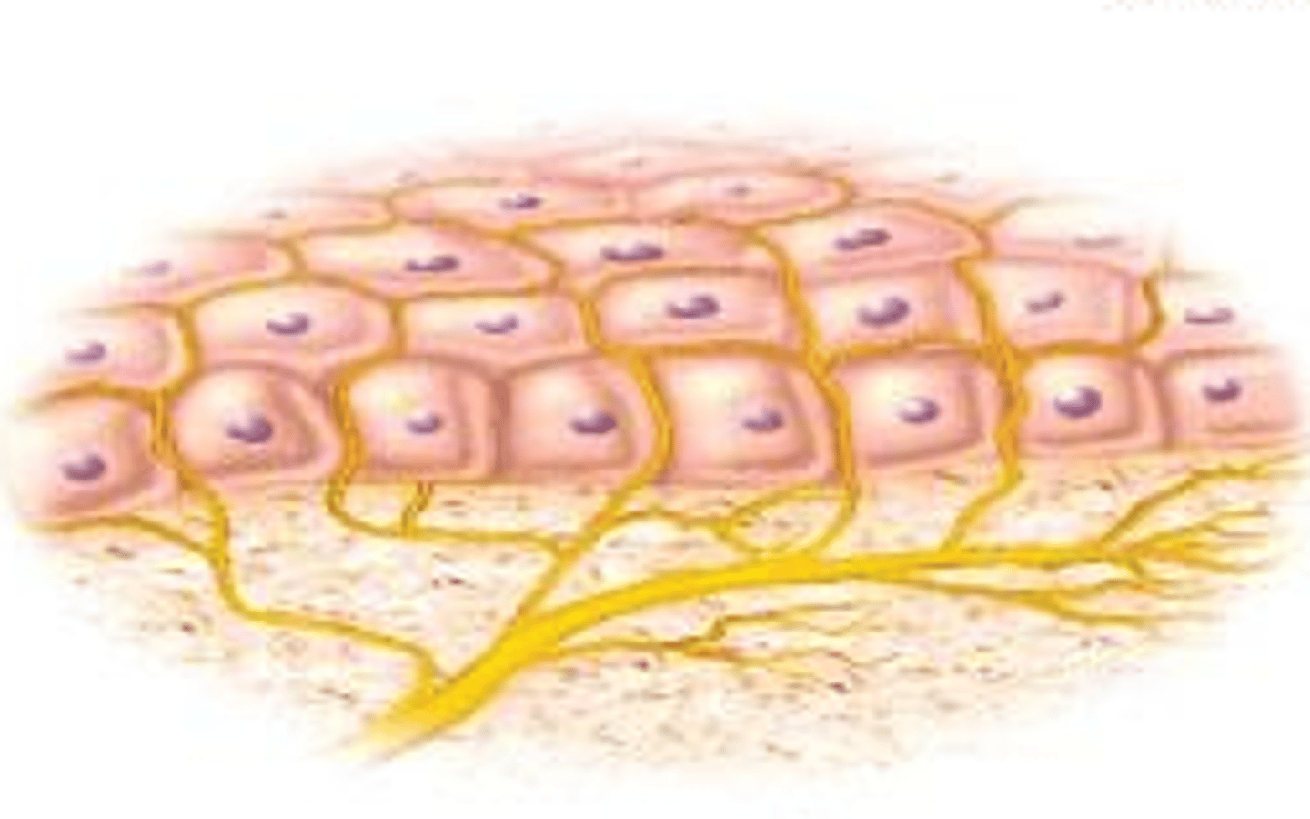
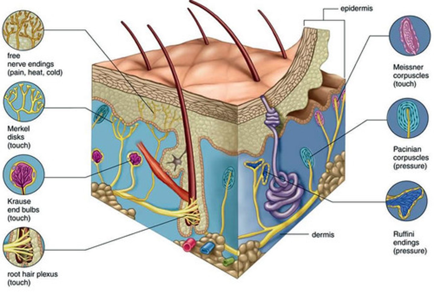
Free nerve endings
detect touch and pressure
found everywhere in the skin and other tissues
Meissner corpuscles
rapidly adapting (within a fraction of a second) receptors that detect movement of light objects over skin
Found on nonhairy skin (glabrous skin) (fingertips, lips, eyelids) -located in the dermal papillae of hairless skin
Merkels discs
respond rapidly at first and then slowly adapt, detect the "steady state"
found on glabrous and nonglabrous skin (hairy and nonhairy)
stratum basale and then into dermis

Hair end organ
adapts rapidly and detects movement over the body
Ruffini's end organ
slowly adapting and respond to continual deformation of skin - tugging
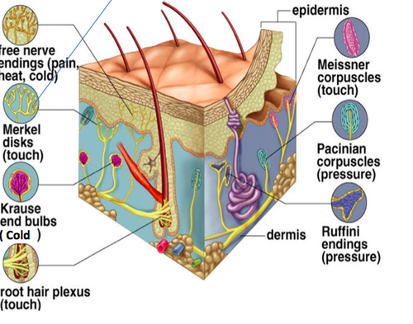
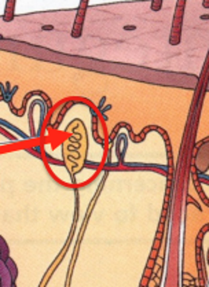
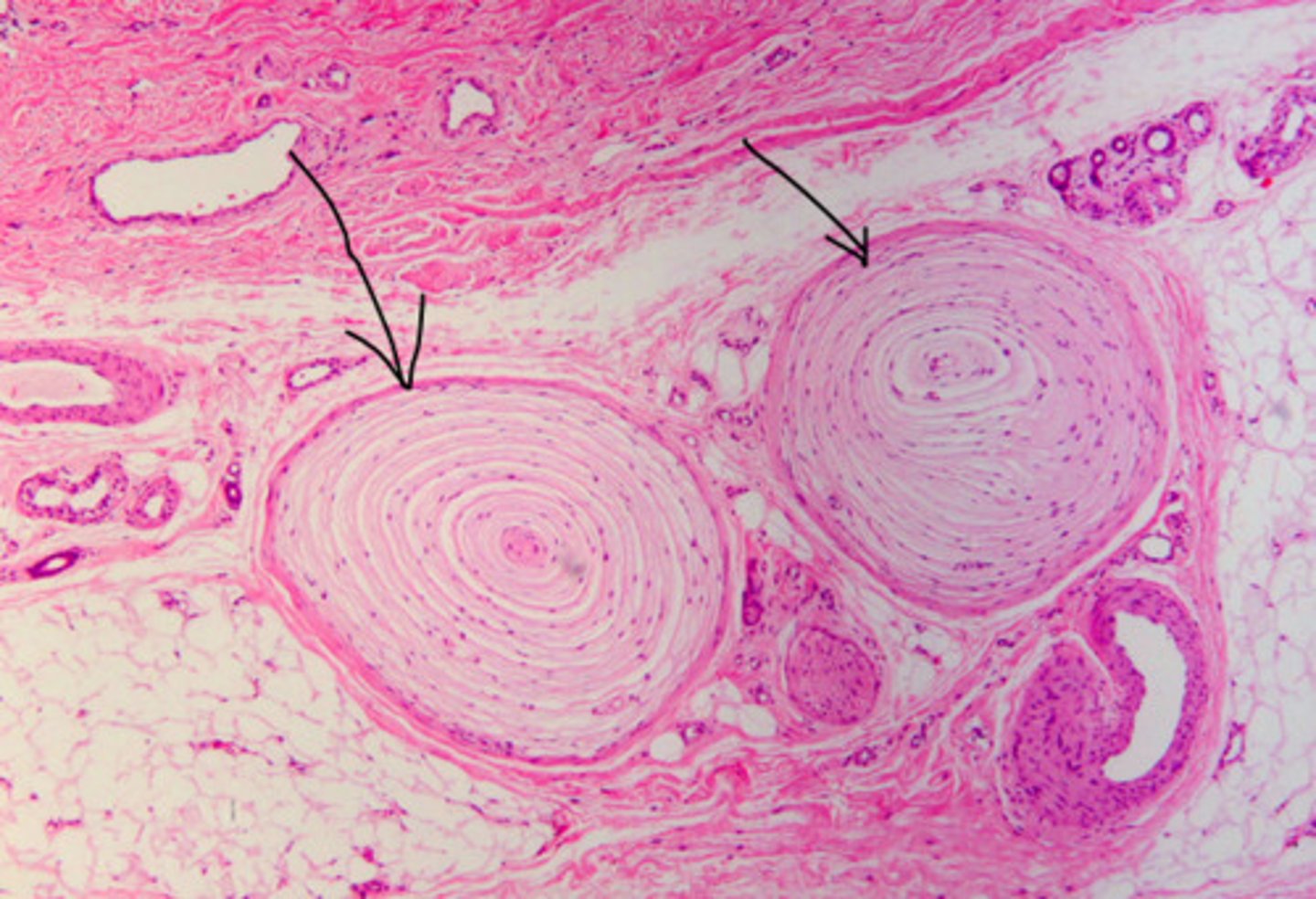
Pacinian corpuscle
very rapidly adapting and is stimulated only by rapid
movement & deep pressure
detects deep pressure and deep vibration
Tactile Receptors
Free nerve endings
Meissner's corpuscles
Merkel's discs
Hair end organ
Ruffini's end organ
Pacinian corpuscle
Tactile Sense Transmission - fast
Meissner's corpuscles, hair receptors, Pacinian
corpuscles and Ruffini's end organs transmit signals
in type Ab nerve fibers FAST
30-70 m/s
Tactile Sense Transmission - slower
Free nerve endings transmit signals in type Ad nerve
fibers at 5-30 m/sec, some by type C unmyelinated
fibers at
Tactile Sense Transmission - The more critical the information
the faster the rate of transmission
Almost all sensory information enters the spinal
cord through
dorsal roots of spinal nerves
Pathways for the Transmission of Sensory Information
2 pathways for sensory information:
1. dorsal column - medial leminscal system
2. anterolateral system
Dorsal Column System
◻ Contains large myelinated nerve fibers for FAST TRANSMISSION
◻ High degree of spatial orientation maintained
throughout the tract
◻ Transmits information rapidly
Dorsal Column System - examples
1. touch
2. vibration
3. position
4. fine pressure
Anterolateral System
• Smaller myelinated & unmyelinated fibers for SLOW TRANMISSION
• Low degree of spatial orientation
• Transmits a broad spectrum of modalities
Anterolateral System - examples
1. pain
2. temp
3. crude touch
4. tickle and itch
5. sexual sensation
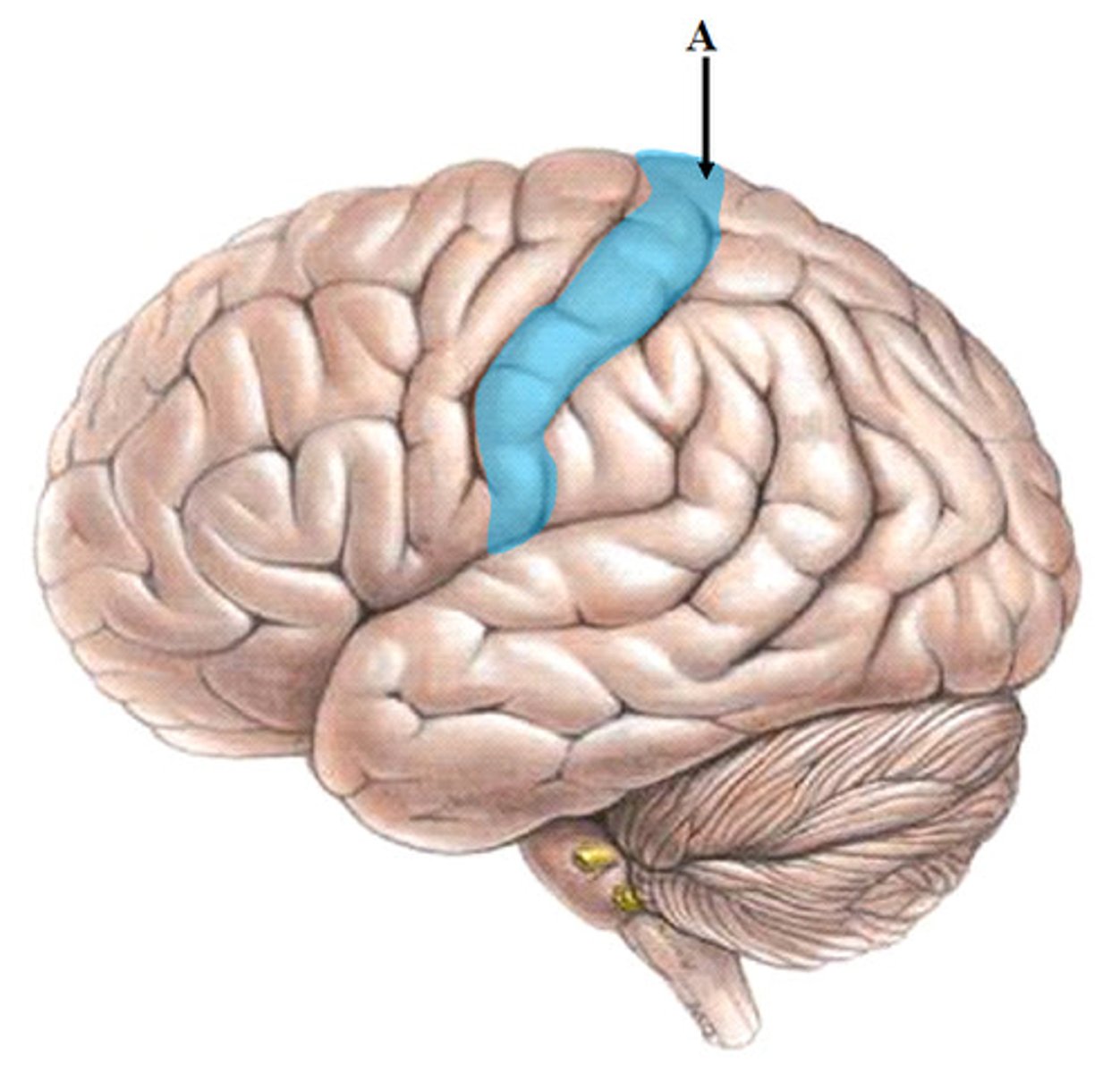
Somatic Sensory Cortex location
postcentral gyrus
Somatic Sensory Cortex
receives sensory input that gives rise to our sensations of heat, touch, and cold and to our senses of balance and body movement
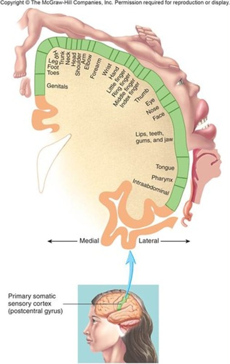
Somatic Sensory Cortex organization
• Highly organized distinct spatial orientation
• Each side of the cortex receives information from opposite side of the body
Unequal representation of the body - Somatic Sensory Cortex
lips have greatest area of representation followed by the face and the thumb - trunk and lower body have the least area
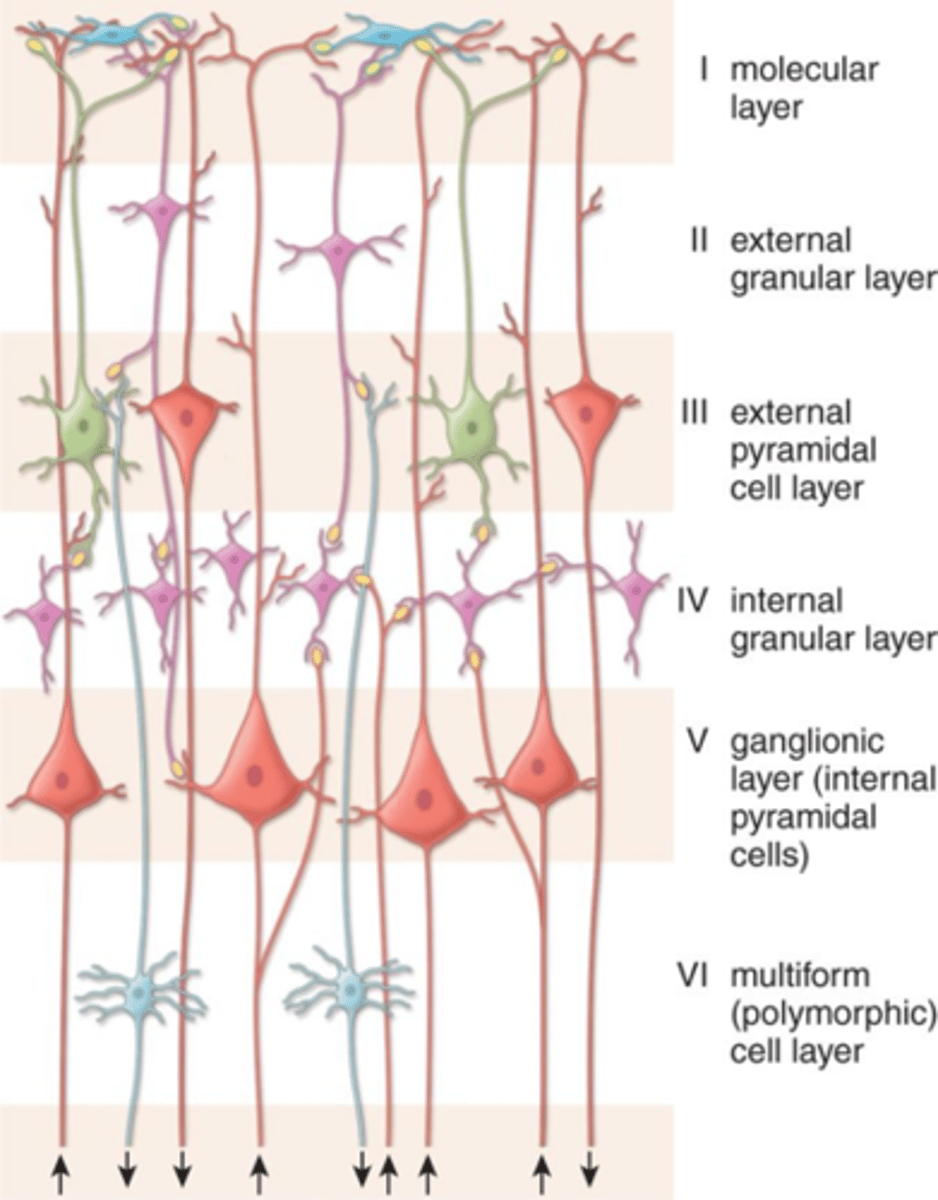
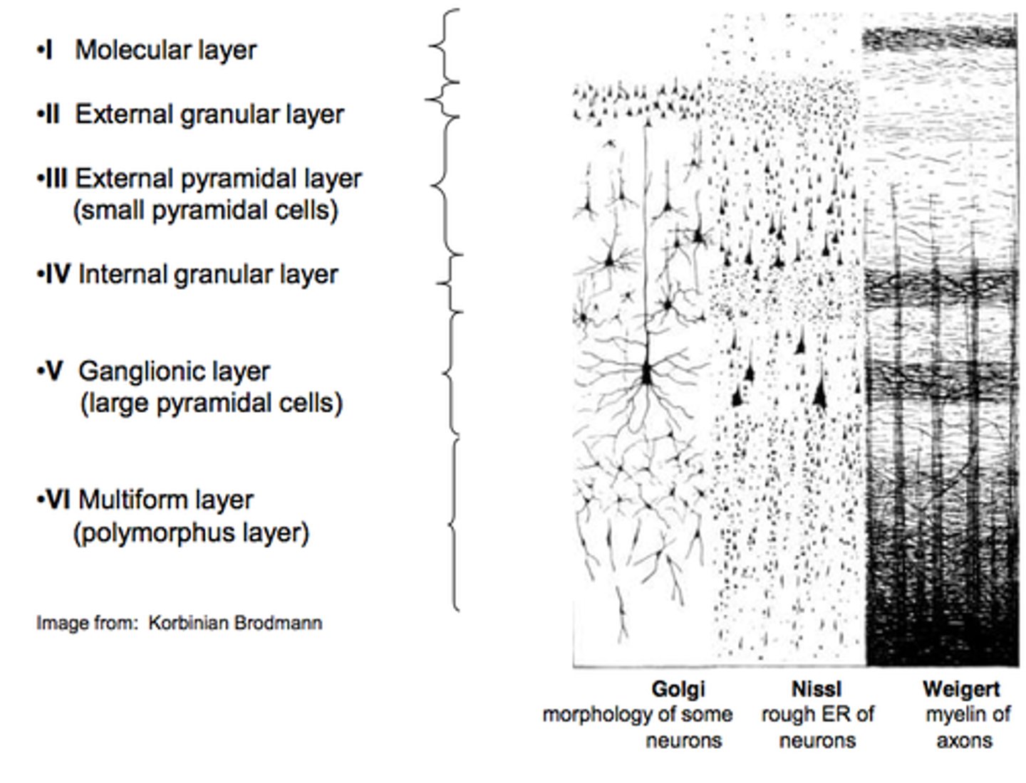
Cellular Organization of the Cortex
- Six separate layers of neurons
layer I is near the surface of cortex
layer VI is deep within the cortex
cortex incoming signals enter what layer
layer IV (4)
Layers I and II receive
diffuse input from lower brain centers
Layer II and III neurons send axons to
closely related areas of cortex (similar function)
layer V and VI send axons to
distant parts
VI to thalamus
V to brainstem and cord
Further cellular organization of Cortex
- Within the layers the neurons are also arranged in
columns
- Each column serves a specific sensory modality (ex. strength, pressure, touch)
- Different columns interspersed among each other (may have 2 strength, then 2 pressure, than 3 touch, then 1 pressure, etc.)
Cellular Organization of the Cortex - Layer I
near the surface of the cortex

Cellular Organization of the Cortex - layer VI
deep in the cortex

Cellular Organization of the Cortex - IV
incoming signals enter

Cellular Organization of the Cortex - Layer I & II
receive input from lower brain centers

Cellular Organization of the Cortex - layer II & III
closely regulated cortex nearby areas
send axons to communicate to areas receiving similar signals
Cellular Organization of the Cortex - layer V and VI
more distant parts
V - brainstem and spinal cord
VI - thalamus
Function of the Somatic Sensory Cortex: Destruction of Somatic area I results in
1. lose discrete localization ability (touch)
2. lose ability to judge degree of pressure
3. lose ability to determine weight of object
4. asterognosia
5. inability to judge texture
asterognosia
inability to determine shape of an object by touch
Graphesthesia
ability to "read" a number by having it traced on the skin
Somatic Association Areas - location
Located behind the somatic sensory cortex - parietal area of cortex
Somatic Association Areas - receive input from where?
Association areas receive input from somatic sensory
cortex, ventrobasal nuclei of the thalamus, visual and
auditory cortex
Somatic Association Areas - function
decipher sensory meaning
Somatic Association Areas - loss fo these areas
results in loss of ability to recognize complex objects - smell, feel, etc
also even loss of self (self awareness)
pain occurs when
tissue is being damaged
Pain function
protection
two types of pain
fast pain and slow pain
fast pain
felt within 0.1 sec of stimulation (type A fiber)
described as sharp
easily localized by patients
slow pain
can take up to 1 sec or more
- throbbing or aching pain
- general areas of pain, less localized
All pain receptors are
free nerve endings
Pain Receptors can be stimulated by
- can be stimulated by: mechanical stretch, thermal (hot/cold) and chemical (bradykinin, serotonin, histamine, prostygandins, substance P)
Bradykinin
chemical that causes the most pain and most responsible chemical for causing pain