Brigs anatomy exam 2
1/361
There's no tags or description
Looks like no tags are added yet.
Name | Mastery | Learn | Test | Matching | Spaced | Call with Kai |
|---|
No analytics yet
Send a link to your students to track their progress
362 Terms
origin of the external abdominal oblique m
ribs 5-12
insertion of the external abdominal oblique m
aponeurosis to the linea alba
iliac crest
pubic crest
innervation of the external abdominal oblique m
ventral rami of the lower 6 thoracic spinal nerves
iliohypogastric n
ilioinguinal n
same an internal abdominal oblique and transversus abdominus
action of the external abdominal oblique m
support and compress abdominal contents
flexion and rotation of trunk
same as internal abdominal oblique
origin of the internal abdominal oblique m
lumbar fascia
iliac crest
lateral 2/3 of the inguinal ligament
insertion of the internal abdominal oblique m
aponeurosis to ribs 10-12
xiphoid process
linea alba
pubic symphysis
innervation of internal abdominal oblique
ventral rami of lower 6 thoracic spinal nerves
iliohypogastric n.
ilioinguinal n.
same as external abdominal oblique and transversus abdominus
action of the internal abdominal oblique m
support and compress abdominal contents
flexion and rotation of the trunk
same as external abdominal oblique
origin of the transversus abdominis m
ribs 7-12
lumbar fascia
iliac crest
lateral 1/3 of inguinal ligament
insertion of transversus abdominis m
xiphoid process
linea alba
pubic symphysis
same as internal oblique minus ribs
innervation of the transversus abdominis m
ventral rami of lower 6 thoracic spinal nerves
iliohypogastric n
ilioinguinal n
same as both obliques
action of the transversus abdominus m
support and compress abdominal contents
-acts as a girdle to abdomen, helps with IAP
origin of the rectus abdominis m
pubic crest
pubic symphysis
insertion of the rectus abdominis m
costal cartilage of ribs 5-7
note: these are 3 tendonous structures in the belly muscles
innervation of rectus abdominis m
ventral rami of the lower 6 thoracic spinal nerves
note: this is lacking ilio muscles seen in obliques and transversus abdominis
action of the rectus abdominis m
support and compress abdominal contents
flex the trunk
2 planes in the quadrant method
median
transumbilical
RUQ organs
liver
colon
R kidney
gallbladder
pancreas
LUQ organs
liver
colon
L kidney
stomach
spleen
pancreas
RLQ organs
colon
small intestine
appendix
ureter
major a and v to R leg
R kidney
LLQ organs
colon
small intestine
L kidney
ureter
major a and v to L leg
9 regions
umbilical
hypogastric
epigastric
L and R iliac
L and R lumbar
L and R hypochondriac
3 planes for regions
subcostal
transtubercular
midclavicular
Umbilicus level
L3-L4 in someone lean
pubic crest
bony ridge lateral from pubic symphysis to pubic tubercle
pubic tubercle
small, rounded elevation where inguinal ligament attaches
iliac crest
bony ridge coming posteriorly from anterior superior iliac spine
iliac tubercle
thickened part of iliac spine 6cm posterior to anterior superior iliac spine
inguinal groove
separates abdomen and leg
-site of inguinal ligament which goes from ASIS to pubic tubercle
epigastric fossa
pit under xiphoid process
linea alba
from fascial planes surrounding abdominal muscles
-xiphoid process to pubic symphysis
-location of medial borders of rectus abdominis
linea semilunaris
curved groove from 9th costal cartilage to pubic tubercle
-location of lateral borders of rectus abdominis
midinguinal point
midpoint of line between ASIS and pubic symphysis
superior border of abdominal wall
L and R costal margins
inferior border of abdominal wall
horizontal line connecting anterior and superior iliac spines
lateral border of abdominal line
vertical line through anterior superior iliac spines
Horizontal planes (4)
1. transpyloric @ L1
2. subcostal @ L3
3. transumbilical @ L3-L4
4. transtuberculear @ L5
transpyloric plane
L1
midway between suprasternal notch and pubic symphysis
-passes pylorus of stomach
two layers of superficial fascia in the abdominal wall
1. camper's fascia - fat (fat little campers)
2. scarpa's fascia - deep fascia blends with fascia lata from thigh
Anterior wall of the rectus sheath
formed of the aponeuroses of the internal and external oblique muscles
posterior wall of the rectus sheath
formed from the aponeuroses of the internal oblique and transversus abdominis muscles
what do the aponeuroses form in the middle
linea alba
arcuate line
posterior wall of the rectus sheath between umbilicus and pubic rami
-formed as posterior and anterior rectus sheaths fuse
below arcuate line
no posterior rectus sheath, just a thin layer known as transversalis fascia
Inguinal ligament
-formed by aponeuroses of external abdominal oblique as it reflects inwards
-attached laterally to anterior superior iliac spine
-attached medially to pubic tubercle
lacunar ligament
-also EAO aponeuroses, but it is reflected anteriorly
-attaches to pectineal ligament and line on innominate bone
-is an extension of the inguinal ligament
-medial border of inguinal triange
Location of the inguinal canal
medial 1/2 of the inguinal ligament, directly superior
-is ~5cm long and connects the abdominal cavity to the external abdominal wall w/ reproductive structures
inguinal canal contents
males = spermatic cord
females = round ligament
significance of an oblique course in the inguinal canals
hernia
borders of inguinal canal
anterior - aponeurosis of the external abdominal oblique
posterior - transversalis fascia and conjoint tendon
inferior - inguinal and lacunar ligaments
superior - internal abdominal oblique and transversus abdominis muscles
superficial inguinal ring
medial end of the inguinal canal
-superior and lateral to the pubic tubercle
-arched opening in the IAO aponeurosis
-medial and lateral crus
medial and lateral crus attachments of the inguinal ring
medial crus - pubic symphysis
lateral crus - pubic tubercle
deep inguinal ring
lateral end of inguinal canal
-goes through the transversalis fascia superior to the external iliac artery
-courses medially to the external iliac artery
-runs lateral to the inferior epigastric artery
function of the round ligament
smooth muscle that anchors the labia majora
reflected ligament
is the inguinal ligament reflected superiorly to the linea alba
conjoint tendon
aponeuroses of the IAO and transversus abdominis
-reinforce posterior wall of inguinal canal
direct inguinal hernia
older males
-peritoneal bulge through the posterior wall of the inguinal canal due to thin transversalis vascia
hasselbach's triange - direct inguinal hernias - borders
lateral - inferior epigastric artery
inferior - inguinal ligament
medial - rectus abdominis muscle
indirect inguinal hernias
most common, usually R sided
-peritoneal contents push through the deep inguinal ring into inguinal canal
femoral hernia
more in females
-peritoneal bulge passes through femoral ring to femoral canal
2 types of umbilical hernias
congenital and acquired
congenital umbilical hernias
when the midgut does not return to abdominal cavity during fetal development
acquired umbilical hernias
infancy: weakness in scar of umbilicus in linea alba
adult: more in women, umbilical region of linea alba
epigastric hernia
high intra-abdominal pressure
-in the large portion of the linea alba between the xiphoid and umbilicus
-middle-aged laborers
superior border of the abdominal cavity
diaphragm
inferior border of the abdominal cavity
none b/c it is continuous with the pelvic cavity
which muscular and fascial structures form the anterior, posterior, lateral walls of the abdominal cavity
external abdominal oblique
internal abdominal oblique
transversus abdominis
rectus abdominis
transversalis fascia
ribs
Peritoneum
-thin, serous membrane of abdominal and pelvic cavity
-mesothelium as "middle" layer
-thin basement layer of alveolar CT
Structure of the peritoneum
folds back on itself to create 2 layers: visceral and parietal
parietal peritoneum
lines the peritoneal cavity
visceral peritoneum
covers the external surfaces of abdominal organs
2 specializations of the peritoneum
omentum and mesothelium
omentum
greater and lesser
-double-layered peritoneum with adipose, blood vessels, and lymphatic cells
-connects the stomach to body wall or other viscera
greater omentum
has a free edge
is connected to one organ, greater curvature of stomach
3 ligaments that bind the stomach to other structures (formed by the greater omentum)
gastrophrenic ligament
gastrosplenic ligament
gastrocolic ligament
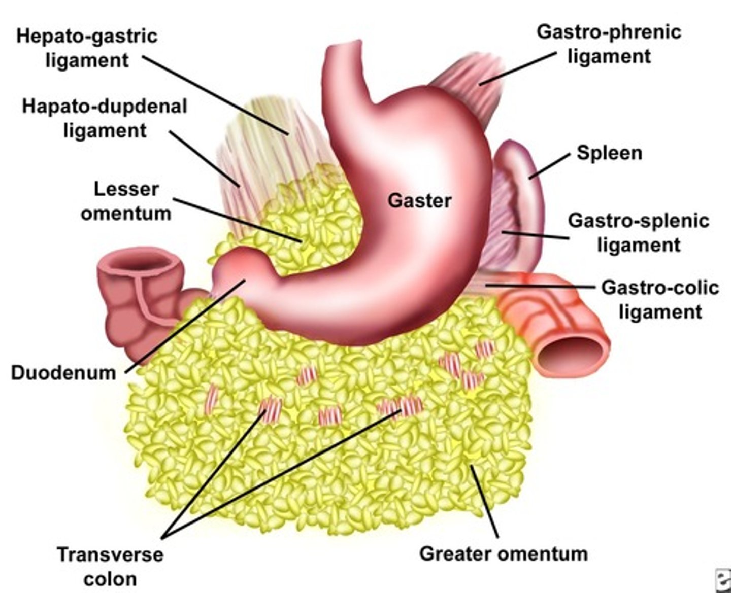
lesser omentum
attaches one organ to another
connects the lesser curvature of stomach to the duodenum
what 2 ligaments are formed by the lesser omentum connection the stomach to the duodenum
1. hepatogastric ligament
2. hepatoduodenal ligament
what is contained within the hepatoduodenal ligament?
the portal triad
portal vein, hepatic artery, bile duct
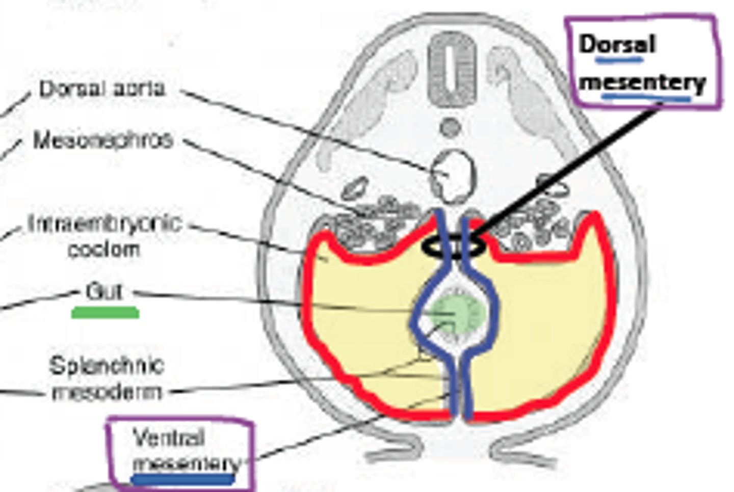
mesentery
double layered peritoneum that anchors organs to the abdominal wall
-contains blood vessels, nerves, and lymph vessels to supply organs
function of omentum
some degree of lymphatic protection against infection spread
Lesser sac
space between the stomach and posterior abdominal wall
-posterior to lesser omentum and superior to transverse colon

greater sac
remainder of the peritoneal cavity, anterior to lesser omentum
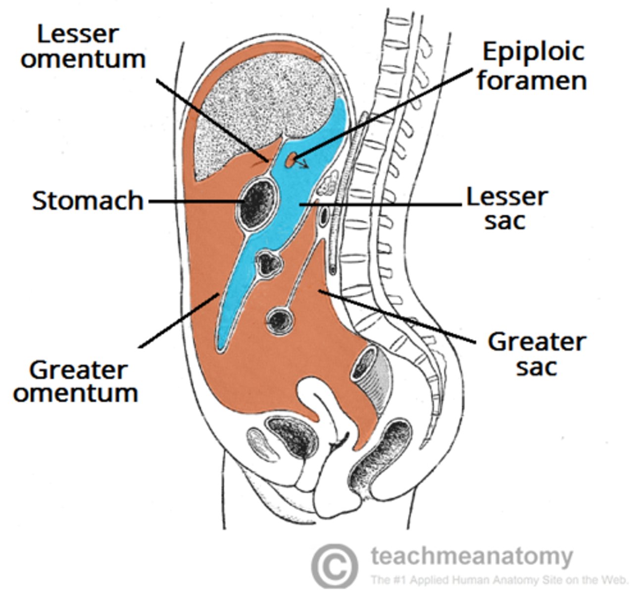
epiploic forament
opening between the greater and lesser sac which allows communication
anterior border of the epiploic foramen
the portal triad on the gastroduodenal ligament
posterior border of the epiploic foramen
inferior vena cava
superior border of the epiploic foramen
caudate lobe of the liver
inferior border of the epiploic foramen
duodenum, portal vein, hepatic artery, bile duct
7 different mesenteries
1. mesentery proper
2. sigmoid mesocolon
3. transverse mesocolon
4. mesoappendix
5. falciform ligament (only ligament to anterior abdominal wall)
6. ligamentum teres (remanent from umbilical vein in fetus)
7. coronary ligament (between liver and diaphragm)
importance of compartments, gutters, and recesses
allow for flow of ascites fluid and infectious material between the abdomen and pelvis
Supracolic compartment
Greater sac above the transverse colon containing the liver, stomach, and spleen
-divided into smaller sections by visceral ligaments
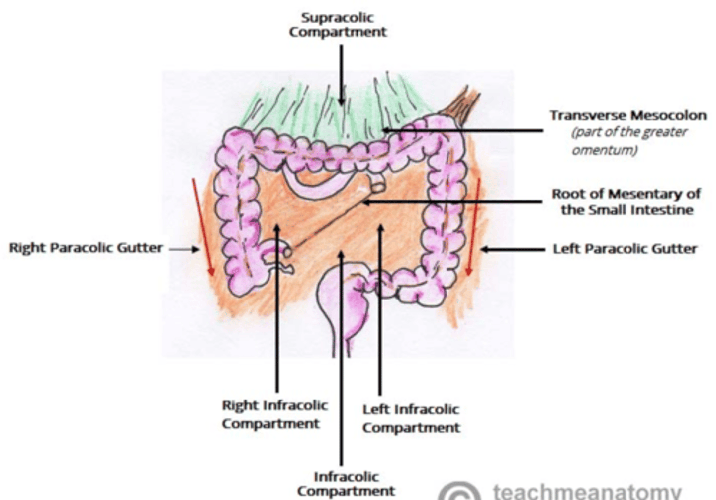
2 subdivisions of the supracolic compartment
1. L and R subphrenic spaces - divided by coronary ligaments
2. hepatorenal pouch
Infracolic compartment
Greater sac below the transverse colon containins small intestine, ascending and descending colon
-divided by mesentery proper
2 infracolic compartment subdivisions
1. R infracolic space
2. L infracolic space
divided by the mesentery proper
peritoneal gutters
a means for the supra and infracolic compartments to communicate
4 peritoneal gutters
1. R lateral paracolic gutter
2. L lateral paracolic gutter
3. Gutter to the right of the mesentery
4. Gutter to the left of the mesentery
Right paracolic gutter
lateral to the ascending colon
-communication between the lesser sac and pelvis
-pathway for infection between hepatorenal pouch and pelvis
left paracolic gutter
lateral to the descending colon
gutter to the right of the mesentery
is bounded both inferiorly and superiorly
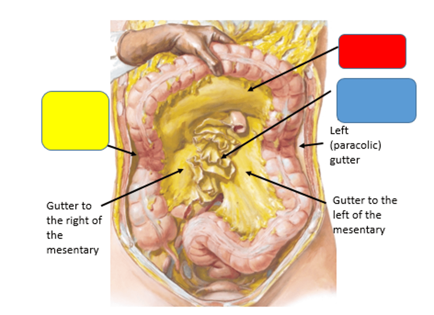
gutter to the left of the mesentery
is open to the pelvis
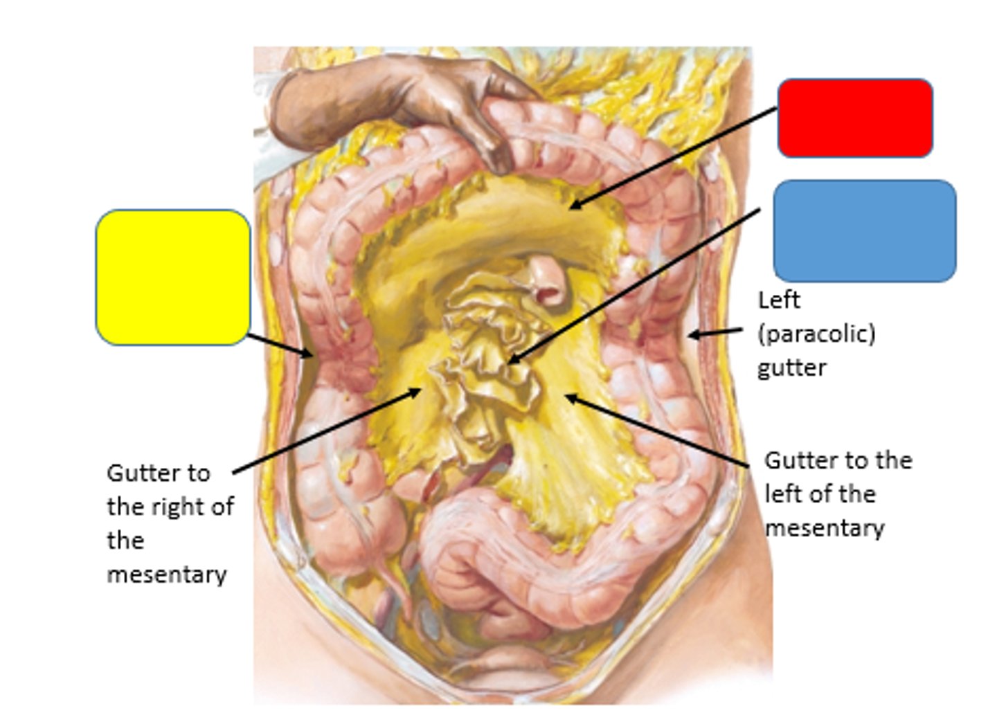
what is the only gutter that does not connect to the pelvis?
the gutter to the right of the mesentery