Heart and great vessels
1/90
There's no tags or description
Looks like no tags are added yet.
Name | Mastery | Learn | Test | Matching | Spaced | Call with Kai |
|---|
No analytics yet
Send a link to your students to track their progress
91 Terms
pericardium
fibrous sac that encloses the heart
3 portions of pericardium
-Fibrous pericardium
-Serous pericardium: parietal layer and visceral layer -pericardial cavity
Fibrous pericardium
outermost layer
function: protection
Serious pericardium: parietal layer
contacts fibrous pericardiu
Serious pericardium: Visceral layer
contacts epicardium (surface of heart)
pericardial cavity
space between visceral and parietal layer (of serous pericardium)
filled with serous fluid (5-30mL)
Inner to outer layers of heart to pericardium
heart epicardium
visceral pericardium (serous pericardium)
pericardial cavity with serous fluid
parietal pericardium (serous pericardium)
Fibrous pericardium
Heart anatomical location
1. intercostal spaces
- right: 2-5
-left: 2-6
right of midline= 1/3 mass
left of midline= 2/3 mass
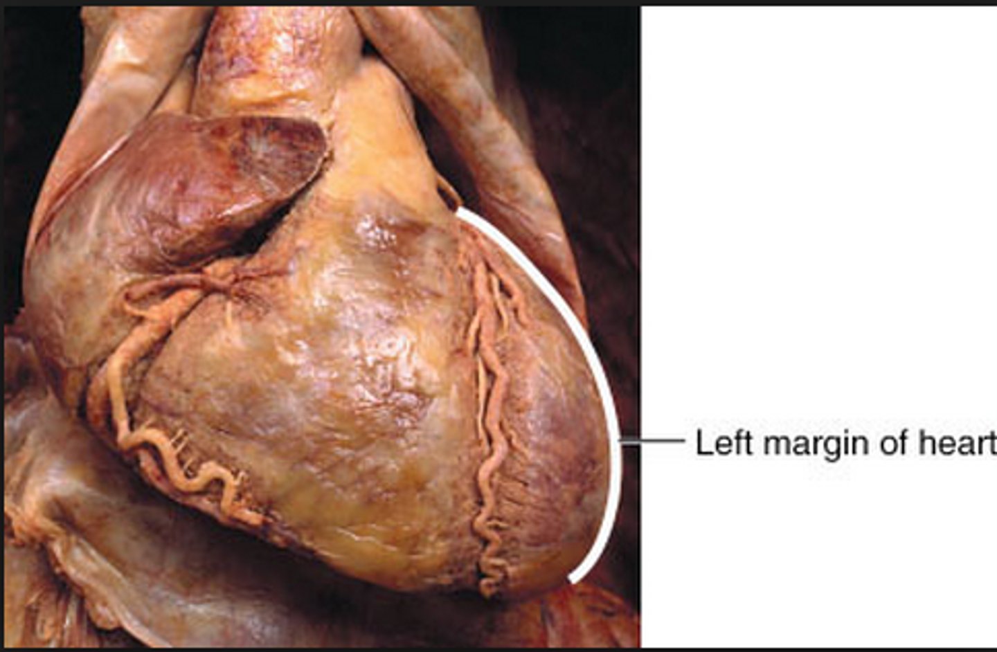
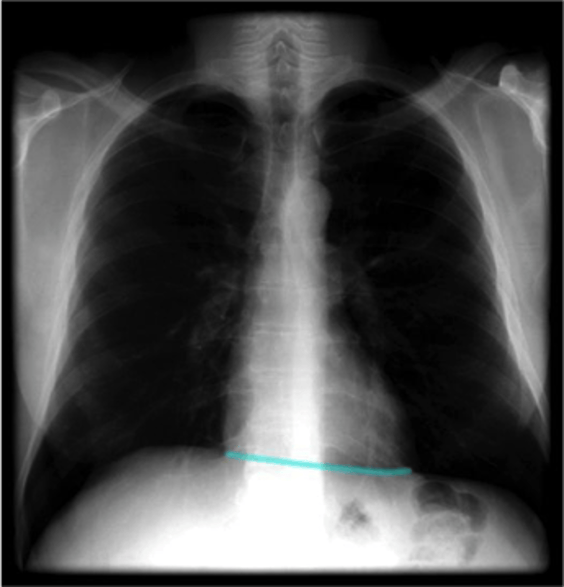
4 heart borders
1. right
2. left
3.superior
4. inferior
Left heart margin
right heart margin
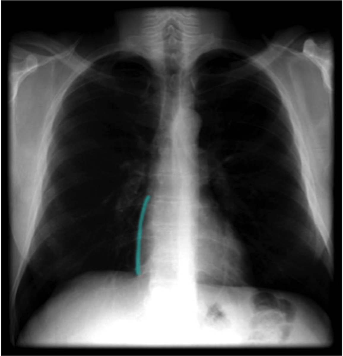
inferior heart margin
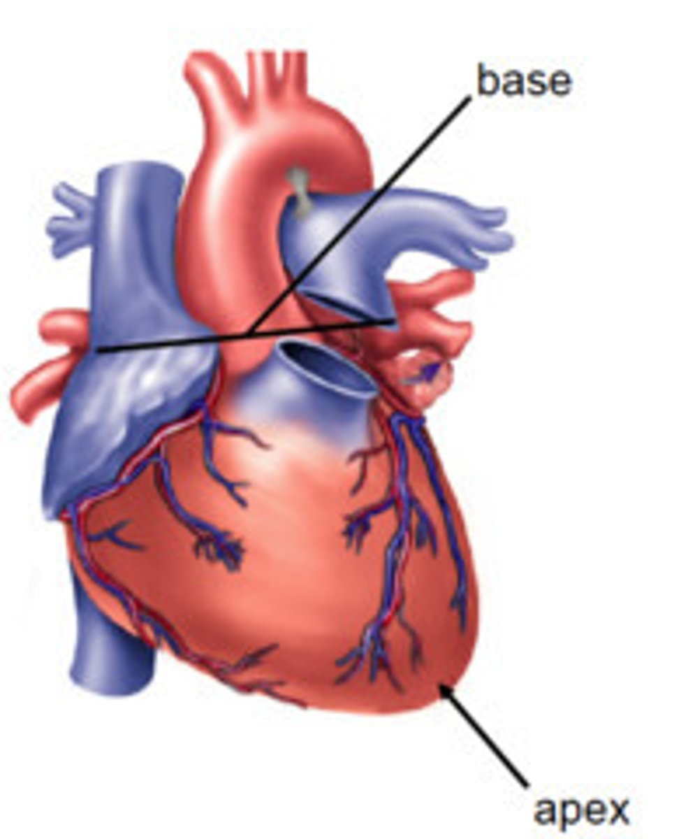
Apex of heart
inferior
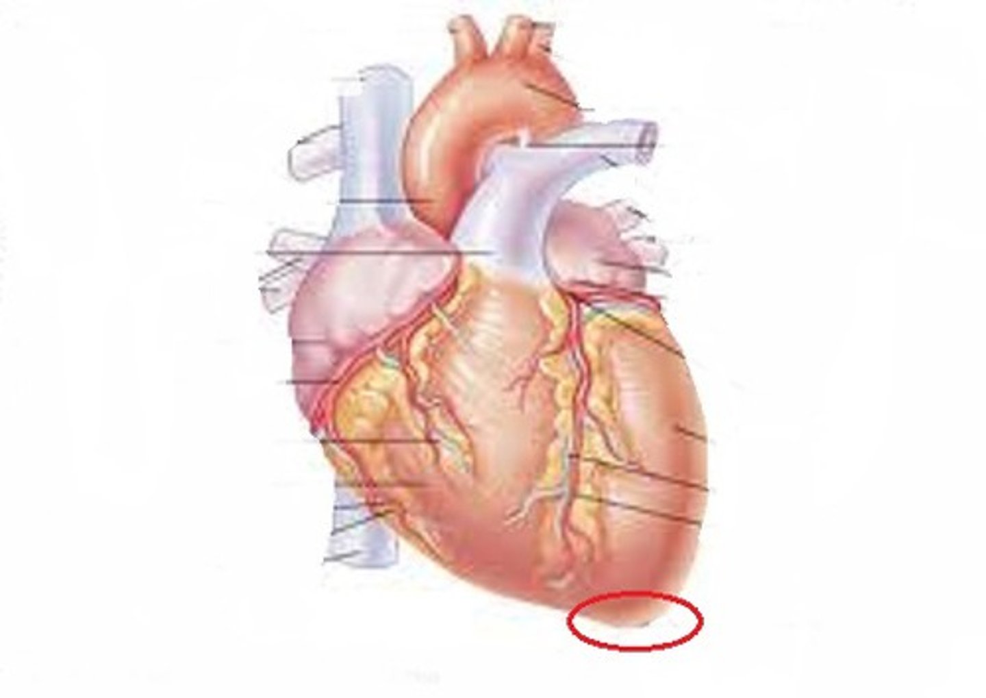
base of the heart
superior (this is where vessels reside)
Auricles of the heart
Flaps that cover the atria
Function: unknown but thought to be evolution remnants
3 layers of the heart wall
Epicardium
myocardium
endocardium
epicardium
composed of mesothelial cells and connective tissue (fat)
contains: fat, vessels, nerves
myocardium
muscle layer (contractile myocytes)
endocardium
lines inner cavities of the heart
comprised of smooth muscle and connective tissue
deep layer of connective tissue contains conduction system
-AKA: subendocardial layer
pericardium and heart wall layers from outside to insude
pericardial sac (fibrous layer then serous layer)
epicardium
myocardium
endocardium
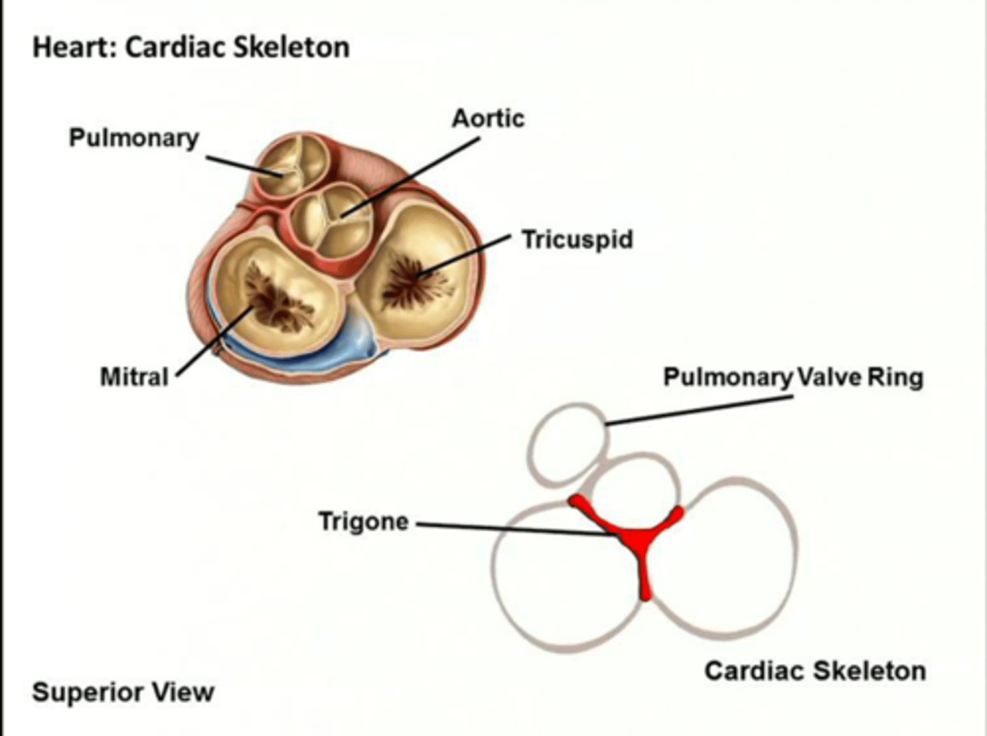
skeleton of the heart
fibrous rings that hold the heart open and provide a substrate for cardiac muscle contraction
rings surround valves and pulmonary orifices (openings)
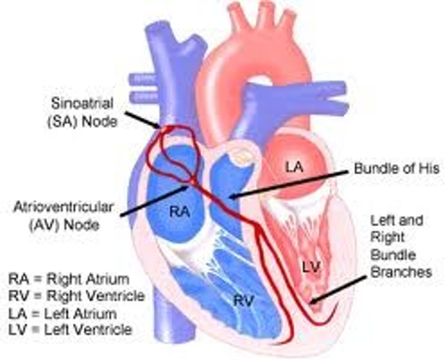
Electrical conduction system
sinoatrial node
atrioventricular node
AV bundles
Left and right bundle branches
purkinje (plexus) fibers
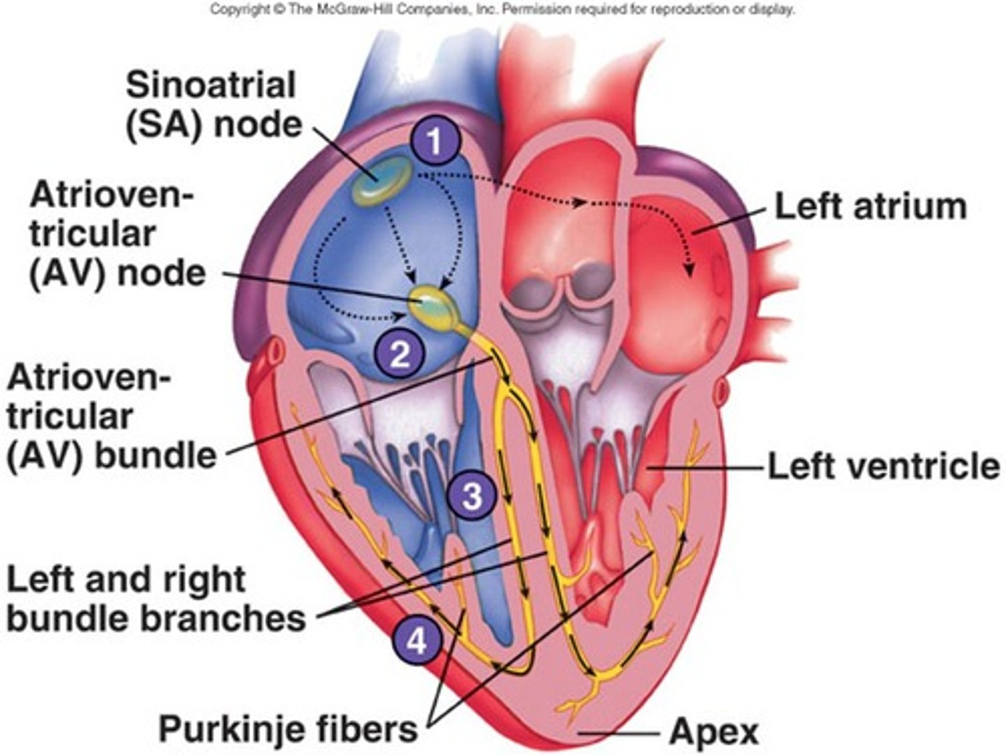
sinoatrical node
pacemaker (in right atrium)
Atrioventricular node
ectopic pacemaker (backup)
in interatrial septum
AV bundles
(bundle of His)
top (superior) of interventricular septum
Left and right bundle branches
run inferiorly along right and left sides of interventricular spetum
purkinje (plexus) fibers
lateral walls of ventricles
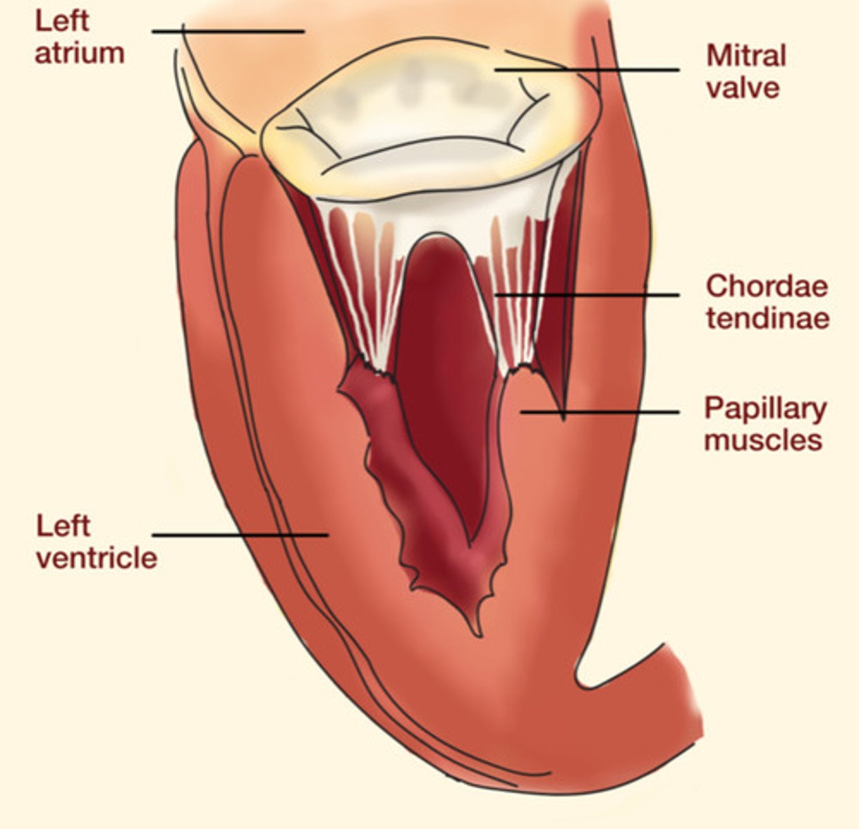
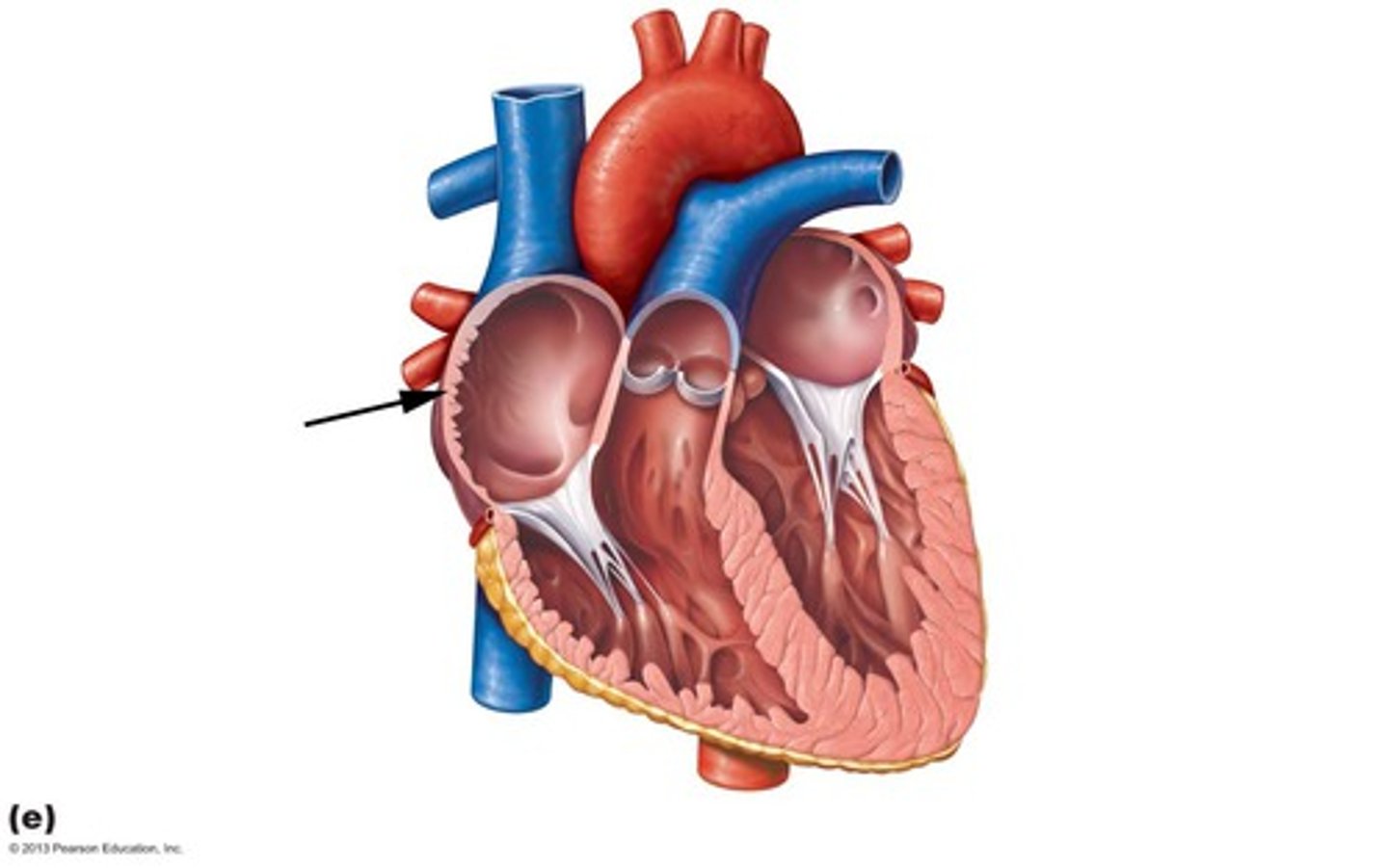
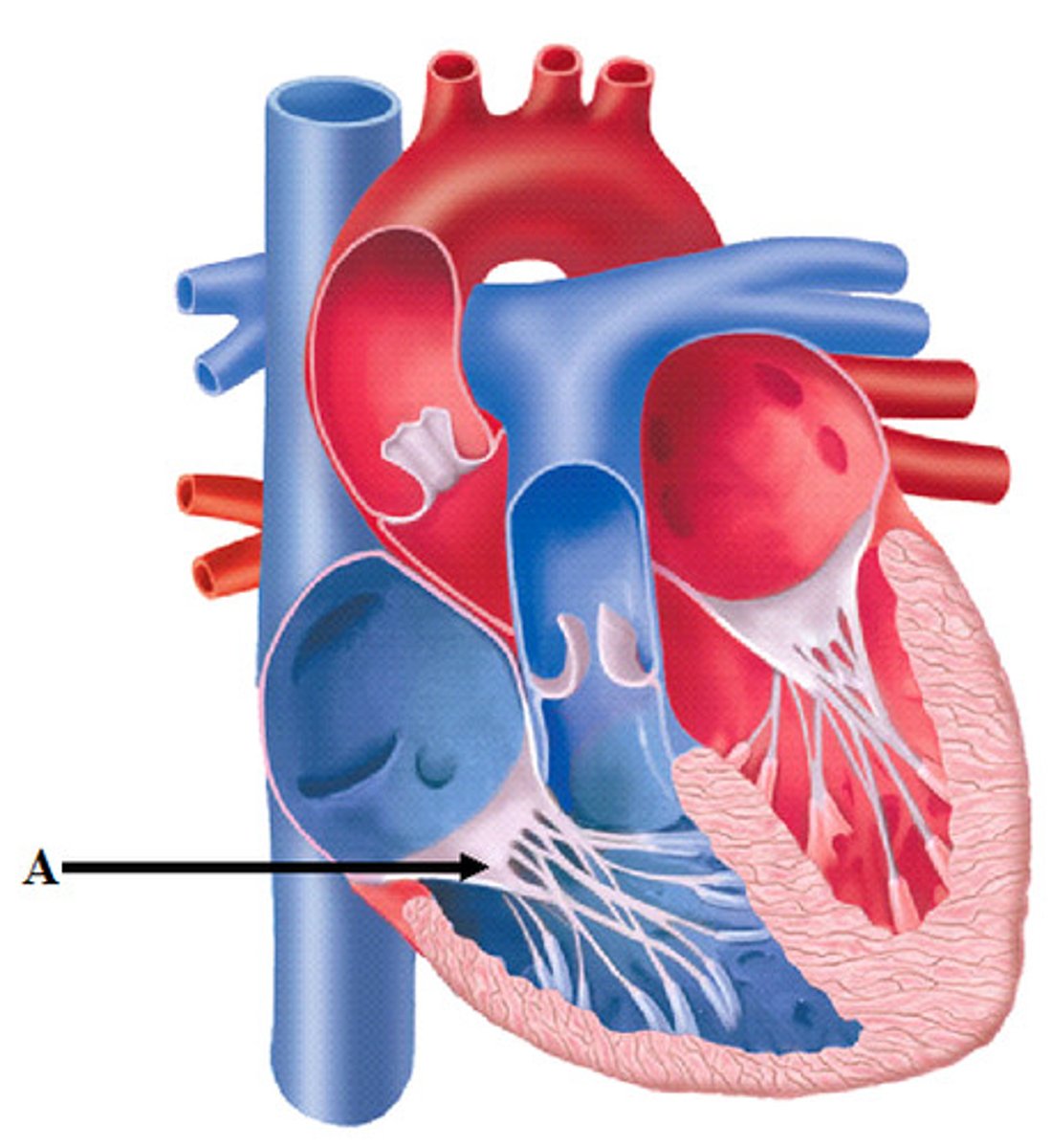
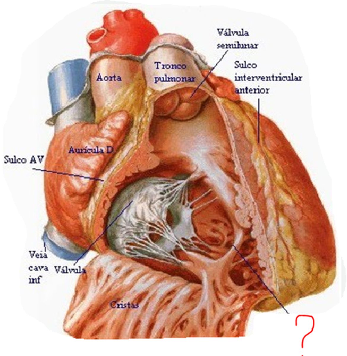
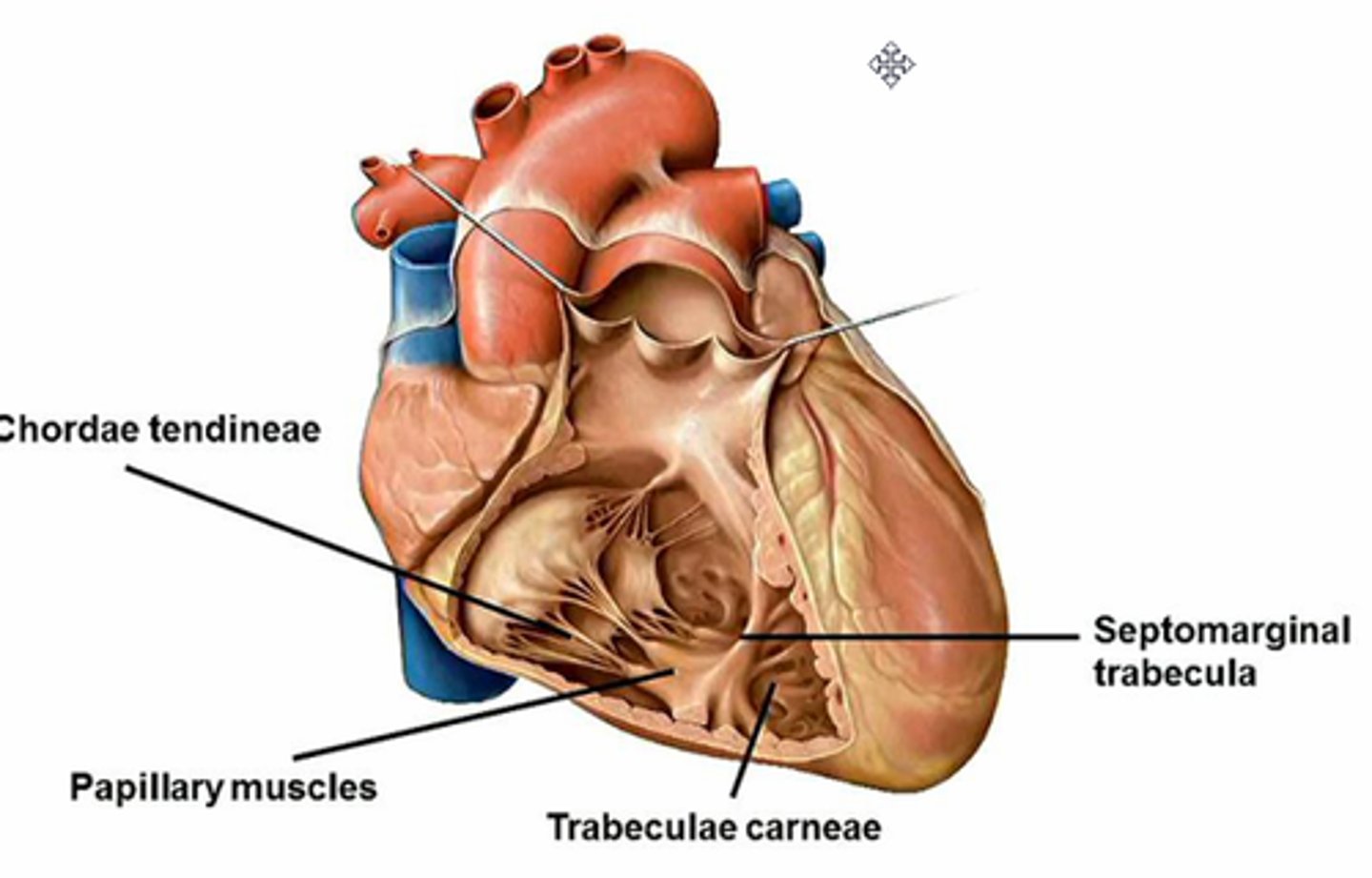
Atrioventricular valves
1. tricuspid (right AV valve)
2. bicuspid (left AV valve) (also called mitral valve)
have chordae tendinae attached
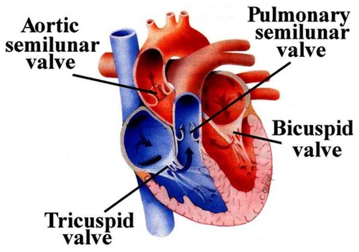
what do chordae tendinae do for AV valves
prevent backflow of blood (from ventricle into atrium)
the papillary muscles contract and that generates tension on the chordae tendinae which hold the AV valves closed
Semilunar valves
pulmonary and aortic
have sinuses

what do sinuses do for semilunar valves
full with blood to prevent backflow (of blood from aorta or pulmonary trunk back into left and right ventricles respectively)
All heart valves lie
in one plane
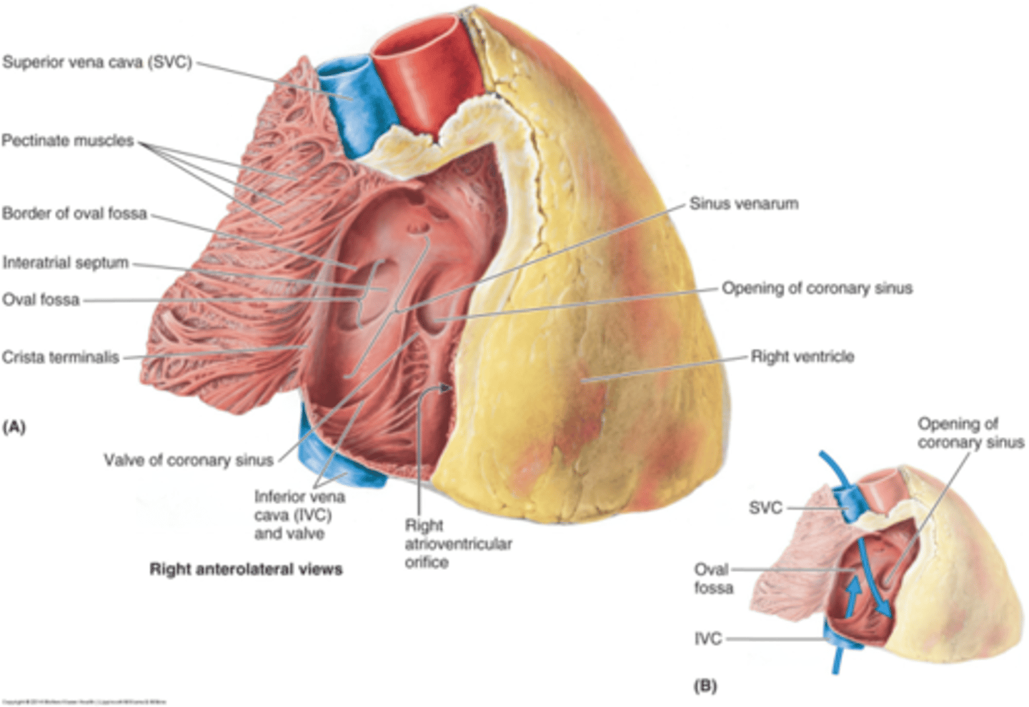
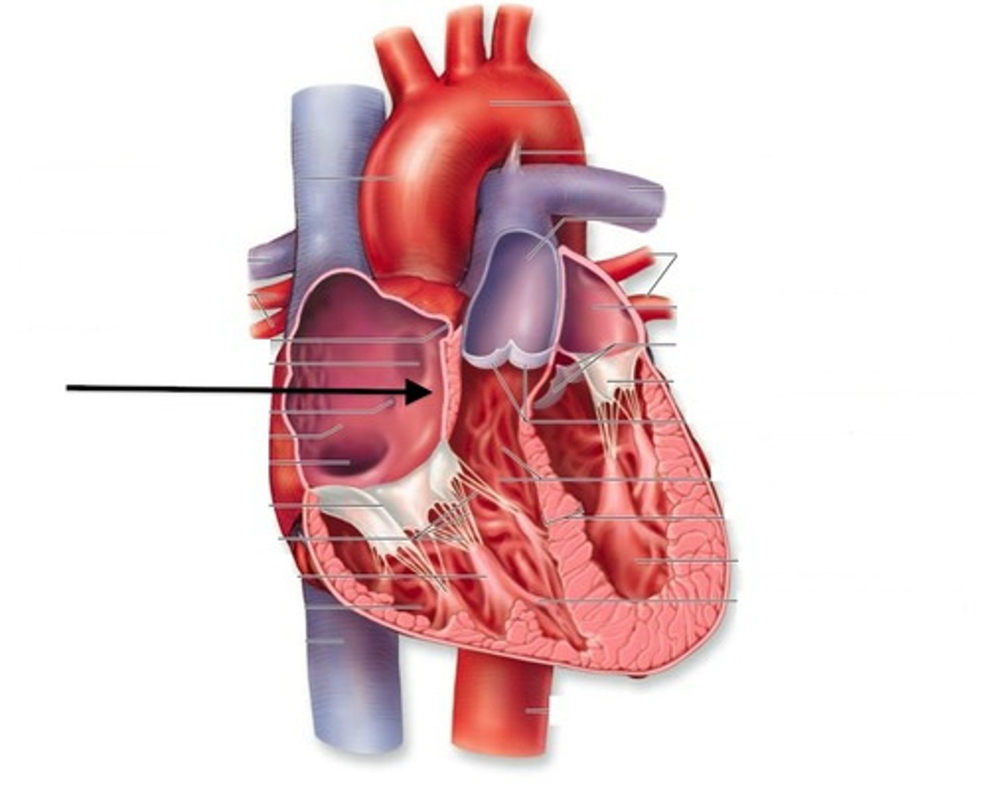
Right atrium
sulcus terminalis (external)- cavity and auricle
crista terminalis- internal ridge
**pectinate muscles- internal ridges
4 major openings of right atrium
Superior vena cava
inferior vena cava
coronary sinus- drains deoxygenated blood from heart into right atrium
right AV orifice - guarded by tricuspid valve
Right atrium: SA node
firing mediated by SNS and PSNS (ANS)
vagal tone (PSNS): heart rate (depolarizations) decreased by vagus nerve
usually depolarize 80-100 times per minute by with vagus nerve, 60-80 time per minute

Right atrium: AV node
regulates impulse conduction from atria to ventricles
Right atrium: interatrial septum
fossa ovalis- depression that is closed
in the fetus it is called foramen ovale (hole)
-connects left and right atrium
-closes within 24 hours of birth
Clinical application of interatrial septum
open back up in post menopausal females
the symptom= feelings bubbles/butterflies
can cause strokes in the post menopausal age (58ish)
Right ventricle communicates with
right atrium via Atrioventricular orifice
Right ventricle characterisitics
-additional openings: pulmonary orifice
-guarded by pulmonary semilunar valve
-thicker walls than right atrium- contain projecting ridges called trabeculae carnae
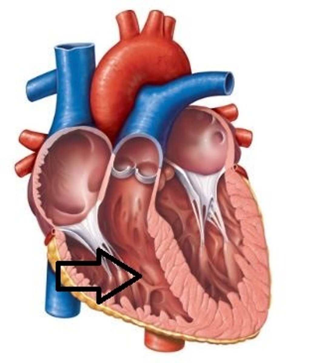
2 types of projecting ridges in right ventricle
1. papillary muscles
2. trabeculae carnae (in right ventricle there is a specific special one)
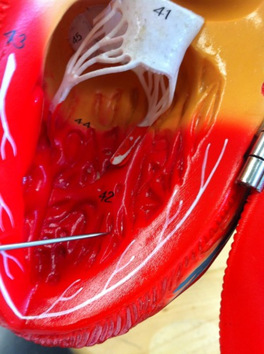
Special trabeculae carnae in right ventricle
septomarginal trabecule (moderator band)
-used during fetal ultrasound to find fetus heart rate...helps orient yourself to heart because you know it is the right ventricle (can find what is what after this)
septomarginal trabecule (moderator band)
-prevent overdistension of ventricle
-spans base anterior papillary muscle to ventriclular septum
-carries part of right branch of AV bundle to anterior papillary muscle (helps with electrical conduction system pathway)
Left atrium
cavity and auricle
AV orifice guarded by mitral valve
4 pulmonary orifices-represent 4 pulmonary veins (number can vary)
Left ventricle communicates with
left atrium via AV orifice guarded by mitral valve
Left ventricle characteristics
additional opening= aortic orifice
walls 3-5 times thicker than right ventricle because more pressure on the left side
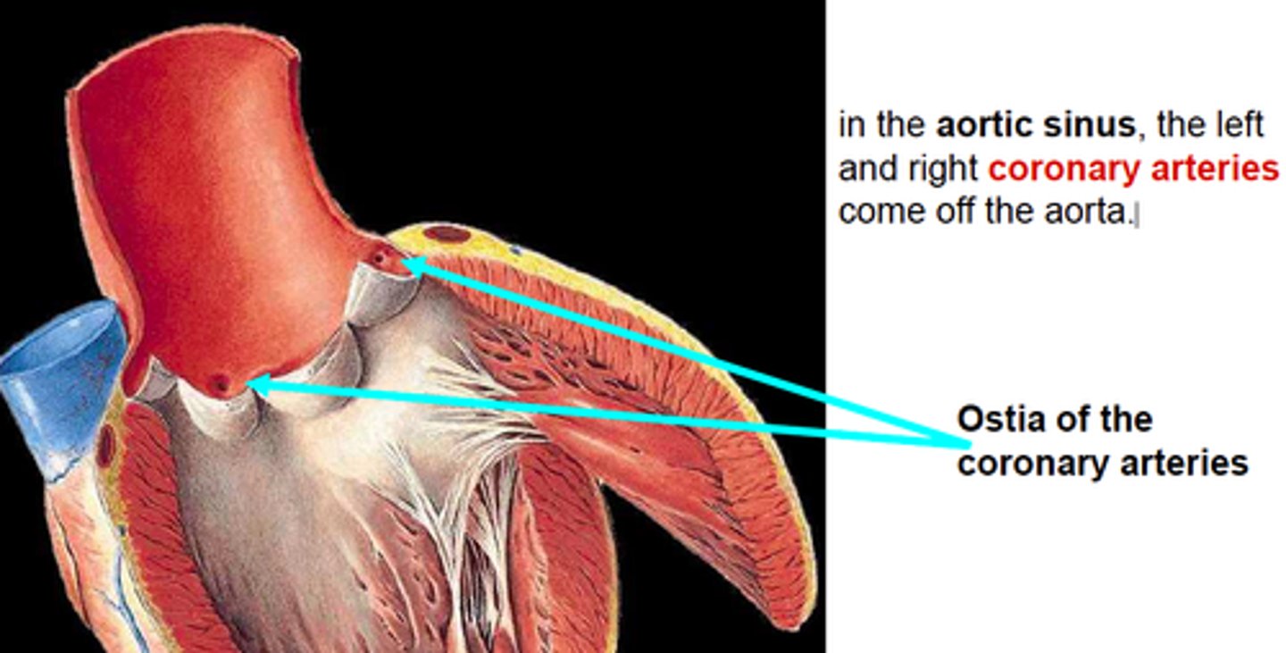
Aortic SL valve
Aortic SL valve
behind each cusp are bulges
-anterior and posterior aortic sinuses that give rise to right and left coronary arteries (feed oxygen rich blood to heart muscle)
ostium
opening of the right and left coronary artery (in aorta)
Blood flow through the heart
Superior/ Inferior vena cava
Right atrium
tricuspid valve
right ventricle
pulmonary semilunar valve
pulmonary trunk (artery)
lungs
The blood picks up oxygen in the lungs, and then flows from the lungs:
pulmonary veins
left atrium
Bicuspid valve
left ventricle
aortic semilunar valve
aorta
to the body
When is blood deoxygenated in that path
from superior vena cava to the lungs
-notice that the pulmonary artery holds deoxygenated blood
When is blood oxygenated in that path
From lungs to aorta
notice that the pulmonary veins have oxygenated blood
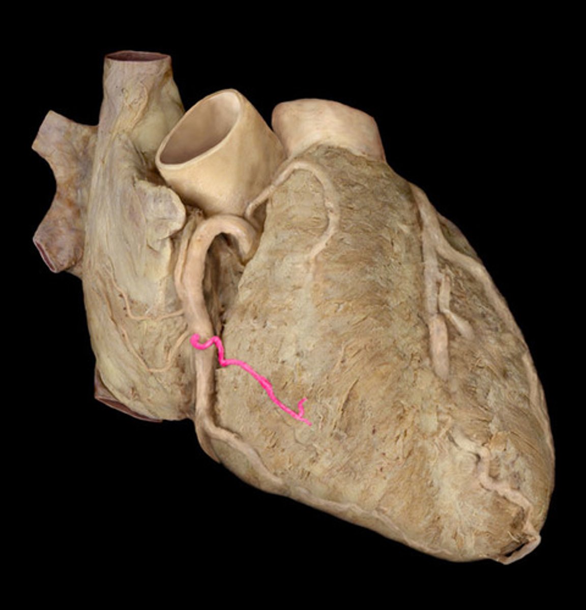
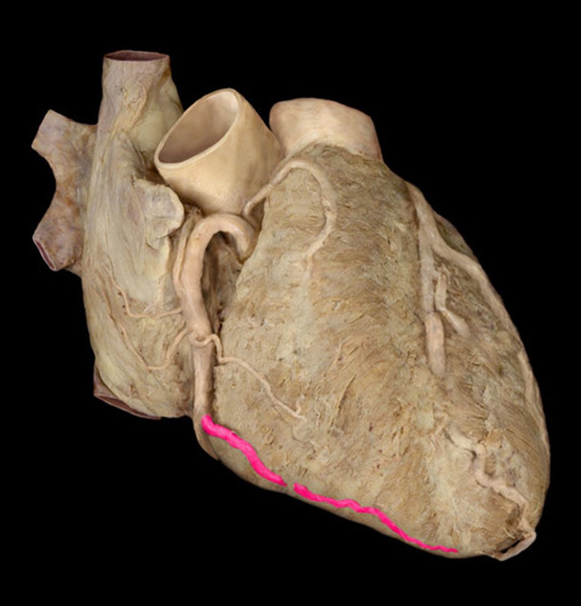
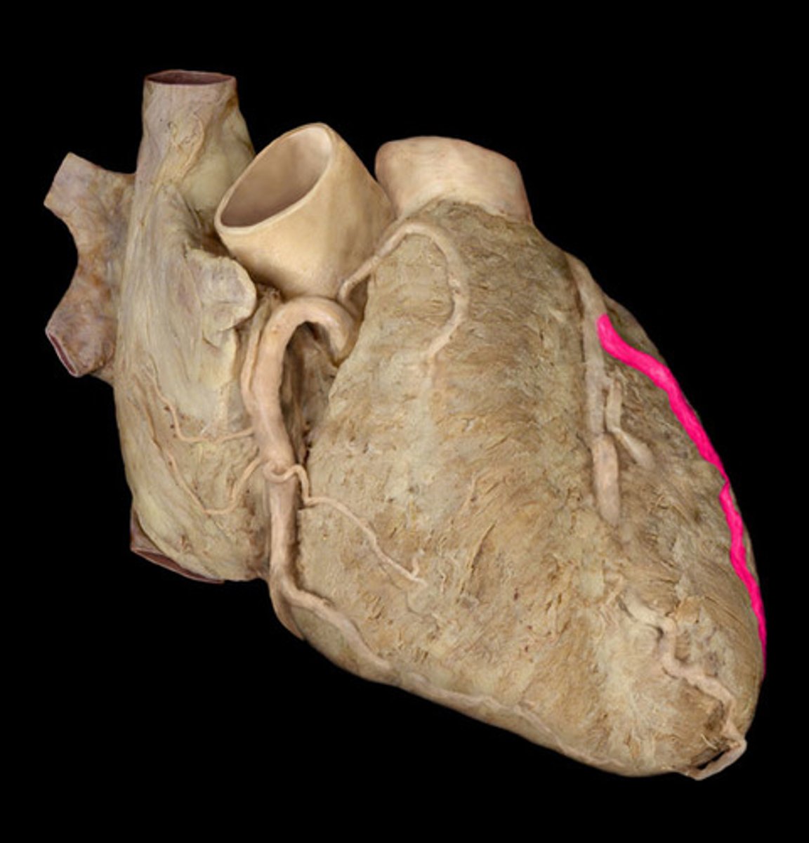
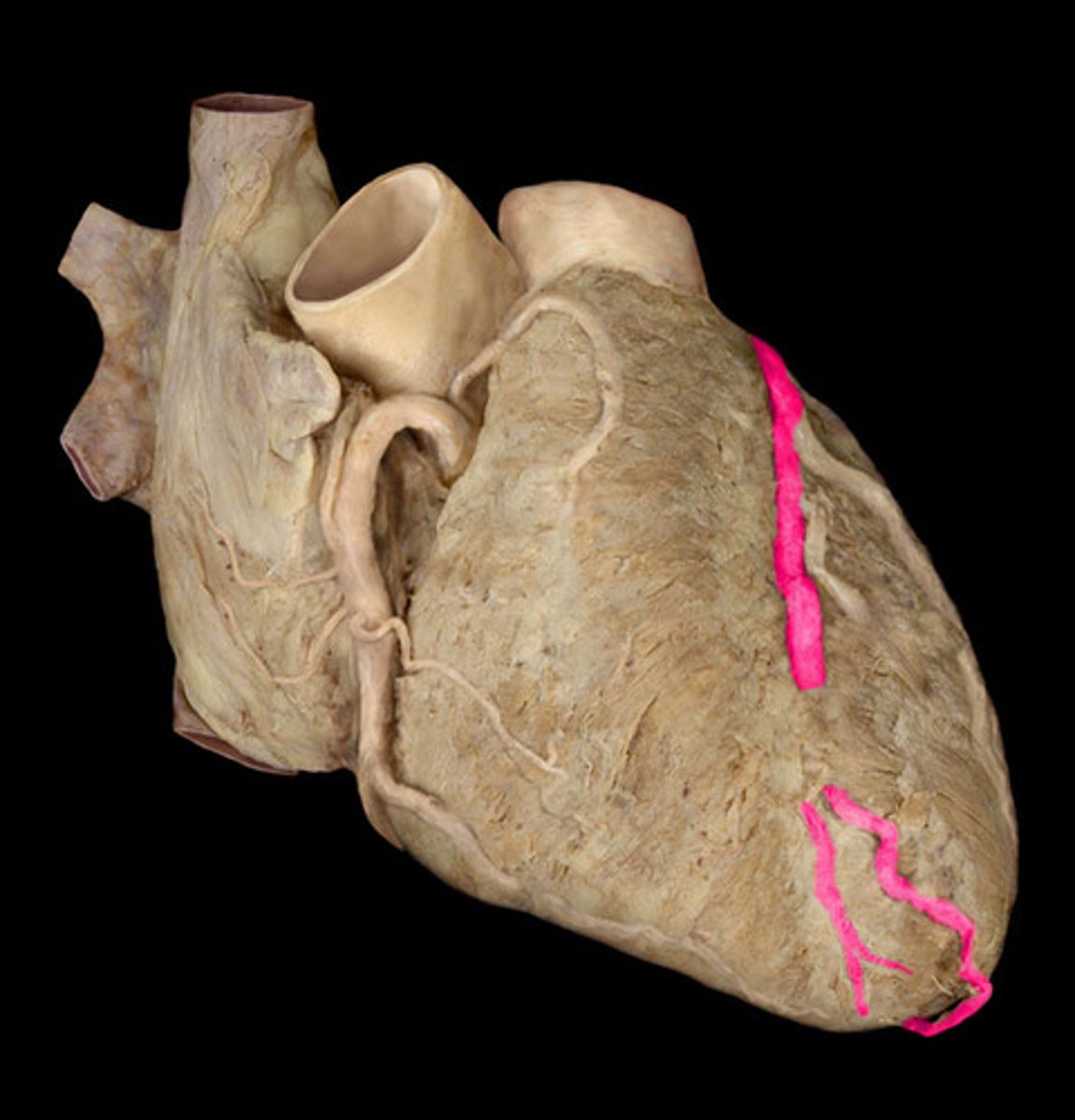
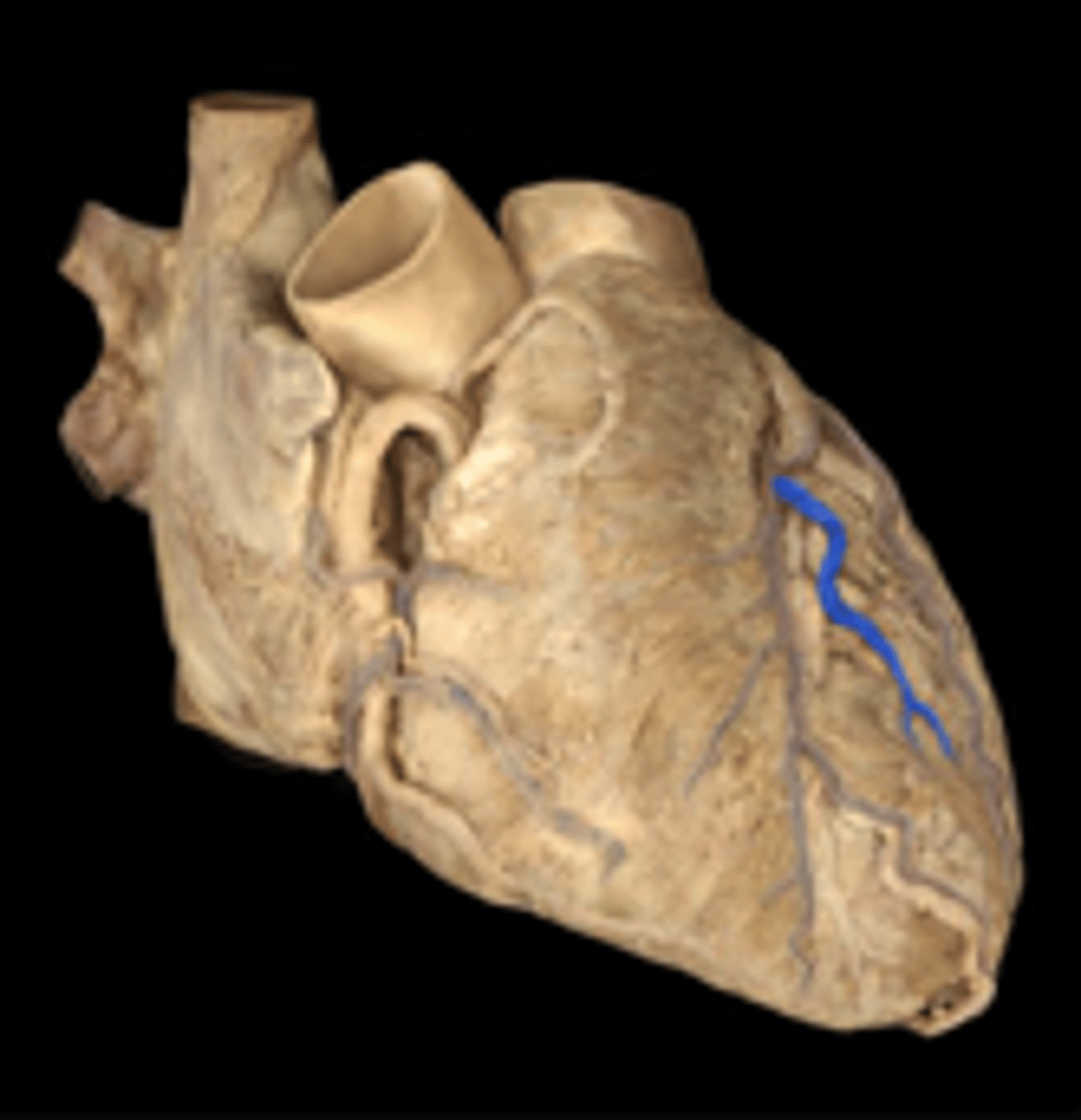
Heart arterial supply
variation!!!
right coronary artery ad left coronary artery
Right coronary artery supplies
right atrium, most right ventricle, part of left ventricle, SA and AV nodes
Right coronary branches
right marginal artery
nodal artery (SA node=60%) and (AV node=80%)
posterior interventricular artery (most commonly)
Left coronary artery supplies
left atrium, most left ventricle, part of right ventricle, SA node= 40% and AV node=20%
Branches of left coronary artery
circumflex artery
left anterior interventricular artery (AKA: left anterior descending artery...LAD)
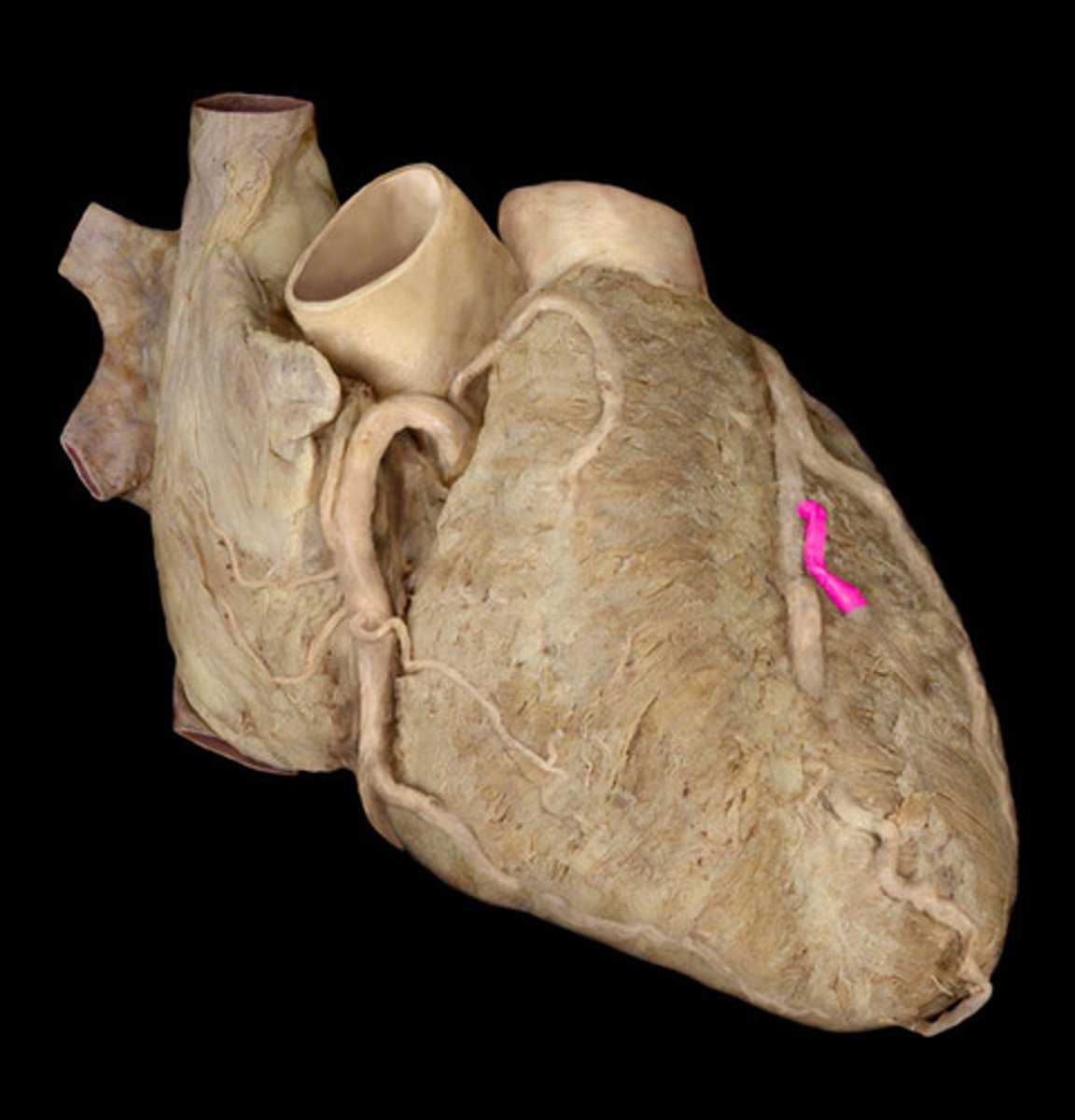
Coronary artery dominance
which coronary artery gives rise to the posterior interventricular artery
most common= RCA (67%)
right and left coronary artery
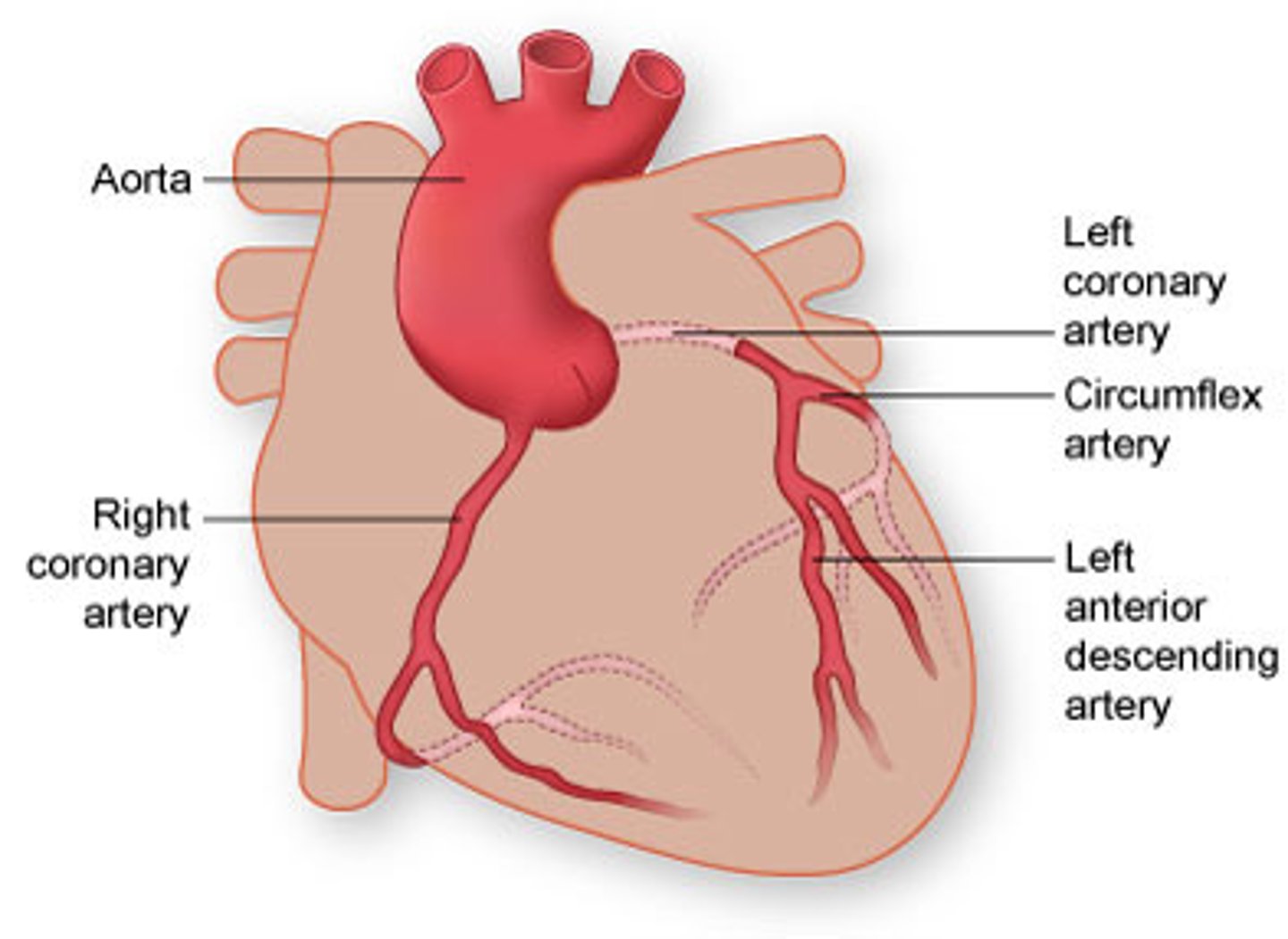
right anterior ventricular artery
right coronary artery branch
right marginal artery
right coronary artery branch
right atrial artery
right coronary artery branch
right conus artery
right coronary artery branch
left marginal artery
left coronary artery branch
left interventricular artery
left coronary artery branch
left anterior ventricular artery
left coronary artery branch
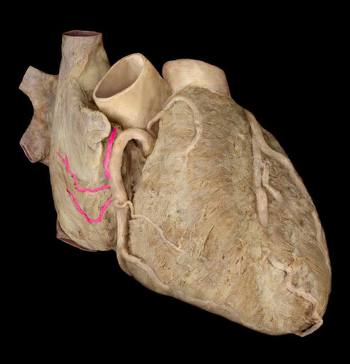
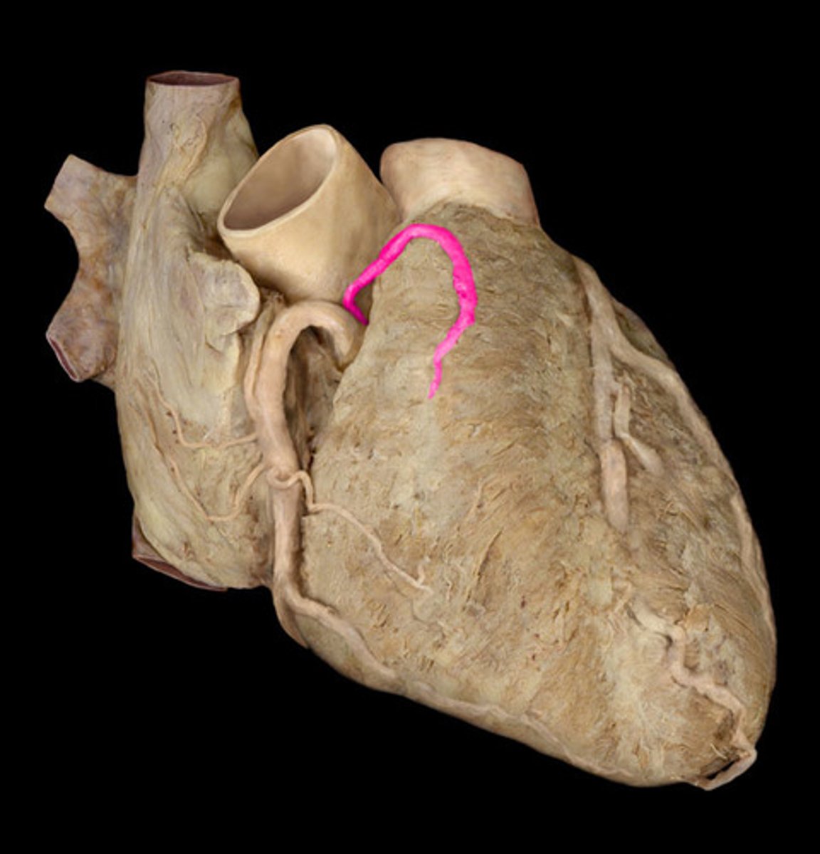
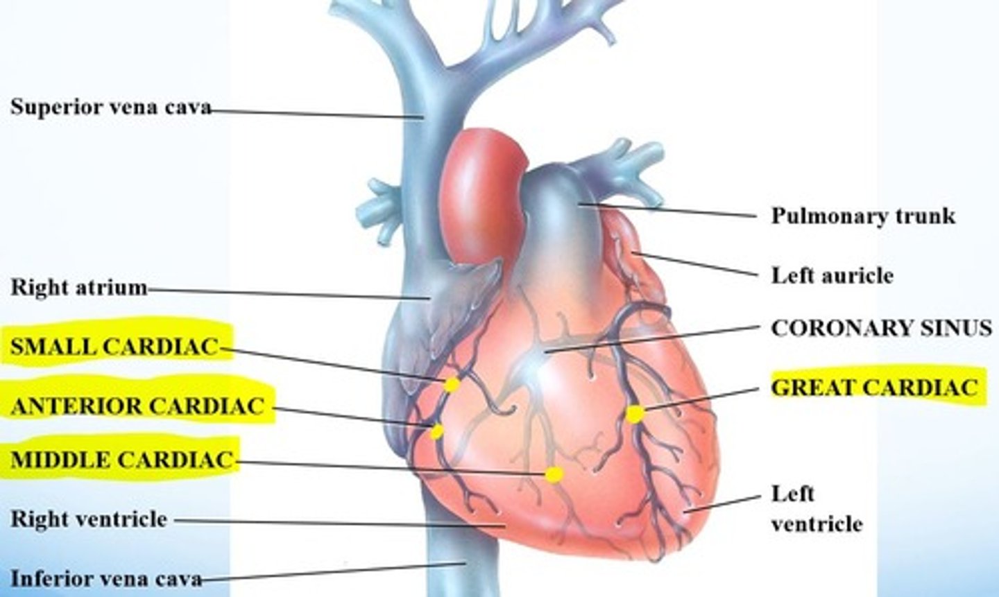
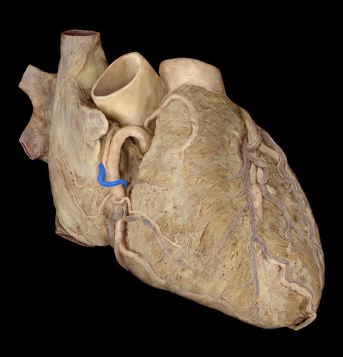
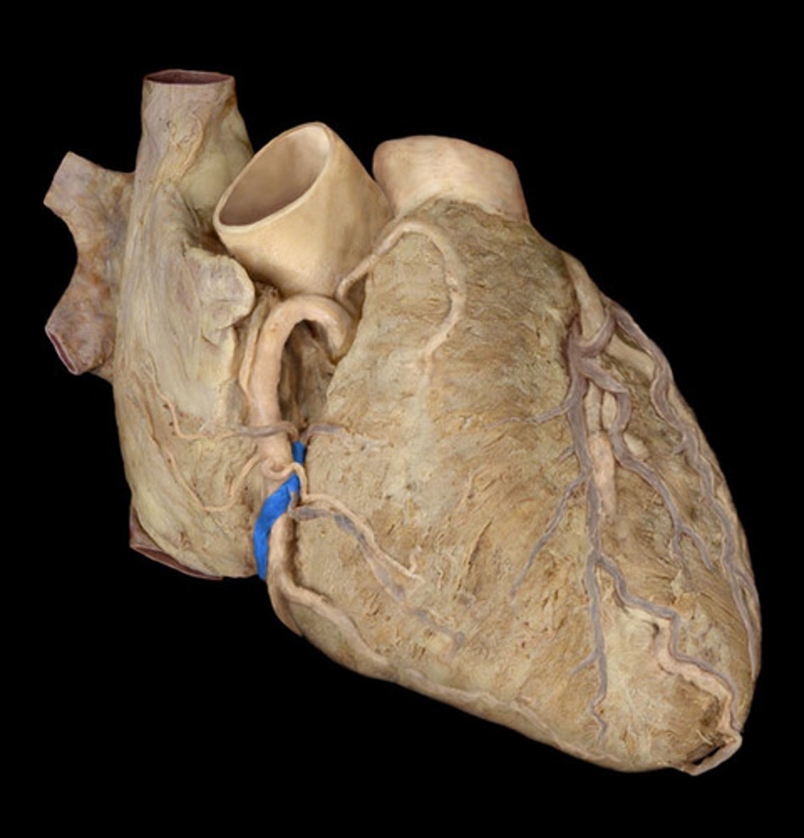
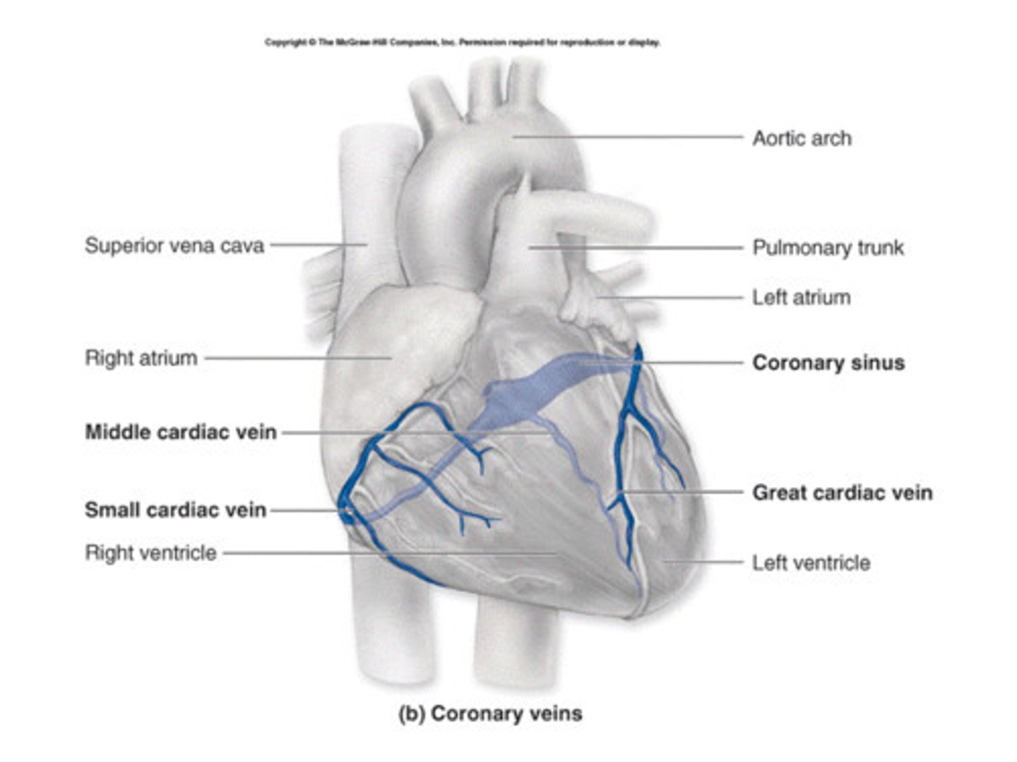
venous drainage of the heart
primarily via coronary sinus
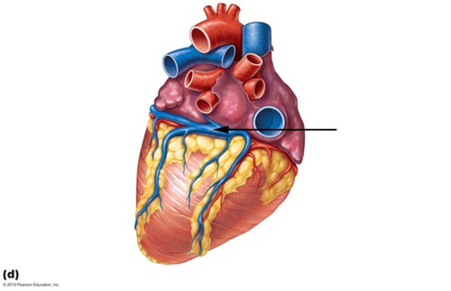
coronary sinus is a continuation of
great cardiac vein
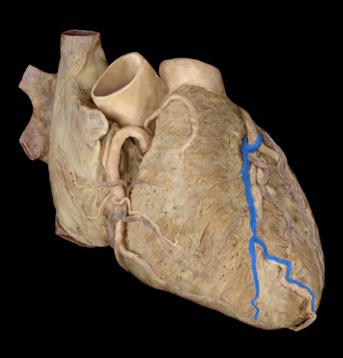
tributaries of coronary sinus
small and middle cardiac veins
AKA: coronary veins (coronary is usually just used for arteries)
right anterior coronary vein branches
right atrial vein
small cardiac vein
right anterior ventricular vein
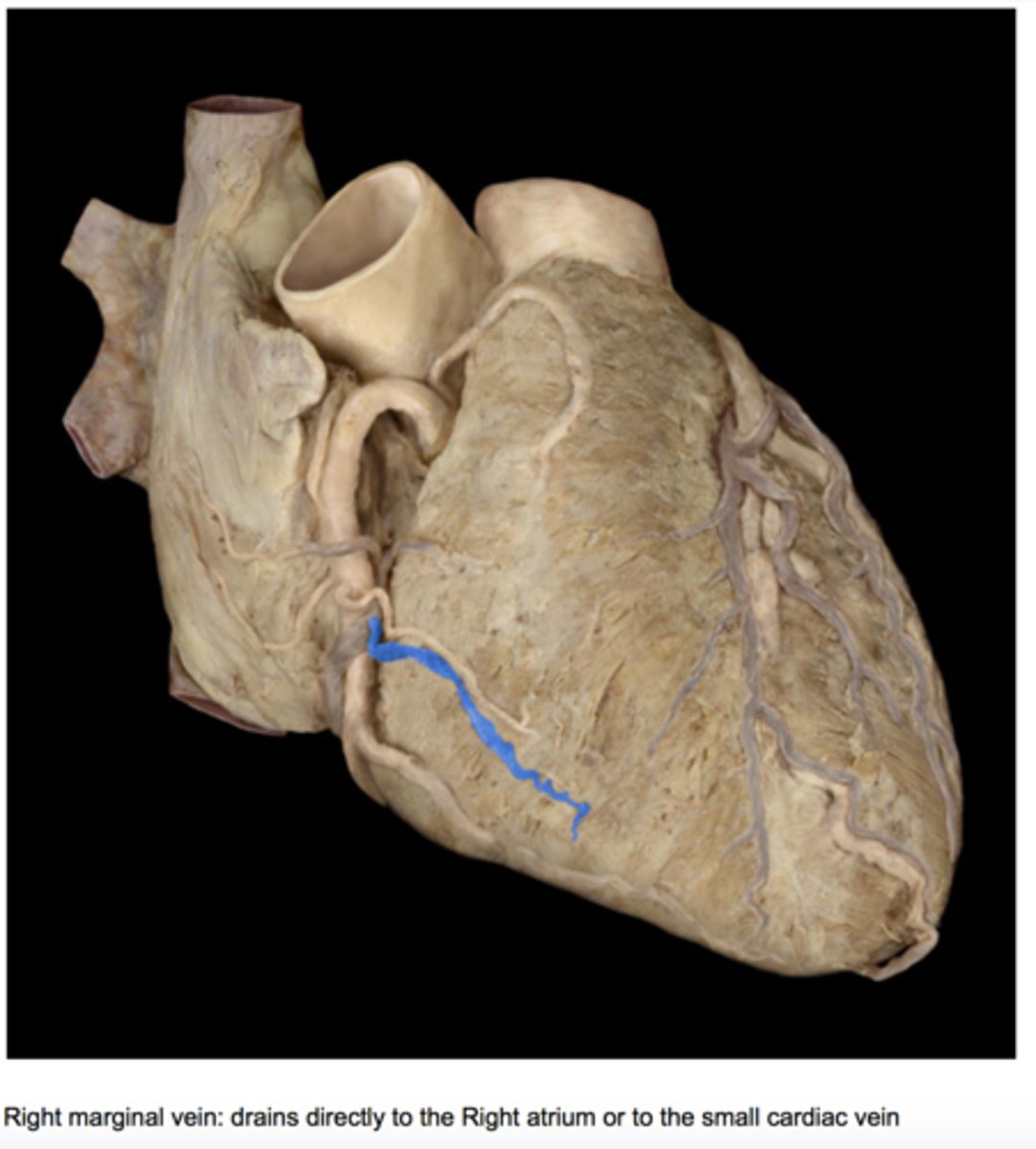
right marginal vein
right atrial vein
small cardiac vein
right anterior ventricular vein
right marginal vein
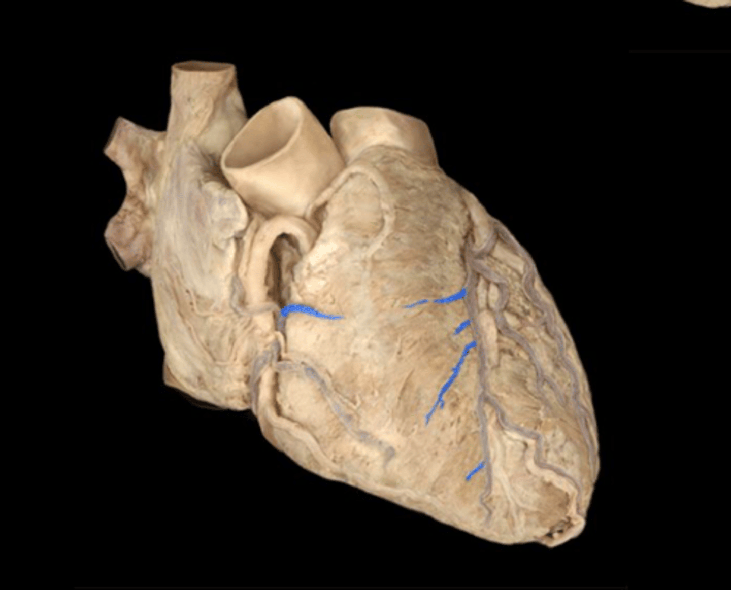
left anterior coronary vein branches
great cardiac vein
left marginal vein
left anterior ventricular vein
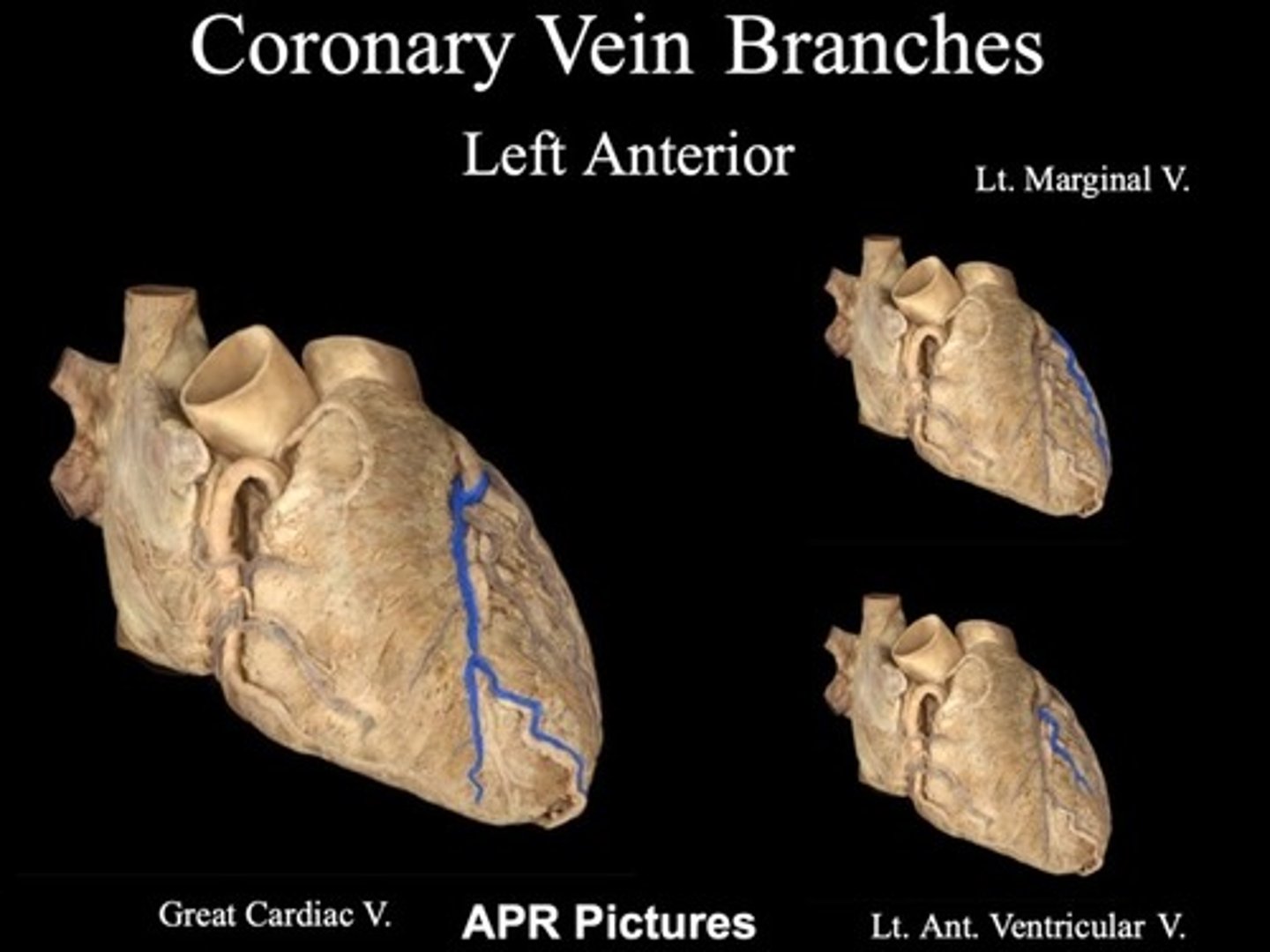
great cardiac vein

left marginal vein
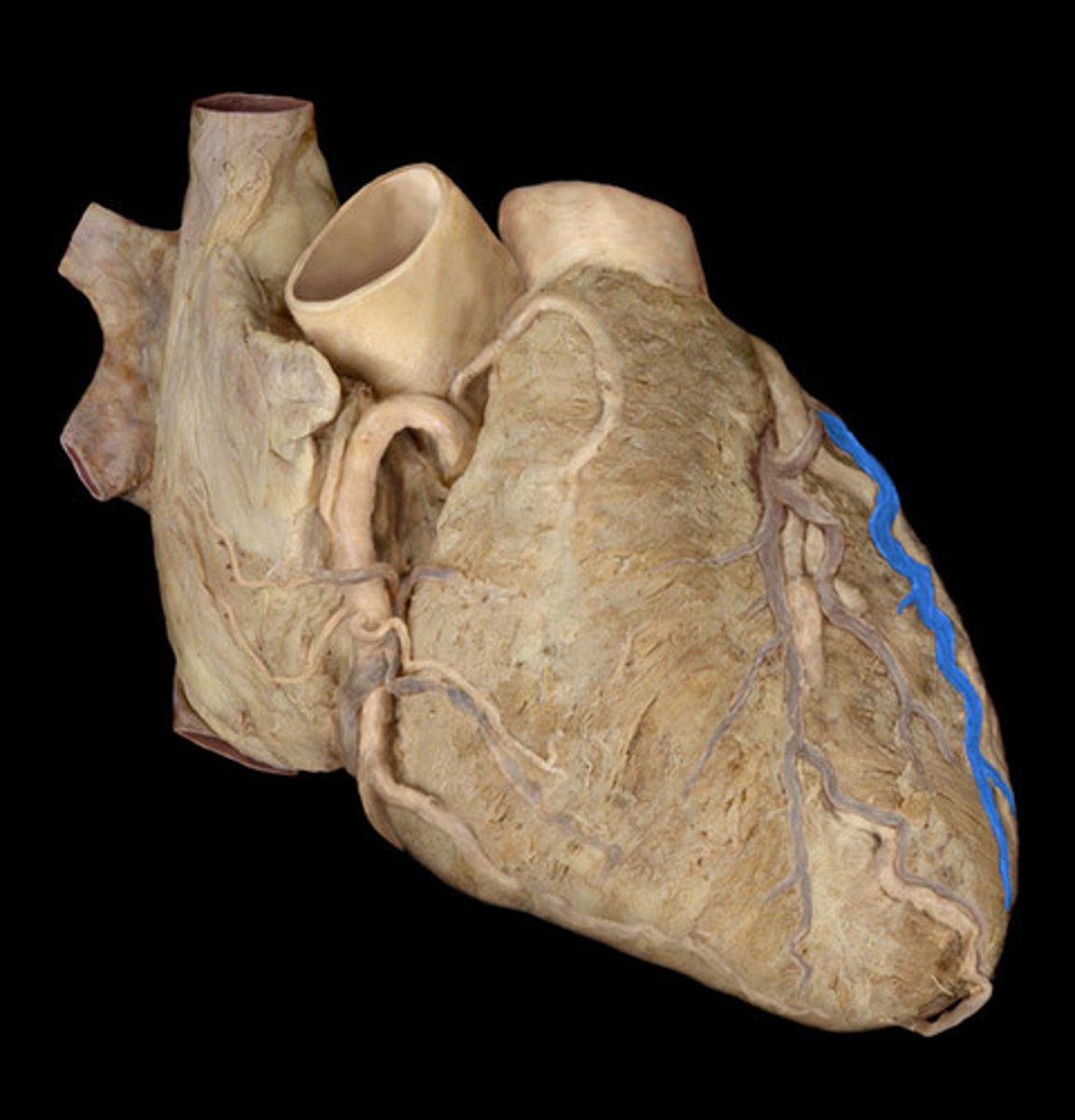
left anterior ventricular vein
coronary/cardiac veins
small cardiac vein, great cardiac vein, middle cardiac vein all drain into coronary sinus
rate of contraction set by
SA node but you can control with NS
innervation of heart sympathetic
1. cervical sympathetic trunks
2. upper thoracic sympathetic trunks
3. effect on rate= increase heart rate
innervation of heart parasympathetic
1. vagus nerve
2. effect on rate= decrease heart rate (vagal tone)
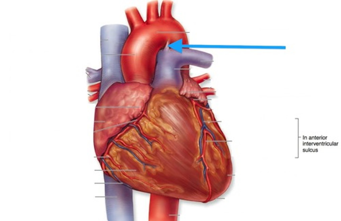
great vessels of the heart
aorta
pulmonary trunk
superior and inferior vena cavae
aorta
ascending
aortic arch
descending
branches of ascending aorta
right and left coronary arteries
branches of aortic arch
brachiocephalic artery, left common carotid artery, left subclavian artery
pulmonary trunk composed of
right and left pulmonary arteries
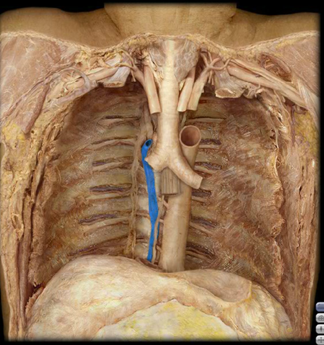
ligamentum arteriosum
location: pulmonary trunk and aorta- closed
fetus-ductus arteriosus (open) (fetal lungs do not work so used to bypass the lungs to aorta)
takes 1 week from birth to close
superior and inferior vena cavae
superior vena cava joined by azygous vein before entering heart
azygous drains thoracic body wall
layers of arteries and veins
tunics externa, tunica media, and tunica intima
arteries vs vein
arteries have thicker tunica media
veins have valves
veins collapse because there is not much muscle to keep up
veins have larger lumen
capillary
1 cell thick and has pores for diffusion (filtration)