Unit 2
1/294
There's no tags or description
Looks like no tags are added yet.
Name | Mastery | Learn | Test | Matching | Spaced | Call with Kai |
|---|
No analytics yet
Send a link to your students to track their progress
295 Terms
A thickened endometrium may be a sign of:
Early intrauterine pregnancy
Gestational trophoblastic disease
Endometrial hyperplasia
Secretory endometrium
Estrogen replacement therapy
Polyps
Tamoxifen/HRT
Endometrial cancer
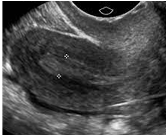
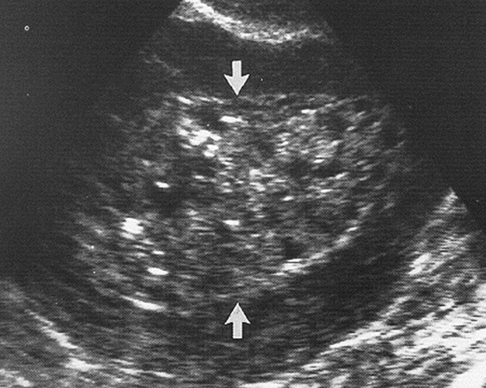
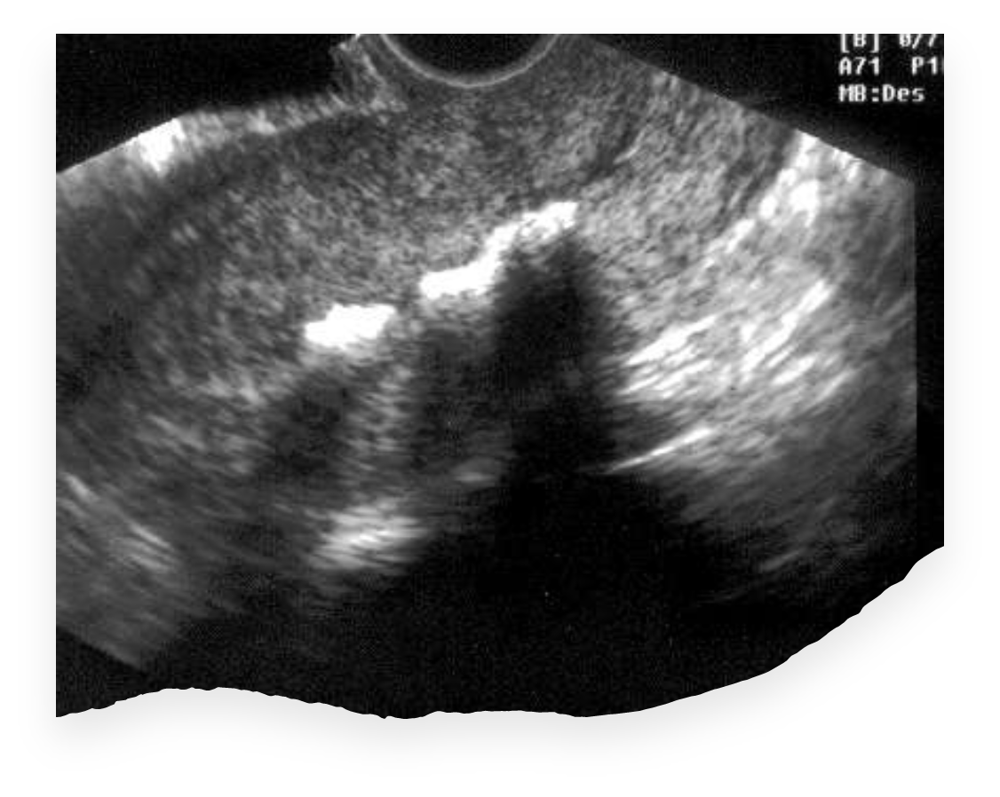
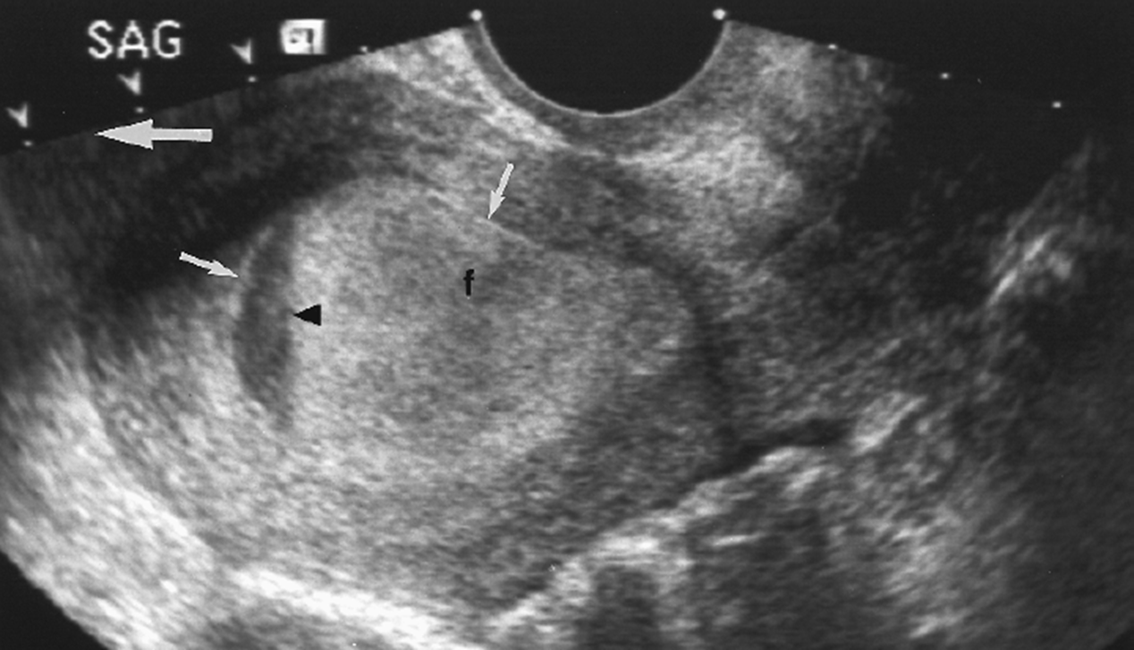
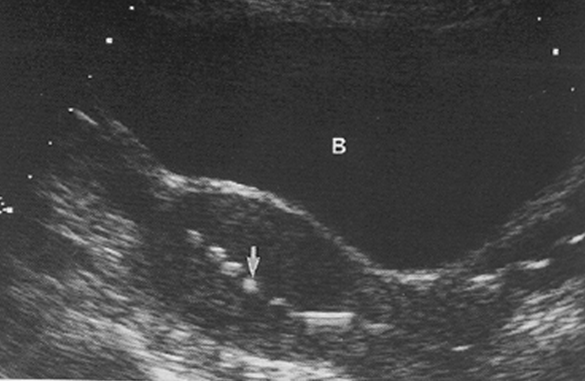
What is this image showing?
Thickened Endometrium
What is this image showing?
Thickened Endometrium
Endometrial hyperplasia is caused by:
Unopposed estrogen stimulation
What is unopposed estrogen?
Estrogen dominance, there isn’t enough progesterone produced to keep up with the amount of estrogen.
A premenopausal patient with endometrial hyperplasia, would have an endometrial thickness of _______.
+14mm
An asymptomatic postmenopausal patient with endometrial hyperplasia, could expect to have an endometrial thickness of _______.
8mm {upper normal limit}
A patient with endometrial hyperplasia on sequential estrogen & progesterone, could expect to have an endometrial thickness of _______.
15mm
What is the most common cause of pre & postmenopausal abnormal bleeding?
Endometrial Hyperplasia
Endometrial Hyperplasia is a possible precursor of ___________ _______.
Endometrial Cancer
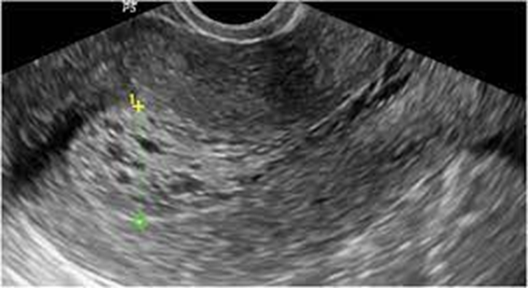
What is this image showing?
Endometrial Hyperplasia
What is this image showing?
Endometrial Hyperplasia
What are endometrial polyps?
Overgrowth of endometrial tissue
What symptom is common with endometrial polyps?
Vaginal bleeding
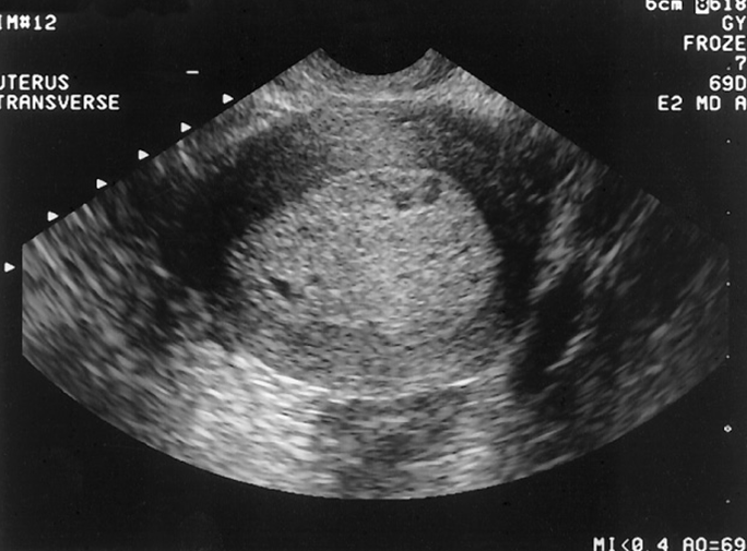
What is this image showing?
Endometrial polyps
What are these images showing?
Endometrial polyps
What is endometritis?
Inflammation / infection of the endometrium
Endometritis most commonly occurs in association with…
PID
Postpartum
Following instrumentation of the uterus
What is the ultrasound appearance of Endometritis?
Thickened endometrium
Irregular endometrium
Endometrial fluid
“Gas”
Retained tissue
Most endometrial carcinomas are _____________ occurring in _____________ patients.
Adenocarcinoma, Perimenopausal
Endometrial carcinoma has a strong association with…
Estrogen replacement therapy
What is the earliest sign of endometrial carcinoma?
Thickened endometrium
What are the advanced signs of endometrial carcinoma?
Uterine enlargement with lobular contour
Mixed echogenicity
Endometrial fluid collections
Abdominal pain
Bleeding (postmenopausal)
If there is uterine enlargement, the carcinoma has invaded the ____________.
Myometrium
What evidence may support the diagnosis of endometrial carcinoma?
Endometrial thickening
TV to measure thickness
Myometrial invasion
Clear evidence for CA
Synechiae/Asherman Syndrome
Bands of endo tissue
What is this image showing?
Endometrial Carcinoma
What is this image showing?
Endometrial Carcinoma
What is synechiae?
Fibrous adhesions across the endometrial cavity
Walls become adhered to each other
Various degrees of adhesions
What is another name for synechiae?
Asherman Syndrome
What may be a result from synechiae?
Infertility
Amenorrhea
Oligomenorrhea
Patients with synechiae may have a history of _____ &/or ______.
D&C, abortion
What is the ultrasound appearance of synechiae?
Bright echoes within the endometrial cavity
What is this image showing?
Synechiae / Asherman Syndrome
What is considered Stage l of endometrial cancer?
Confined to endometrium of body
B, C = myometrial extension
What is considered Stage ll of endometrial cancer?
Endometrium into cervix
What is considered Stage lll of endometrial cancer?
Spread to pelvic area lymph nodes
Extended through the serosal layer
What is considered Stage lV of endometrial cancer?
Mets to other organs
Extension into bladder/bowel
Distal lymph nodes
Differential Considerations for the Uterus: thickened endometrium
Early intrauterine pregnancy
Endometrial hyperplasia
Retained products of conception or incomplete abortion
Trophoblastic disease
Endometritis
Adhesions
Polyps
Inflammatory disease
Endometrial carcinoma
Differential Considerations for the Uterus: endometrial fluid
Endometritis
Retained products of conception
{PID} Pelvic inflammatory disease
Cervical obstruction
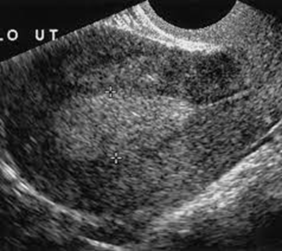
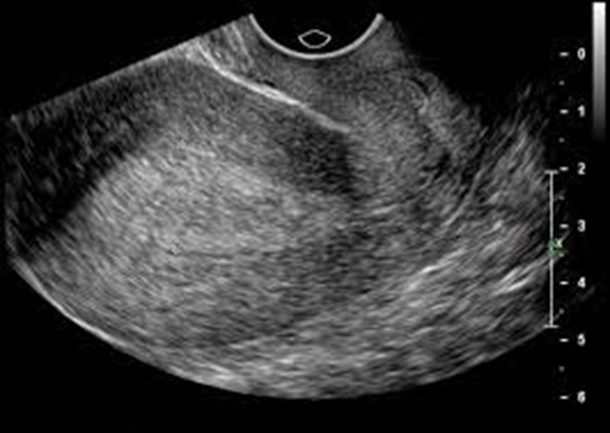
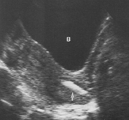
What is this image showing?
A thickened endometrium
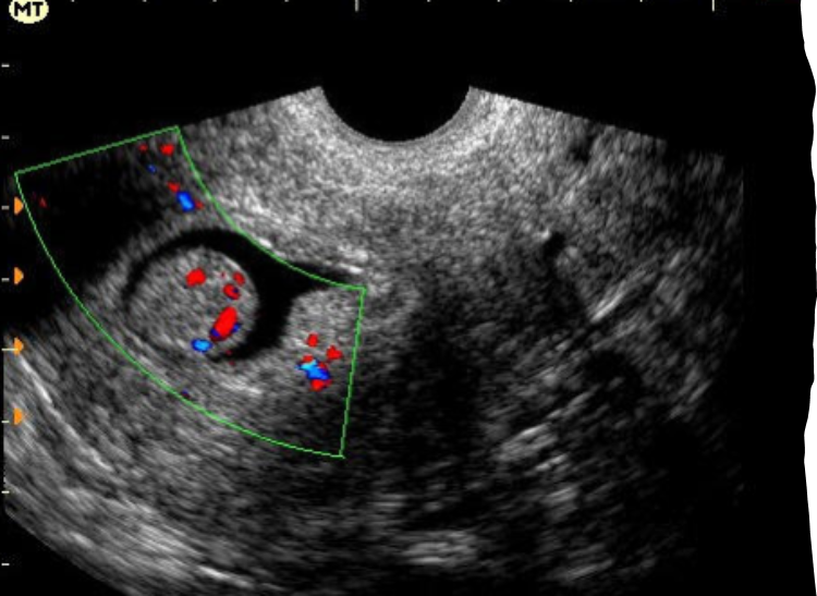
What flow has been seen in patients with endometrial carcinoma?
Low resistance {RI <.4}
What flow has been seen in postmenopausal patients with normal or benign endometrium?
High resistance {RI >.5}
Tumor vascularity is a _______ sensitive indicator than RI alone
More
What is the normal measurement for a small endometrial fluid collection?
< 2 ml
It is normal to find small endometrial fluid collections during the….
Normal menstrual cycle
With postmenopausal patients, when is it normal to see endometrial fluid collections?
Sequential hormones
Endometrial atrophy
Is the fluid collection included in the endometrial measurement?
No, do not measure the fluid collection
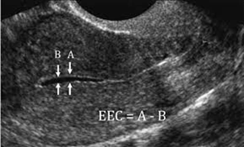
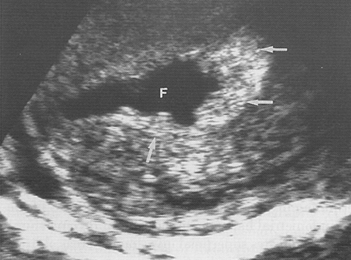
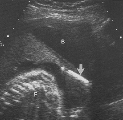
What is this image showing?
Small endometrial fluid collection
Large endometrial fluid collections can be ________, and should be investigated to find a cause.
Suspicious
A large endometrial fluid collection may be a sign of:
PID
Pyometra
Hematometra
CA’s
Cervical stenosis
Congenital anomalies
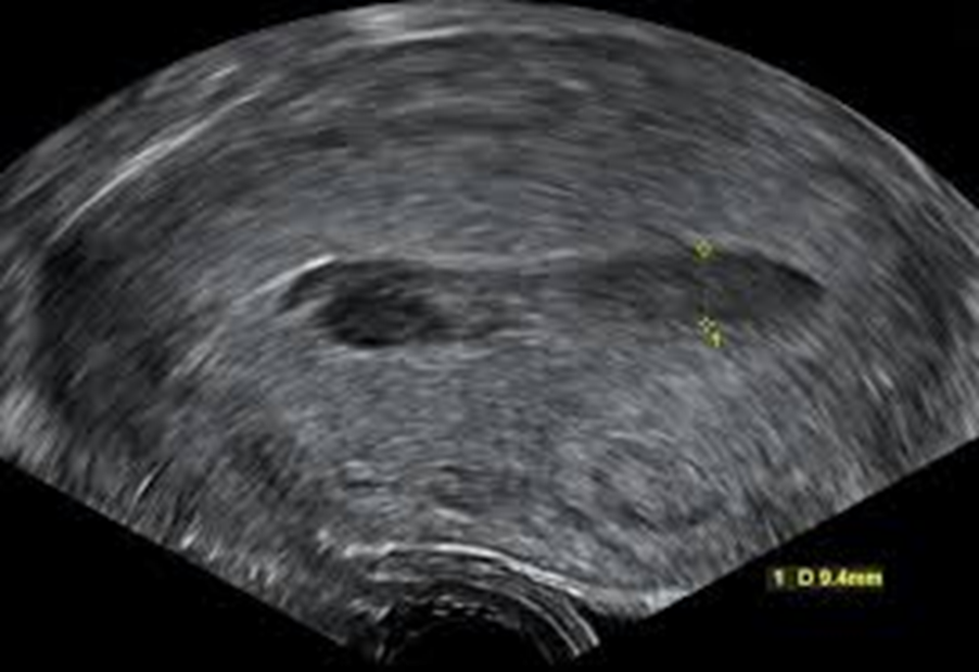
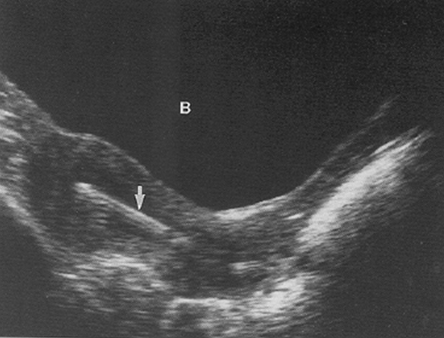
What is this image showing?
Large endometrial fluid collection
What is this image showing?
Cervical stenosis
In a postmenopausal patient
What does IUD stand for?
Intrauterine device
What is an IUD?
Device inserted into the fundal portion of endometrial cavity
An IUD prevents ________ and may act as a _________ _______.
Implantation, spermicidal agent
An IUD placement is verified by the ______ __________.
String identification
If the string goes missing, an investigation for the location is needed?
True
What can be a result of a missing IUD?
Expulsion
Uterine perforation
Most likely at the time of insertion (1:1000)
What are the different types of IUD’s?
Safety coil
Lippe’s loop
Copper T
Dalkon shield
Copper T: ParaGard
Coppler/Plastic
Copper T: Progestasert
Progesterone T
Yearly replacement
Copper T: Mirena
Levonorgestrel-releasing
5 years (50%)
Copper T: Skyla
Levonorgestrel-releasing
3 years
What IUD is no longer used?
Dalkon shield
What is the ultrasound appearance of an IUD?
Perpendicular placement of sound beam
Echogenic linear appearance
Acoustic shadowing
“Double line” of linear shaft
Shape identification
When IUD location identification is needed, where should you look?
Within uterine cavity
Within myometrium
External to uterus
When the IUD is found within the uterine cavity, it can be occasionally obscured by a _______ ___________.
Thick endometrium
What is this image showing?
IUDC identification
What is this image showing?
IUDC identification
What is this image showing?
IUDC identification
What is this image showing?
IUDC identification
With a positive pregnancy test, what is extremely important?
Location
___% of pregnancies lost with extraction of IUD.
50
A transvaginal ultrasound is needed to find the exact location of the ________ ____ & _____.
Gestational sac, IUD
When perforation is suspected, what is needed and what may be the differential diagnosis?
PG test
Neg = possible radiograph
Bowel inflammation
Peritonitis
With an IUD there is an increased risk of:
Ectopic pregnancy (30%)
Pelvic Inflammatory Disease
Tubo-ovarian abscess
Tubo-ovarian abscess may be found __________ but is most commonly found __________.
Unilateral, bilateral
What is a uterine prolapse?
Downward displacement of the uterus into the vaginal canal
Gradual descent of the uterus in the axis of the vagina taking the vaginal wall with it.
Uterine Prolapse: 1st degree
Cervix at lower uterine
Uterine Prolapse: 2nd degree
Cervix is at vaginal opening
Uterine Prolapse: 3rd degree
Uterus protrudes through the vagina
What is the etiology & precursors for prolapse?
Stretching of muscle/fibrous tissue
Increased intra-abdominal/pelvic pressure
Menopause/Aging
What can cause: stretching of muscle / fibrous tissue.
Pregnancy
Childbirth
What can cause: increased intra-abdominal/pelvic pressure.
Lifting
Obesity
Chronic cough
‘Chronic’ upright position
Menopause/Aging causes an _________ in ________.
Decrease, estrogen
A type of treatment for uterine prolapse is a _______ ________.
Vaginal pessary
Soft, flexible device placed into the vagina to reduce the symptoms of prolapse
What are the surgery options for uterine prolapse?
Repair supporting tissue for organ
Repair the tissue around vagina
Close the vaginal opening
Hysterectomy
What are the preventative measures for uterine prolapse?
Early detection
Kegel exercises
What are differential considerations for an enlarged uterus?
Pregnancy
Postpartum
Leiomyoma
Adenomyosis
Bicornuate or didelphic uterus
What are the differential considerations for a uterine tumor?
Leiomyoma
Carcinoma
What are Leiomyomas also known as?
Fibroids or Myomas
What is the most common benign gynecological tumor?
Leiomyomas
What is the US appearance of a Leiomyoma?
Whorled, spherical shaped mass of myometrial tissue
Degeneration may occur or can enlarge
Multiple
Pseudocapsule
What are the clinical symptoms of a leiomyoma?
Enlarged uterus
Pelvic Pressure
Pain
Irregular Bleeding
Menorrhagia
A leiomyoma can cause infertility issues due to what location?
Obstruction of tubes
Obstruction of birth canal
During what process can an increased growth of a leiomyomas occur and what happens?
Pregnancy and bleeding withing fibroid may cause pain
What is a leiomyoma stimulated by?
Estrogen- receptive to hormones
What is the submucosal classification of a leiomyoma?
Into the endometrial canal
Deforms the endometrial canal
May cause irregular or heavy bleeding
What is the intramural classification of a leiomyoma?
Within the myometrium
Most common
What is the subserosal classification of a leiomyoma?
Protrudes outward
Pedunculated = extrauterine mass
Exophytic or Interligamentous
May effect adjacent organs