17. Locomotion and Movement
1/234
Earn XP
Description and Tags
Almost all the stuff from NCERT. Skipped the obvious stuff like "what do we need locomotion for?" and stuff like that. Suitable revision for IAT, NEST, NEET, etc. Question mode: Flashcards only. Answer mode: Answer with definition. Recommended: Spaced Repetition. Good luck with exams!
Name | Mastery | Learn | Test | Matching | Spaced | Call with Kai | Chat |
|---|
No analytics yet
Send a link to your students to track their progress
235 Terms
What is meant by streaming of protoplasm?
Movement of intracellular substances inside the cell.
Some of the movements result in a change of place or location. Such voluntary movements are called ___________.
Some of the movements result in a change of place or location. Such voluntary movements are called locomotion.
All locomotions are movements but all movements are not locomotions.
True or false?
True
Cells of the human body exhibit three main types of movements. What are these?
Amoeboid
Ciliary
Muscular
Cells of the human body exhibit three main types of movements.
Give an example of amoeboid movement in the human body.
Some specialised cells in our body like macrophages and leucocytes in blood exhibit amoeboid movement. It is effected by pseudopodia formed by the streaming of protoplasm.
Cells of the human body exhibit three main types of movements.
Give two examples of ciliary movement in the human body.
Ciliary movement occurs in most of our internal tubular organs which are lined by ciliated epithelium. The coordinated movements of cilia in the trachea help us in removing dust particles and some of the foreign substances inhaled alongwith the atmospheric air.
Passage of ova through the female reproductive tract is also facilitated by the ciliary movement.
Muscle is a specialised tissue of _________al origin.
Muscle is a specialised tissue of mesodermal origin.
What per cent of the body weight of a human adult is contributed by muscles?
about 40-50%
What are the two special properties of muscles (that are not present in other forms of movement and locomotion)?
excitability
elasticity (contractibility and extensibility)
Based on their location, three types of muscles are identified, which are?
skeletal
visceral
cardiac
What is the location of skeletal muscles?
Skeletal muscles are closely associated with the skeletal components of the body.
What is the appearance of skeletal muscles?
They have a striped appearance under the microscope.
Skeletal muscles have striped appearance under the microscope, hence they are called _______ muscles.
Skeletal muscles have striped appearance under the microscope, hence they are called striated muscles.
Skeletal muscles’ activities are under the voluntary control of the nervous system, hence they are known as _________ muscles.
Skeletal muscles’ activities are under the voluntary control of the nervous system, hence they are known as voluntary muscles.
What is the main purpose of skeletal muscles?
They are primarily involved in locomotory actions and changes of body postures.
What is the location of visceral muscles?
Visceral muscles are located in the inner walls of hollow visceral organs of the body like the alimentary canal, reproductive tract, etc.
What is the appearance of visceral muscles?
They do not exhibit any striation and are smooth in appearance.
Visceral muscles do not exhibit any striation and are smooth in appearance. Hence, they are called ______ muscles, or __________ muscles.
Visceral muscles do not exhibit any striation and are smooth in appearance. Hence, they are called smooth muscles, or unstriated muscles.
Visceral muscles’ activities are not under the voluntary control of the nervous system and are therefore known as ___________ muscles.
Visceral muscles’ activities are not under the voluntary control of the nervous system and are therefore known as involuntary muscles.
What is the main purpose of visceral muscles?
Assisting in automatic body movements. They assist, for example, in the transportation of food through the digestive tract and gametes through the genital tract.
What is the location of cardiac muscles?
Cardiac muscles are the muscles of heart.
What is the structure of cardiac muscles?
Many cardiac muscle cells assemble in a branching pattern to form a cardiac muscle.
What is the appearance of cardiac muscles?
Based on appearance, cardiac muscles are striated.
Cardiac muscles are ___________ in nature as the nervous system does not control their activities directly.
Cardiac muscles are involuntary in nature as the nervous system does not control their activities directly.
Each organised skeletal muscle in our body is made of a number of ________ ________ or _________.
Each organised skeletal muscle in our body is made of a number of muscle bundles or fascicles.
Each organised skeletal muscle in our body is made of a number of muscle bundles or fascicles held together by a common collagenous connective tissue layer called ________.
Each organised skeletal muscle in our body is made of a number of muscle bundles or fascicles held together by a common collagenous connective tissue layer called fascia.
Each organised skeletal muscle in our body is made of a number of muscle bundles or fascicles held together by a common connective tissue layer called fascia.
What is this tissue layer made of?
collagen
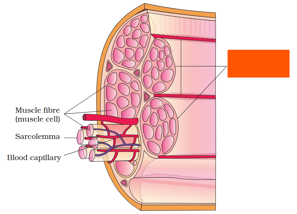
Fascicle (muscle bundle)
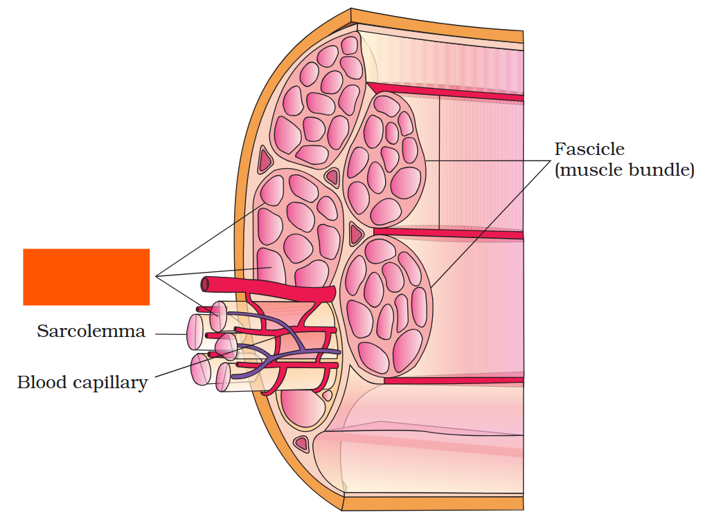
Muscle fibre (muscle cell)
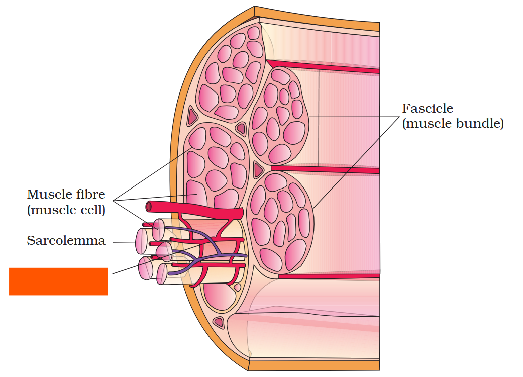
Blood capillary
Each skeletal muscle fibre is lined by the plasma membrane called ___________.
Each skeletal muscle fibre is lined by the plasma membrane called sarcolemma.
Each skeletal muscle fibre is lined by the plasma membrane called sarcolemma enclosing the __________.
Each skeletal muscle fibre is lined by the plasma membrane called sarcolemma enclosing the sarcoplasm.
Muscle fibre is a syncitium. Why is it called that?
Muscle fibre is a syncitium as the sarcoplasm contains many nuclei.
The __________ __________ of the muscle fibres is the store house of calcium ions.
The sarcoplasmic reticulum of the muscle fibres is the store house of calcium ions.
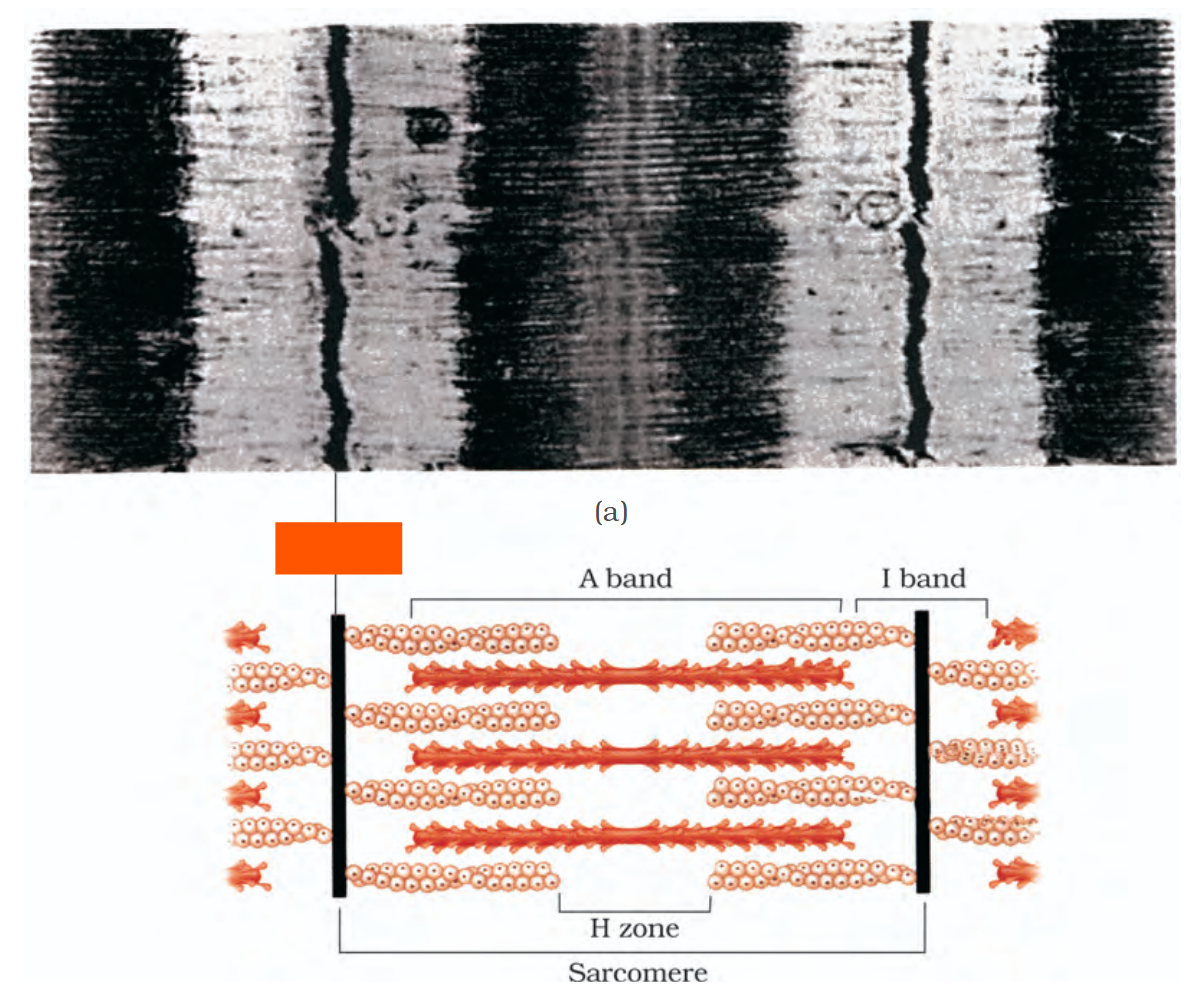
A characteristic feature of the muscle fibre is the presence of a large number of parallelly arranged filaments in the sarcoplasm called ____________ or _____________.
A characteristic feature of the muscle fibre is the presence of a large number of parallelly arranged filaments in the sarcoplasm called myofilaments or myofibrils.
A characteristic feature of the muscle fibre is the presence of a large number of parallelly arranged filaments in the sarcoplasm called myofilaments or myofibrils.
Each myofibril has alternate dark and light bands on it, due to the distribution pattern of two important proteins, which are?
Actin
Myosin
A characteristic feature of the muscle fibre is the presence of a large number of parallelly arranged filaments in the sarcoplasm called myofilaments or myofibrils.
Each myofibril has alternate dark and light bands on it.
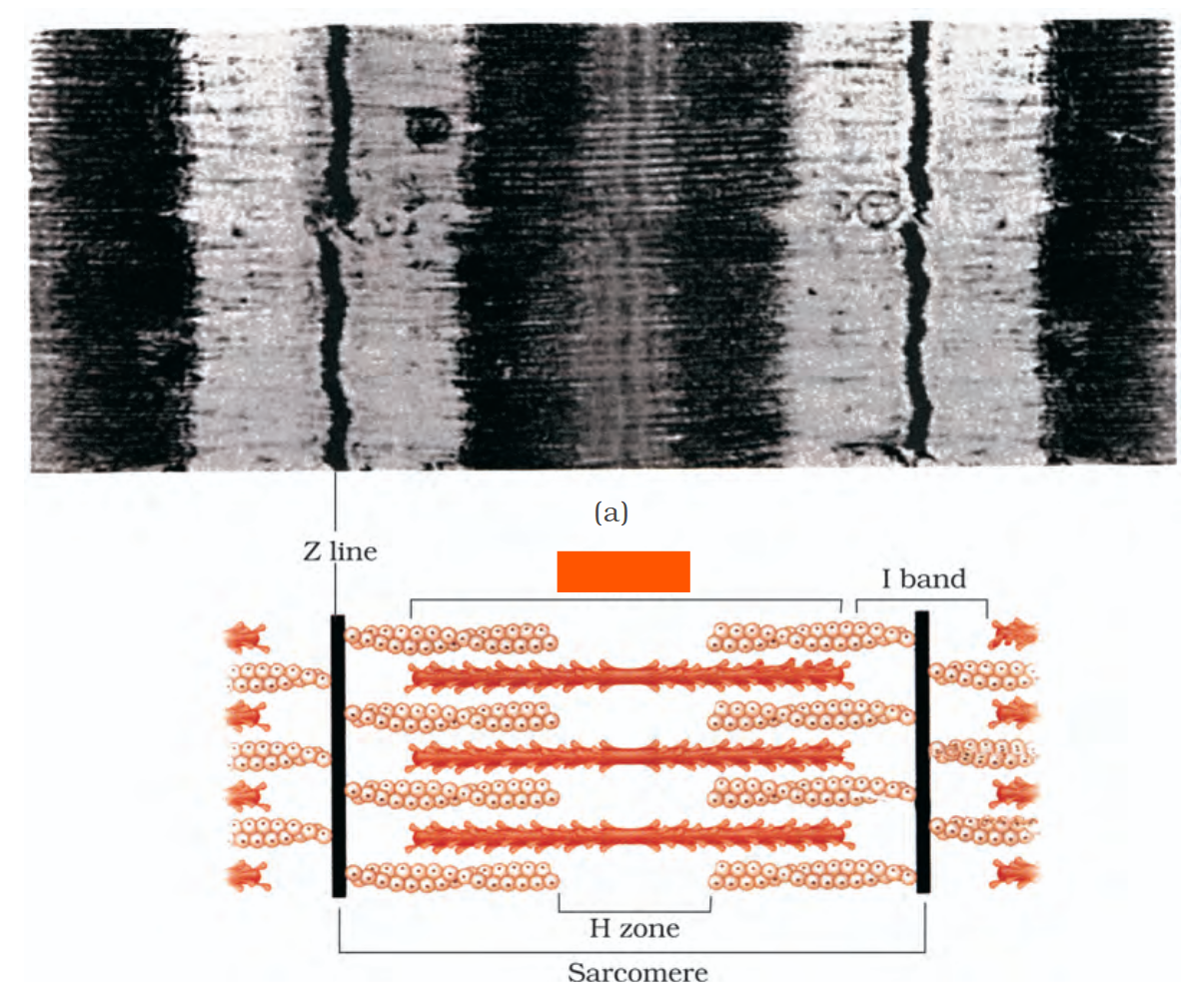
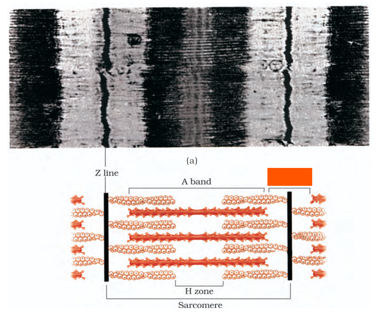
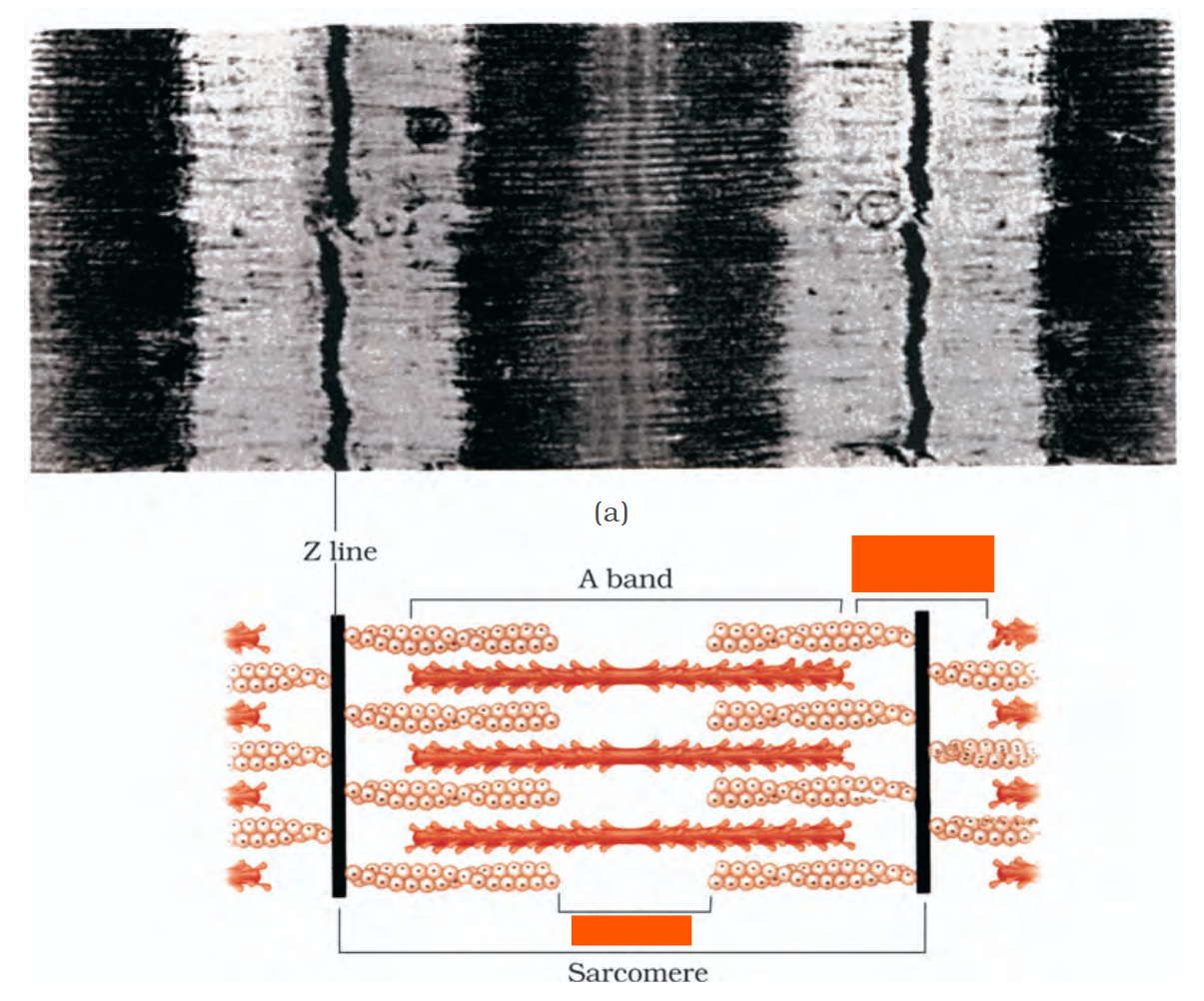
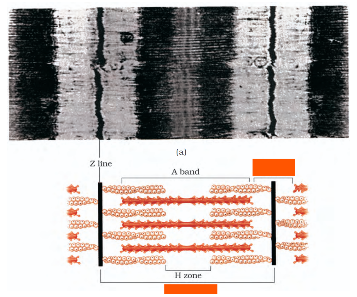
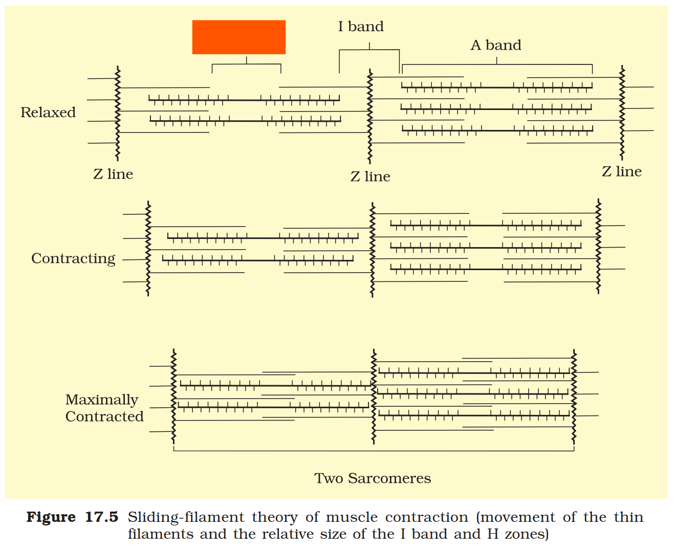
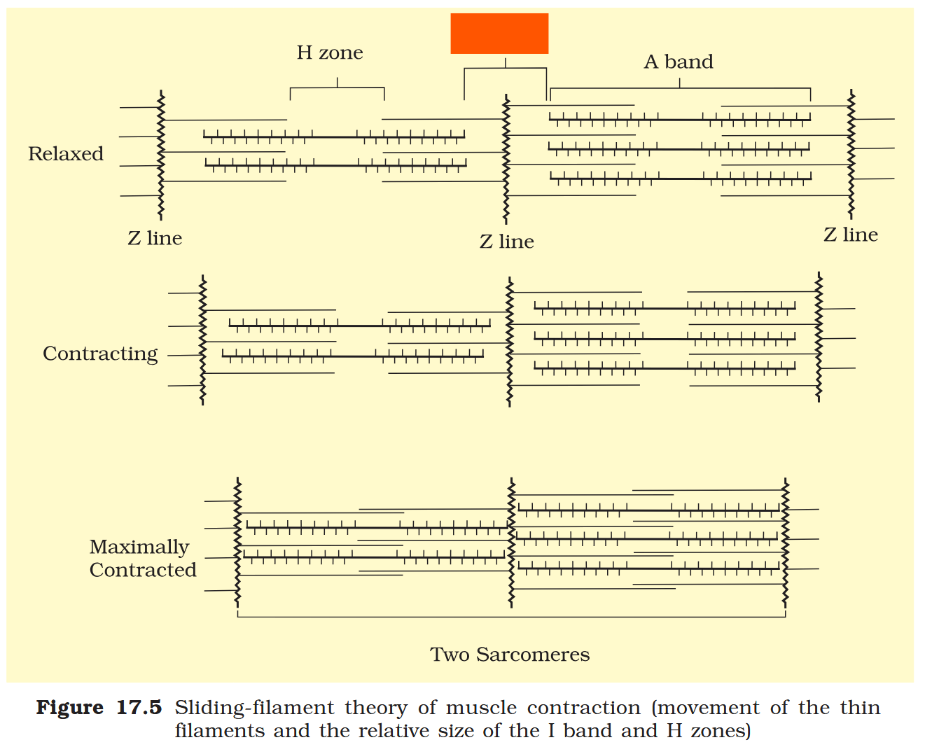
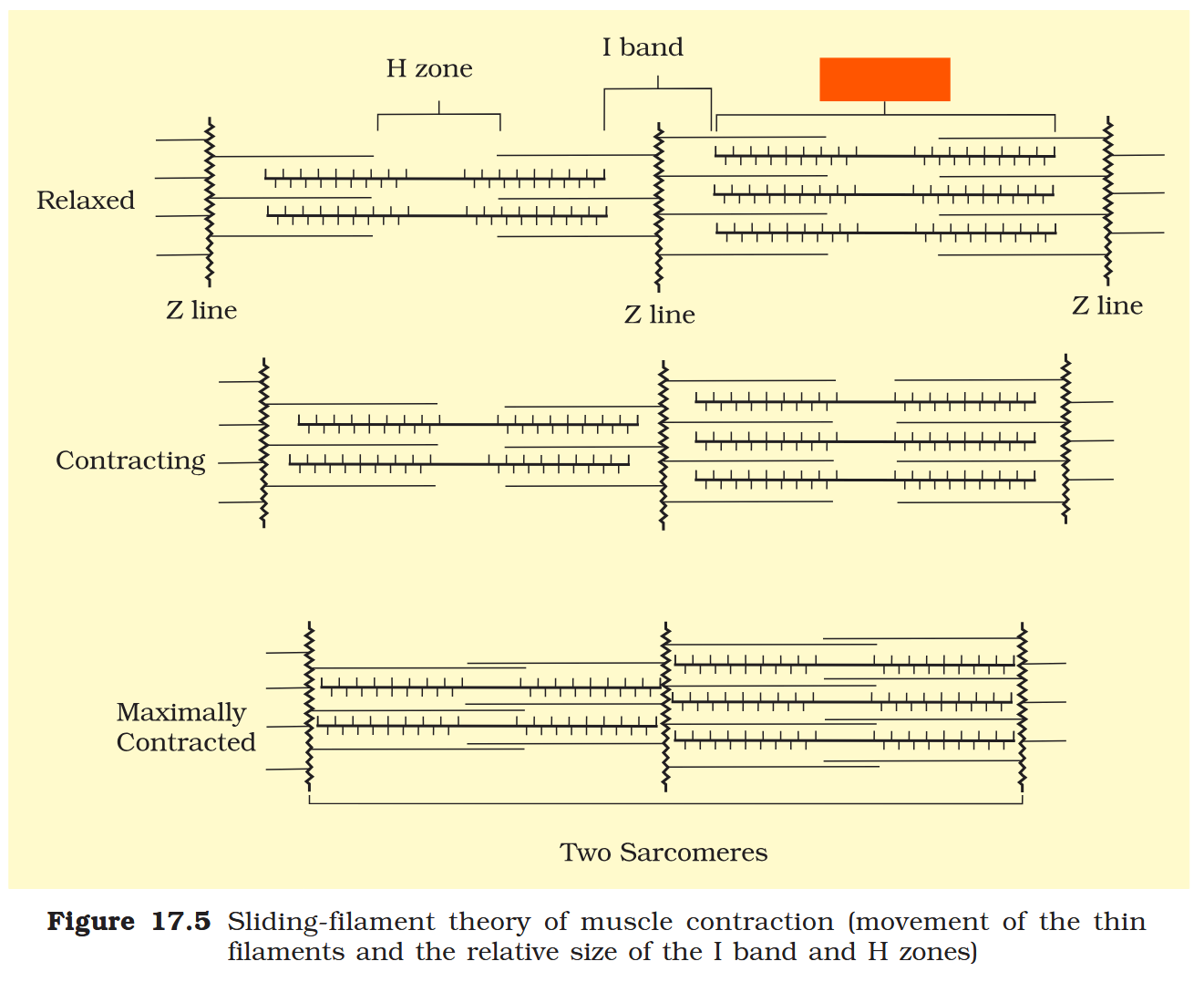
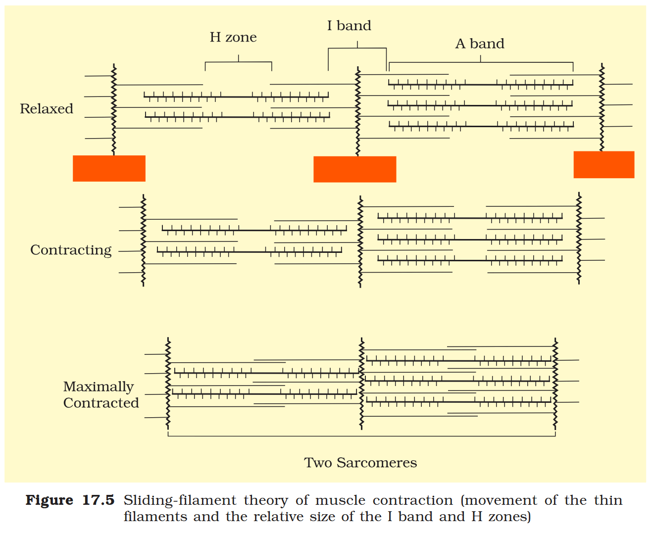
What is the I-band or Isotropic band?
The light bands contain ONLY actin and are called I-band or Isotropic band.
A characteristic feature of the muscle fibre is the presence of a large number of parallelly arranged filaments in the sarcoplasm called myofilaments or myofibrils.
Each myofibril has alternate dark and light bands on it.
What is the A-band or Anisotropic band?
the dark band called ‘A’ or Anisotropic band contains Myosin (as well as actin in some parts).
How are the proteins arranged in the myofibrils of muscle fibres?
Both the proteins are arranged as rod-like structures, parallel to each other and also to the longitudinal axis of the myofibrils.
Which is thicker, actin filaments or myosin filaments?
Actin filaments are thinner as compared to the myosin filaments.
In the centre of each ‘I’ band is an elastic fibre called ________ which bisects it. The thin filaments are firmly attached to it.
In the centre of each ‘I’ band is an elastic fibre called ‘Z’ line which bisects it.
The thick filaments in the ‘A’ band are held together in the middle of this band by a thin fibrous membrane called _______.
The thick filaments in the ‘A’ band are held together in the middle of this band by a thin fibrous membrane called ‘M’ line.
What is the functional unit of contraction in a skeletal muscle fibre?
The portion of the myofibril between two successive ‘Z’ lines is considered as the functional unit of contraction.
What is a sarcomere?
The functional unit of contraction in a skeletal muscle fibre.
What is the structure of actin and myosin in myofibrils in resting state?
In a resting state, the edges of thin filaments on either side of the thick filaments partially overlap the free ends of the thick filaments leaving the central part of the thick filaments.
In a resting state of skeletal muscles, the edges of thin filaments on either side of the thick filaments partially overlap the free ends of the thick filaments leaving the central part of the thick filaments.
This central part of thick filament, not overlapped by thin filaments is called the ________.
This central part of thick filament, not overlapped by thin filaments is called the ‘H’ zone.
What is the ‘H’ zone in myofibrils?
In a resting state of skeletal muscles, the edges of thin filaments on either side of the thick filaments partially overlap the free ends of the thick filaments leaving the central part of the thick filaments.
This central part of thick filament, not overlapped by thin filaments is called the ‘H’ zone.
What is the ‘Z’ line in myofibrils?
In the centre of each ‘I’ band is an elastic fibre called ‘Z’ line which bisects it. The thin filaments are firmly attached to it.
What is the ‘M’ line in myofibrils?
The thick filaments in the ‘A’ band are held together in the middle of the band by a thin fibrous membrane called ‘M’ line.
‘Z’ line
A band
‘I’ band
H zone
sarcomere
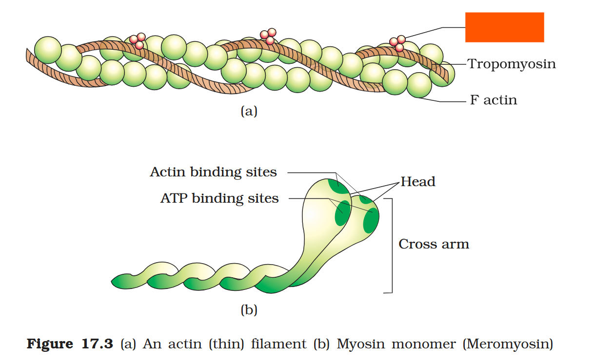
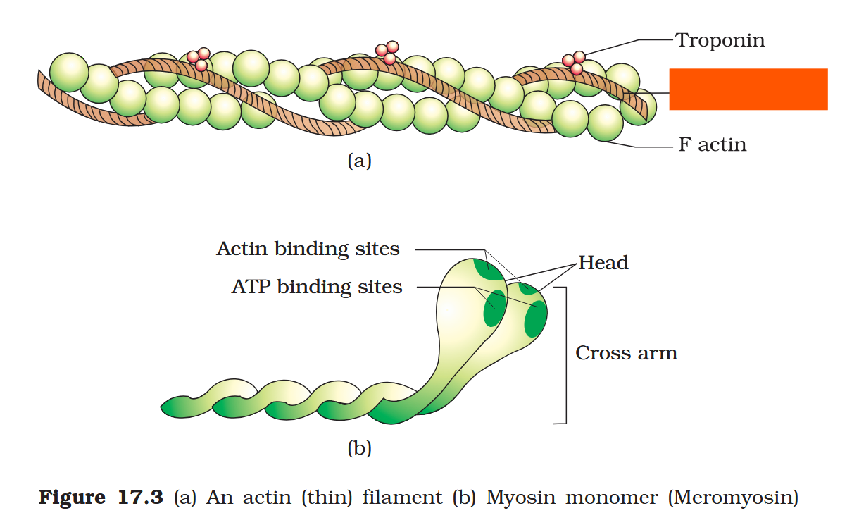
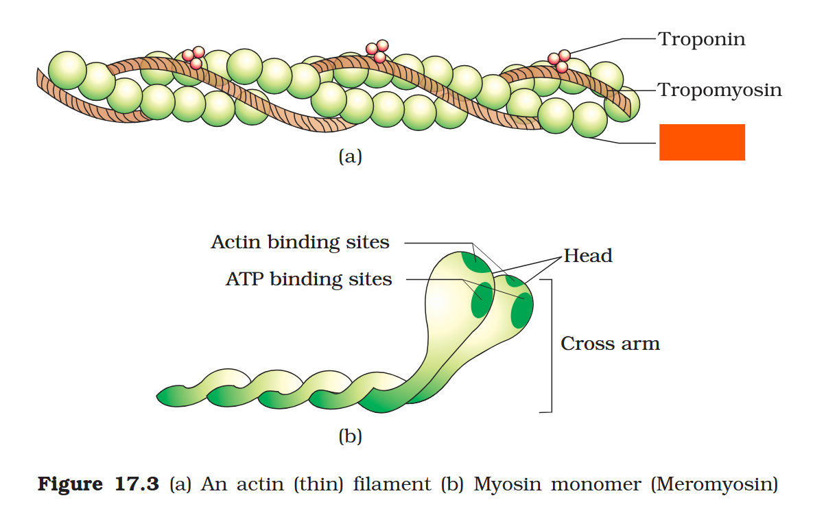
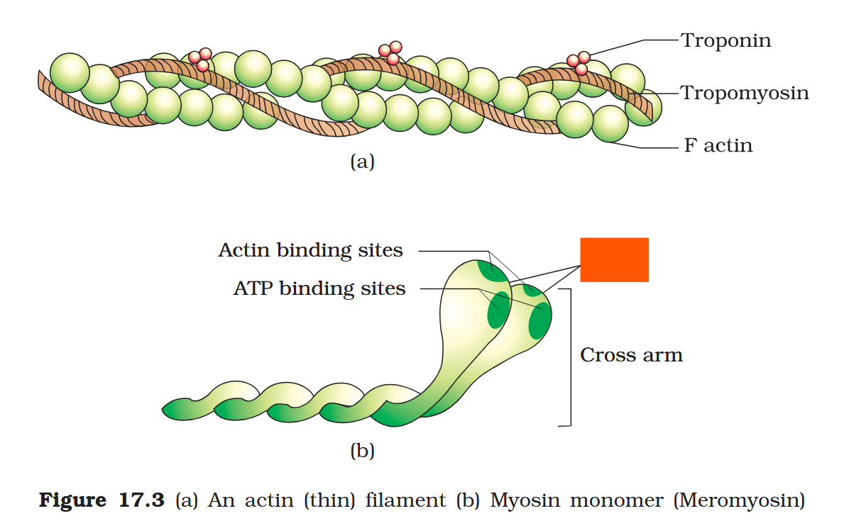
What is the structure of actin filaments?
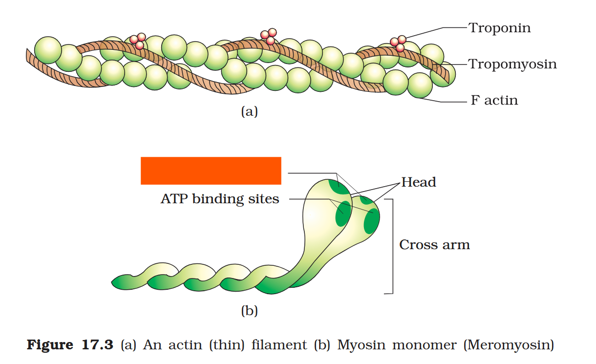
Each actin (thin) filament is made of two ‘F’ (filamentous) actins helically wound to each other. Each ‘F’ actin is a polymer of monomeric ‘G’ (Globular) actins.
What are tropomyosin proteins?
Two filaments of the protein tropomyosin run close to the ‘F’ actins throughout the length of actin filaments.
Each actin (thin) filament is made of two ‘F’ (filamentous) actins helically wound to each other. Each ‘F’ actin is a polymer of monomeric ‘G’ (Globular) actins. Two filaments of another protein, ___________ also run close to the ‘F’ actins throughout its length.
Each actin (thin) filament is made of two ‘F’ (filamentous) actins helically wound to each other. Each ‘F’ actin is a polymer of monomeric ‘G’ (Globular) actins. Two filaments of another protein, tropomyosin also run close to the ‘F’ actins throughout its length.
A complex protein ___________ is distributed at regular intervals on the tropomyosin present close to actin filaments.
A complex protein Troponin is distributed at regular intervals on the tropomyosin present close to actin filaments.
What is the function of the protein Troponin in muscle fibres?
In the resting state a subunit of troponin masks the active binding sites for myosin on the actin filaments.
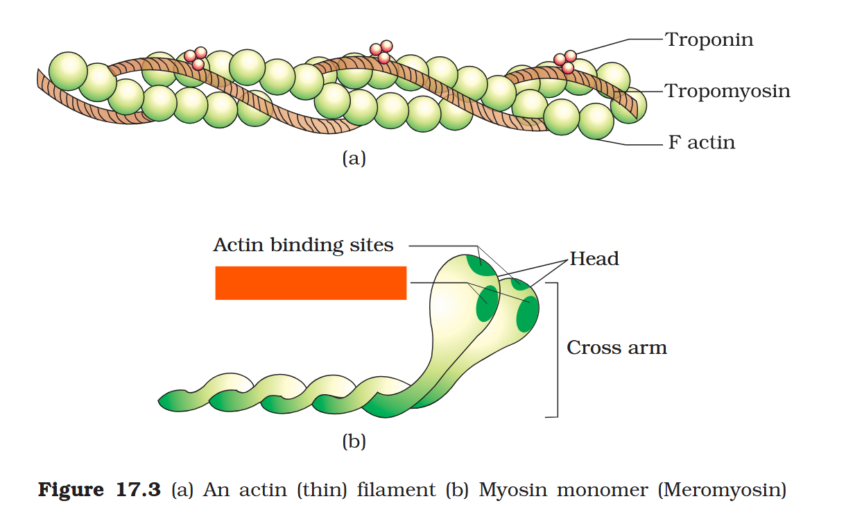
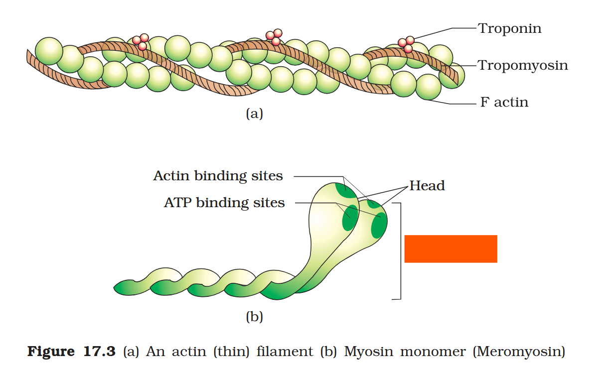
What is the structure of myosin filaments?
Each myosin (thick) filament is a polymerised protein. Many monomeric proteins called Meromyosins constitute one thick filament.
What is the structure of meromyosin?
Each meromyosin has two important parts, a globular head with a short arm and a tail.
The head and short arm projects outwards at regular distance and angle from each other from the surface of a polymerised myosin filament.
What is ‘heavy meromyosin’ (HMM)?
Each meromyosin has two important parts, a globular head with a short arm and a tail, the former being called the heavy meromyosin (HMM).
What is ‘light meromyosin’ (LMM)?
Each meromyosin has two important parts, a globular head with a short arm and a tail, the latter being called the light meromyosin (LMM).
Each meromyosin has two important parts, a globular head with a short arm and a tail.
The head and short arm projects outwards at regular distance and angle from each other from the surface of a polymerised myosin filament, and is known as __________.
The head and short arm projects outwards at regular distance and angle from each other from the surface of a polymerised myosin filament, and is known as cross arm.
What is a cross arm, inside skeletal muscle fibres?
Each meromyosin has two important parts, a globular head with a short arm and a tail.
The head and short arm projects outwards at regular distance and angle from each other from the surface of a polymerised myosin filament, and is known as cross arm.
The globular head of meromyosin is an enzyme, which?
The globular head is an active ATPase enzyme.
The globular head of meromyosin is an enzyme, which active sites does it have?
The globular head is an active ATPase enzyme and has binding sites for ATP and active sites for actin.
troponin
Tropomyosin
F actin
Head
Actin binding sites
ATP binding sites
Cross arm
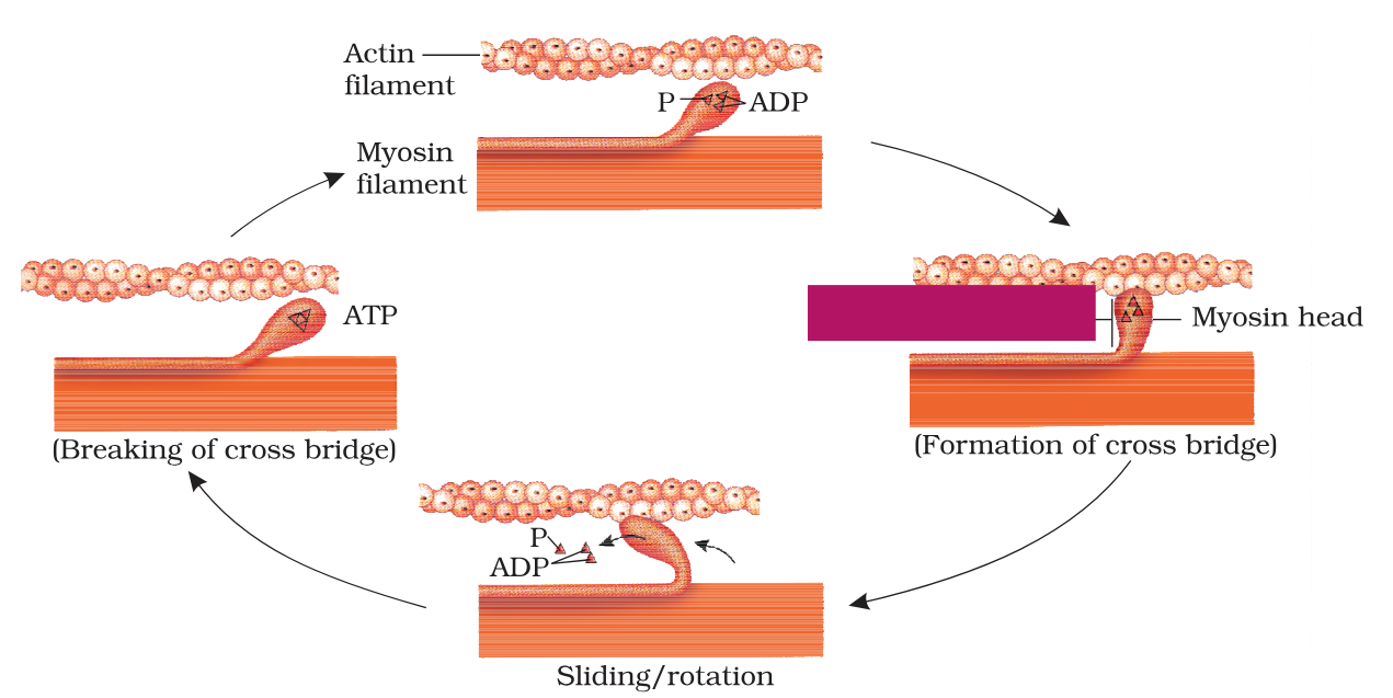
Mechanism of muscle contraction is best explained by which theory?
Sliding Filament Theory
Mechanism of muscle contraction is best explained by the sliding filament theory which states what?
Contraction of a muscle fibre takes place by the sliding of the thin filaments over the thick filaments.
Muscle contraction is initiated by a signal sent by the __________________________ via a motor neuron.
Muscle contraction is initiated by a signal sent by the central nervous system (CNS) via a motor neuron.
Muscle contraction is initiated by a signal sent by the central nervous system (CNS) via a _____________.
Muscle contraction is initiated by a signal sent by the central nervous system (CNS) via a motor neuron.
What is a motor unit?
A motor neuron alongwith the muscle fibres connected to it constitute a motor unit.
The junction between a motor neuron and the sarcolemma of the muscle fibre is called the _____________ _______ or ______-_____ _______.
The junction between a motor neuron and the sarcolemma of the muscle fibre is called the neuromuscular junction or motor-end plate.
The junction between a motor neuron and the sarcolemma of the muscle fibre is called the neuromuscular junction or motor-end plate.
A neural signal reaching this junction releases which neurotransmitter?
Acetyl choline
The junction between a motor neuron and the sarcolemma of the muscle fibre is called the neuromuscular junction or motor-end plate.
A neural signal reaching this junction releases a neurotransmitter (Acetyl choline) which generates an ________ ________ in the sarcolemma.
The junction between a motor neuron and the sarcolemma of the muscle fibre is called the neuromuscular junction or motor-end plate.
A neural signal reaching this junction releases a neurotransmitter (Acetyl choline) which generates an action potential in the sarcolemma.
A neural signal reaching a neuromuscular junction releases a neurotransmitter (Acetyl choline) which generates an action potential in the sarcolemma.
What does this cause?
This spreads through the muscle fibre and causes the release of calcium ions into the sarcoplasm.
A neural signal reaching a neuromuscular junction releases a neurotransmitter (Acetyl choline) which generates an action potential in the sarcolemma.
This spreads through the muscle fibre and causes the release of calcium ions into the sarcoplasm.
What is the consequence of this?
Increase in Ca++ level leads to the binding of calcium with a subunit of troponin on actin filaments and thereby remove the masking of active sites for myosin.
Increase in Ca++ level leads to the binding of calcium with a subunit of troponin on actin filaments and thereby remove the masking of active sites for myosin.
How does myosin take advantage of this?
Utilising the energy from ATP hydrolysis, the myosin head now binds to the exposed active sites on actin to form a cross bridge.
Increase in Ca++ level leads to the binding of calcium with a subunit of troponin on actin filaments and thereby remove the masking of active sites for myosin.
Utilising the energy from _________________, the myosin head now binds to the exposed active sites on actin to form a cross bridge.
Utilising the energy from ATP hydrolysis, the myosin head now binds to the exposed active sites on actin to form a cross bridge.
cross bridge
Increase in Ca++ level leads to the binding of calcium with a subunit of troponin on actin filaments and thereby remove the masking of active sites for myosin.
Utilising the energy from ATP hydrolysis, the myosin head now binds to the exposed active sites on actin to form a cross bridge.
What is the general consequence of this?
This pulls the attached actin filaments towards the centre of ‘A’ band. The ‘Z’ line attached to these actins are also pulled inwards thereby causing a shortening of the sarcomere, i.e., contraction.
During muscle contraction, he ‘Z’ line attached to these actins are pulled inwards thereby causing a shortening of the sarcomere.
How does this reflect in the lengths of the ‘I’ bands and the ‘A’ bands?
During shortening of the muscle, i.e., contraction, the ‘I’ bands get reduced, whereas the ‘A’ bands retain the length.
Utilising the energy from ATP hydrolysis, the myosin head binds to the exposed active sites on actin to form a cross bridge. This pulls the attached actin filaments towards the centre of ‘A’ band.
What happens after this?
The myosin, releasing the ADP and P1 goes back to its relaxed state. A new ATP binds and the cross-bridge is broken.
Utilising the energy from ATP hydrolysis, the myosin head binds to the exposed active sites on actin to form a cross bridge. This pulls the attached actin filaments towards the centre of ‘A’ band.
The myosin, releasing the ADP and P1 goes back to its relaxed state. A new ATP binds and the cross-bridge is broken.
How, then, does the cycle continue?
The ATP is again hydrolysed by the myosin head and the cycle of cross bridge formation and breakage is repeated causing further sliding.
Utilising the energy from ATP hydrolysis, the myosin head binds to the exposed active sites on actin to form a cross bridge. This pulls the attached actin filaments towards the centre of ‘A’ band.
The myosin, releasing the ADP and P1 goes back to its relaxed state. A new ATP binds and the cross-bridge is broken. The ATP is again hydrolysed by the myosin head and the cycle of cross bridge formation and breakage is repeated causing further sliding.
How long does this cycle continue?
The process continues till the Ca++ ions are pumped back to the sarcoplasmic cisternae resulting in the masking of actin filaments.
H zone
‘I’ band
‘A’ band
Z line
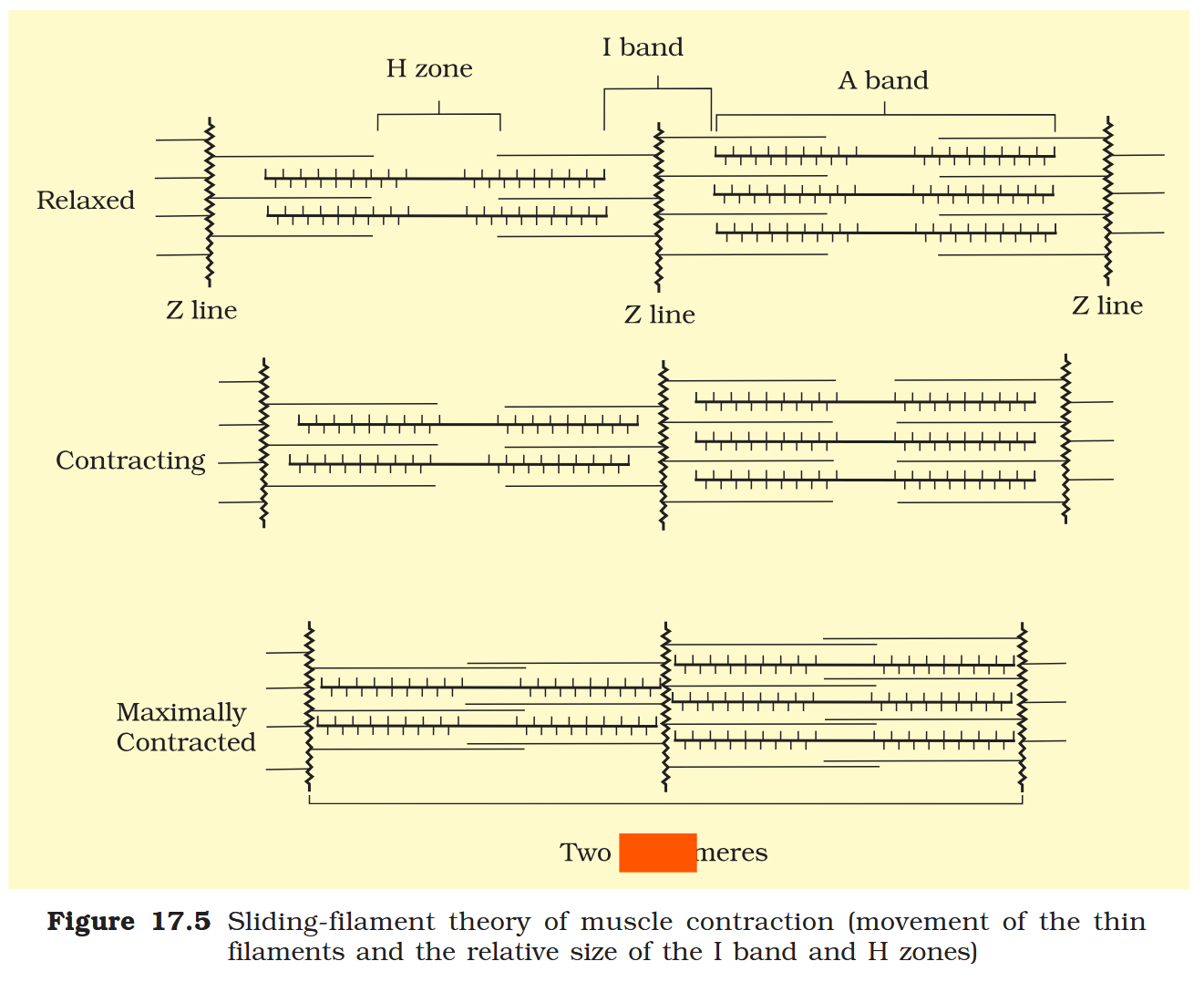
Two Sarcomeres
The process of sarcomere contraction continues till the Ca++ ions are pumped back to the sarcoplasmic cisternae resulting in the masking of actin filaments.
What happens after this?
This causes the return of ‘Z’ lines back to their original position, i.e., relaxation.
The reaction time of the fibres in contraction and relaxation can vary in different muscles.
True or false?
True.
Repeated activation of the muscles can lead to the accumulation of ______________.
Repeated activation of the muscles can lead to the accumulation of lactic acid.