Block 8 Histology-Push Me n Pull You
1/13
There's no tags or description
Looks like no tags are added yet.
Name | Mastery | Learn | Test | Matching | Spaced | Call with Kai |
|---|
No analytics yet
Send a link to your students to track their progress
14 Terms

ID the labeled regions/structures
A. Epithelium
B. Structures
C. Region
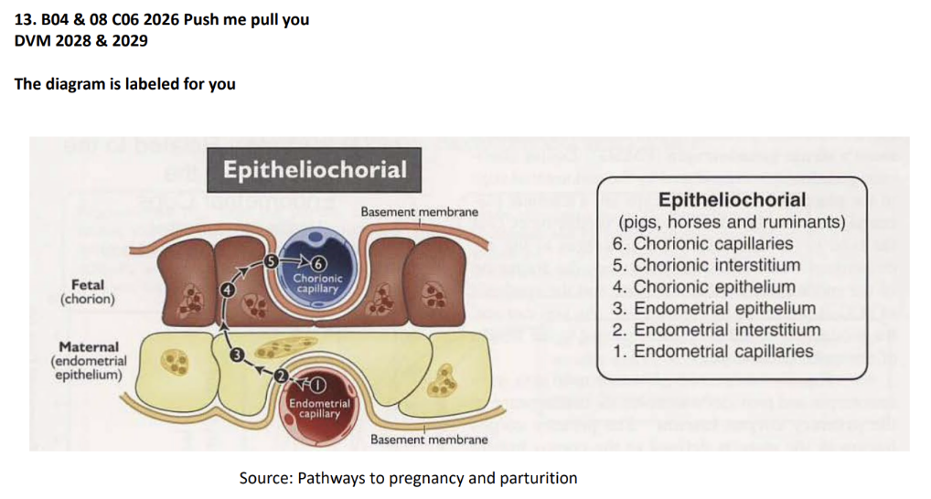
D. Region
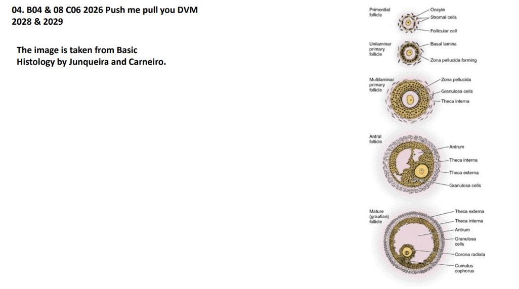
A. germinal/simple cuboidal
B. Follicles
C. Medulla
D. Cortez
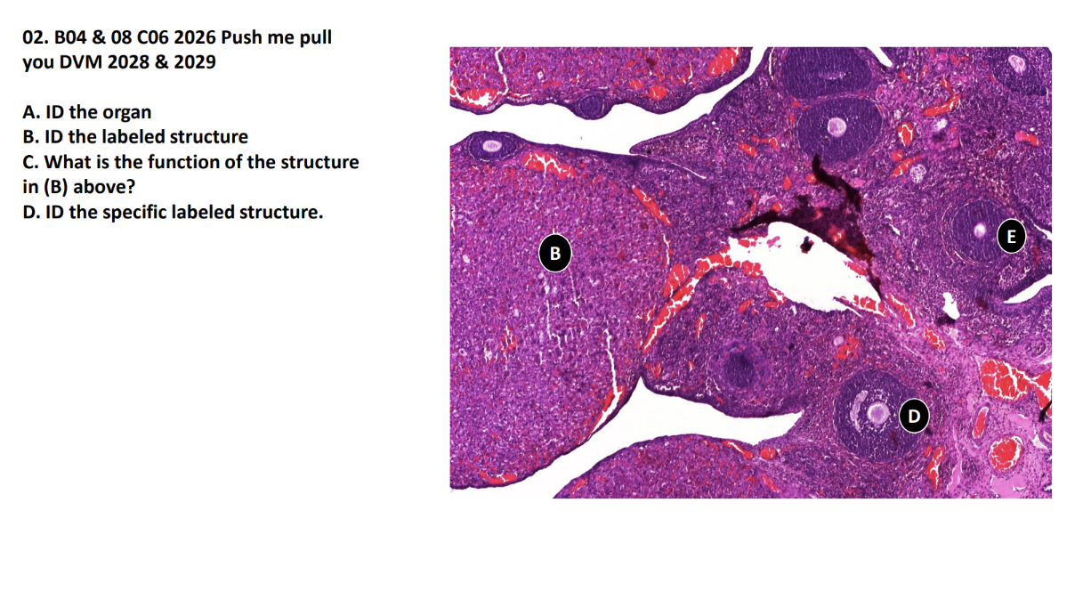
A. ID the organ
B. ID the labeled structure
C. What is the function of the structure
in (B) above?
D. ID the specific labeled structure
A. Ovary
B.Corpus luteum
C.Produce progesteron
D.Secondary Follicle
E. Multilaminar primary follicle
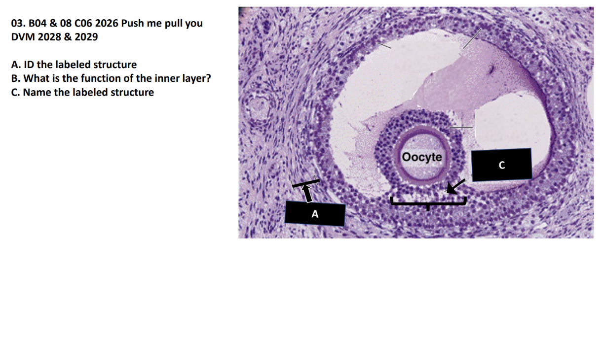
A. ID the labeled structure
B. What is the function of the inner layer?
C. Name the labeled structure
A. Theca layer
B.Secrete androgen hormone androstenedione
C.Cumulus oophorus
Already Labeled
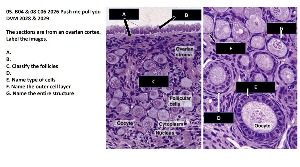
A.
B.
C. Classify the follicles
D.
E. Name type of cells
F. Name the outer cell layer
G. Name the entire structure
A. Tunica albuginea
B. Germinal epithelium (simple
cuboidal)
C. Primary follicles
D. Primary follicle
E. Granulosa cells
F. Stromal (follicular) cells
G. Primordial follicle
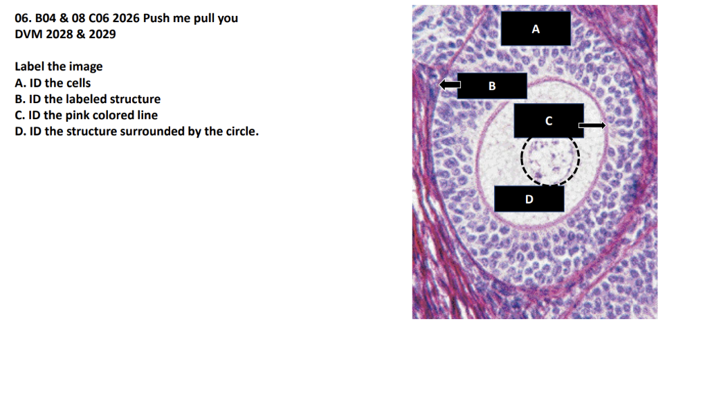
Label the image
A. ID the cells
B. ID the labeled structure
C. ID the pink colored line
D. ID the structure surrounded by the circle
A. Granulosa cells
B. Basement membrane
C. Zona pellucida
D. Oocyte nucleus
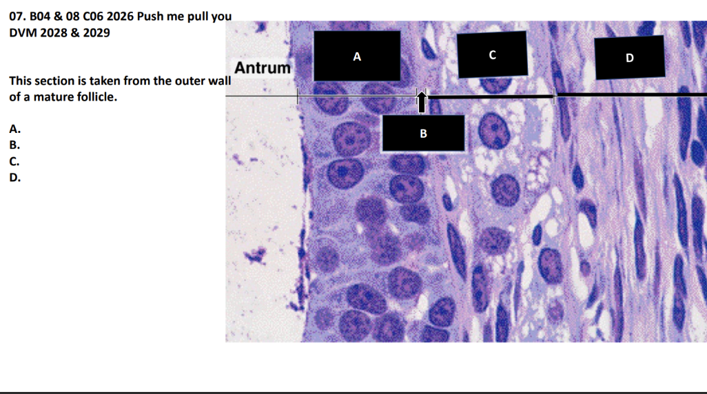
This section is taken from the outer wall
of a mature follicle.
A.
B.
C.
D.
A. Granulosa cells
B. Basement membrane
C. Theca interna
D. Theca externa
08. Image is labeled for you
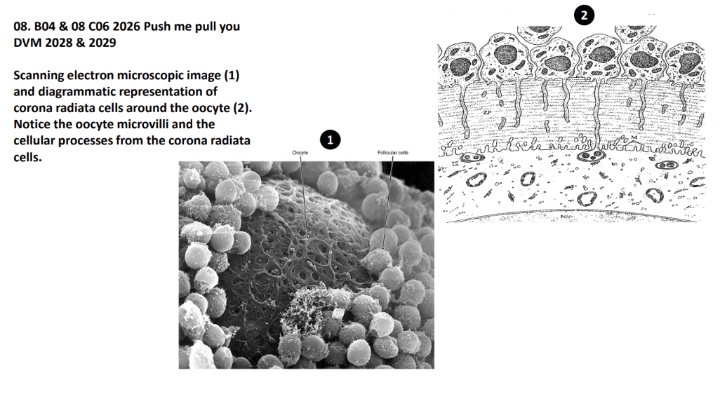
Scanning electron microscopic image (1)
and diagrammatic representation of
corona radiata cells around the oocyte (2).
Notice the oocyte microvilli and the
cellular processes from the corona radiata
cells
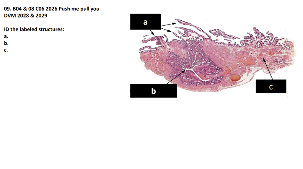
ID the labeled structures:
a.
b.
c.
a. Fimbria of the uterine tube
b. Infundibulum of the uterine tube
c. Mesosalpinx
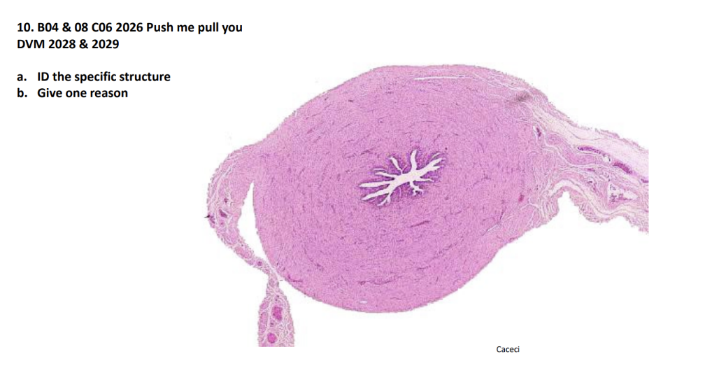
a. ID the specific structure
b. Give one reason
a. Isthmus of the uterine tube
b. Thick muscular layer, low number of fold
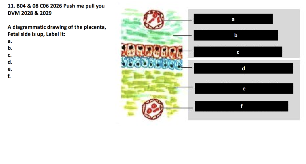
A diagrammatic drawing of the placenta,
Fetal side is up, Label it:
a.
b.
c.
d.
e.
f.
a. Fetal capillary endothelium
b. Fetal connective tissue
c. Chorionic epithelium
d. Maternal (uterine) epithelium
e. Maternal connective tissue
f. Maternal capillary endothelium
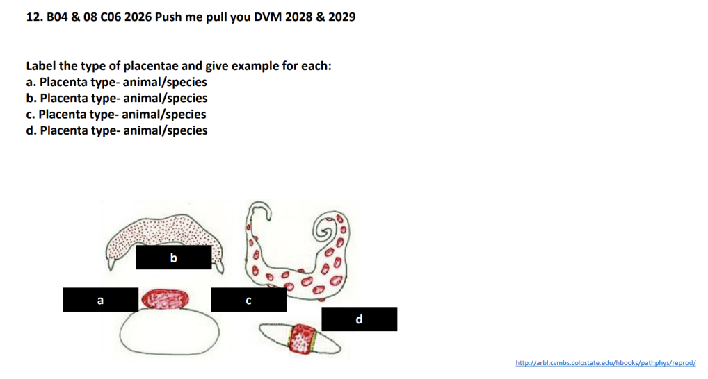
Label the type of placentae and give example for each:
a. Placenta type- animal/species
b. Placenta type- animal/species
c. Placenta type- animal/species
d. Placenta type- animal/species
a. Discoidal - Villi localized in one
area
b. Diffuse - Villi distributed across all
of the chorion
c. Cotyledonary - Villi gathered into
distinct clusters
d. Zonary - Villi gathered in a specific
place on chorion
Labeled
Labeled
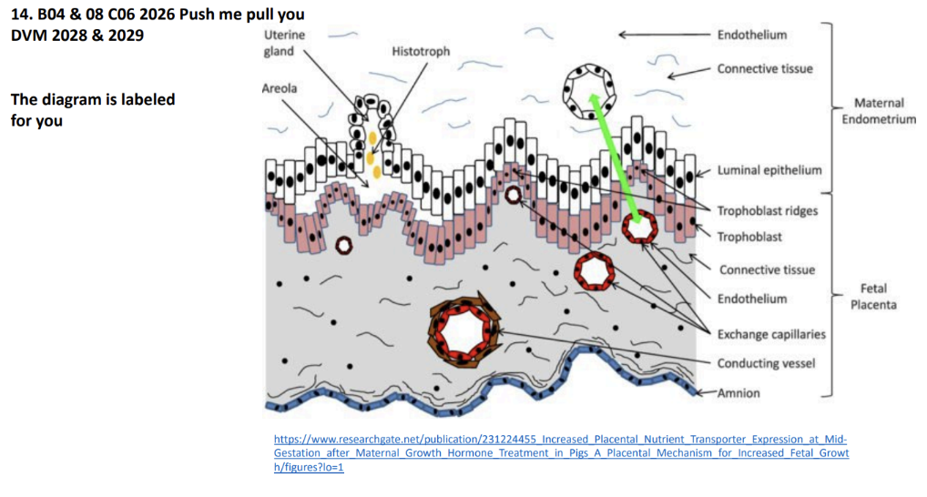
14. Structure of the porcine placenta. Nutrient exchange between
maternal and fetal circulation (green arrow) occurs across six tissue
layers (maternal endothelium, maternal connective tissue, maternal
endometrial epithelium, trophoblast, fetal connective tissues, and fetal
endothelium). Fetal exchange capillaries are located below trophoblast
and in trophoblast ridges. Larger conducting vessels in the fetal placenta
are surrounded by smooth muscle and are located closer to the amnion.
Areloae are specialized exchange areas of fetal placenta that develop
opposite secretory glands of the maternal endothelium. Structure and
nutrient transporter expression were measured in the fetal layers of the
porcine placenta (shaded regions) in this study