Week #9 - Mitochondria and the Chloroplasts
1/40
There's no tags or description
Looks like no tags are added yet.
Name | Mastery | Learn | Test | Matching | Spaced | Call with Kai |
|---|
No analytics yet
Send a link to your students to track their progress
41 Terms
Overview of Cell Signaling
All cells process information (signals) from the environment.
Chemical – a nutrient, waste, ion, hormone
Physical stimulus - light, sound or temperature
Signals can come from outside the organism or from other cells within the organism.
In multicellular organisms, signals may be transmitted over a short or a great distance.
To detect and respond to a signal, a cell (target) must have a specific receptor that can detect it.
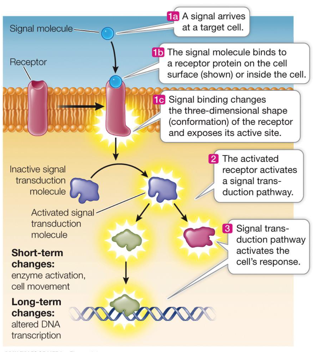
When a cell detects a signal, it initiates a signal trans-duction pathway -- sequence of molecular events and chemical reactions -- that lead to a response to the signal
What are the four key characteristics of signal transduction pathways?
Specificity
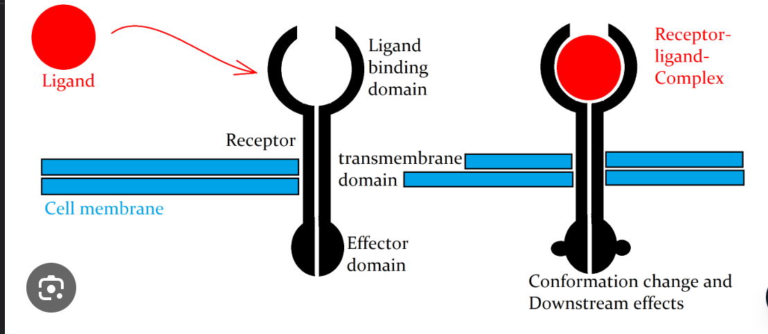
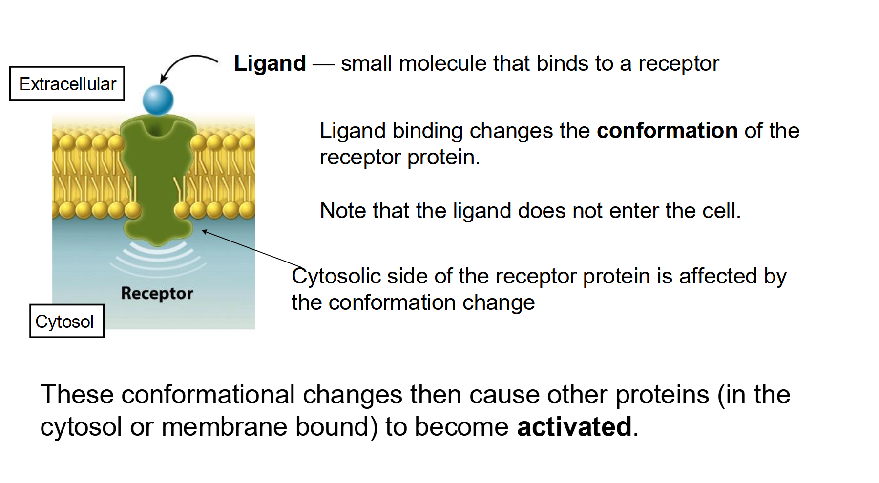
Receptor proteins have very specific binding sites for chemical signal molecules (= ligands).
Ligand binding causes receptor protein to change shape
Shape change activates receptor (e.g., kinase activity)
Activated receptor alters function of a responder protein.
The signal is amplified - more than one responder molecule in cell
Response is executed
Responder activates/deactivates other cellular effectors
Cell activity is altered
What is a ligand in the context of cell signaling, and how does it initiate a response?
A ligand is a specific signaling molecule (such as a hormone, neurotransmitter, or growth factor) that binds to a complementary receptor protein.
Mechanism: The binding of a ligand is highly specific (like a lock and key). When it binds to the extracellular domain of a receptor, it induces a conformational change (shape change) in the receptor.
Function: This shape change "activates" the receptor, allowing it to relay the signal to the interior of the cell to trigger a transduction cascade.
Example from notes: In glycogenolysis, Epinephrine acts as the ligand that binds to the adrenergic receptor on liver cells to start the process of glucose release.
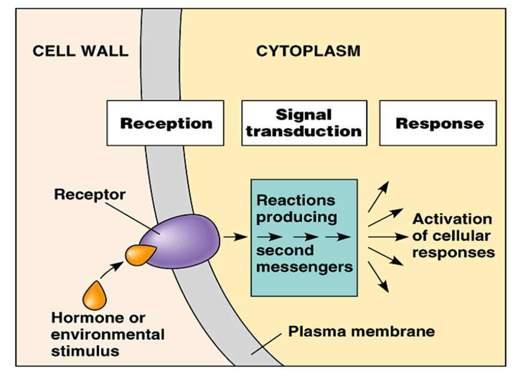
Describe the three sequential stages (and components) of a signal transduction pathway
Components: Signal and Receptor
Reception: Ligand binds to a receptor, causing a conformational change.
Transduction: The signal is relayed through the cell via second messengers or protein cascades (e.g., phosphorylation).
Response: The final cellular activity occurs (e.g., gene expression, enzyme activation, or movement).
Initiated by receptor
Transduced and amplified by other cellular molecules
Ultimately causes change in target cell
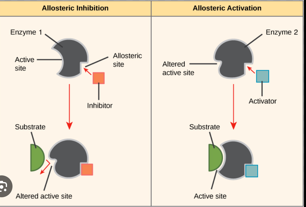
How does signal transduction utilize allosteric regulation and amplification to produce a cellular response?
Allosteric regulation occurs when the binding of a molecule to a specific site on an enzyme—other than the active site—induces a conformational change that either activates or inhibits the enzyme's activity.
Signal transduction often involves enzymes that undergo shape changes.
This is frequently triggered by phosphorylation (adding a phosphate group), which acts as a molecular switch to activate or inhibit the enzyme.
Amplification: A single activated enzyme can catalyze many reactions. This creates a "cascade" effect where one original signal molecule results in a massive internal response.
Response Types:
Short-term: Rapid changes like opening an ion channel.
Long-term: Slower, lasting changes like alteration of gene expression in the nucleus.
What are the primary differences between a ligand and phosphorylation in a signal transduction pathway?
Ligand (The Signal):
Role: The "First Messenger" that initiates the process.
Location: Usually extracellular (outside the cell).
Action: Binds specifically to a receptor to cause a shape change.
Example: Epinephrine (Adrenaline).
Phosphorylation (The Switch):
Role: A modification that acts as a "molecular switch" during transduction.
Location: Intracellular (inside the cytoplasm).
Action: A Kinase (enzyme) adds a phosphate group (PO4) to a protein, causing an allosteric change that activates or inhibits it.
Source: Derived from ATP.
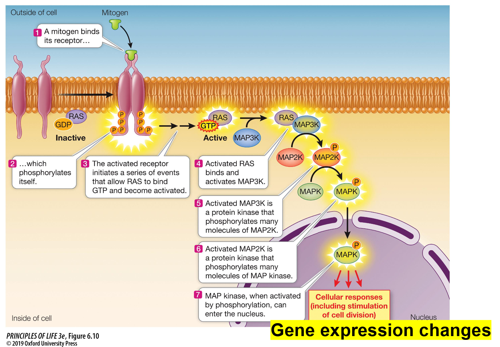
Signal Transduction Cascade Using Kinases
Reception: A mitogen (the ligand) binds to its specific receptor on the cell surface.
Activation: This binding causes the receptor to phosphorylate itself, which initiates a series of events that allow a protein called RAS to bind GTP and become active.
The Cascade (Phosphorylation): Activated RAS binds to and activates MAP3K.
MAP3K is a kinase that phosphorylates many molecules of MAP2K, activating them.
Activated MAP2K then phosphorylates many molecules of MAP kinase (MAPK).
The Response: Once MAPK is activated by phosphorylation, it can enter the nucleus.
Inside the nucleus, it triggers gene expression changes that lead to cellular responses, such as stimulating cell division
Transmembrane Proteins and Signal Transduction
Membrane proteins play a major role in signal transduction by converting an extracellular signal into intracellular signal(s).
Signal transduction allows cells to rapidly respond to events happening in their environment:
Grow
Divide
Survive (or not)
Move
Differentiate (i.e., time to change)
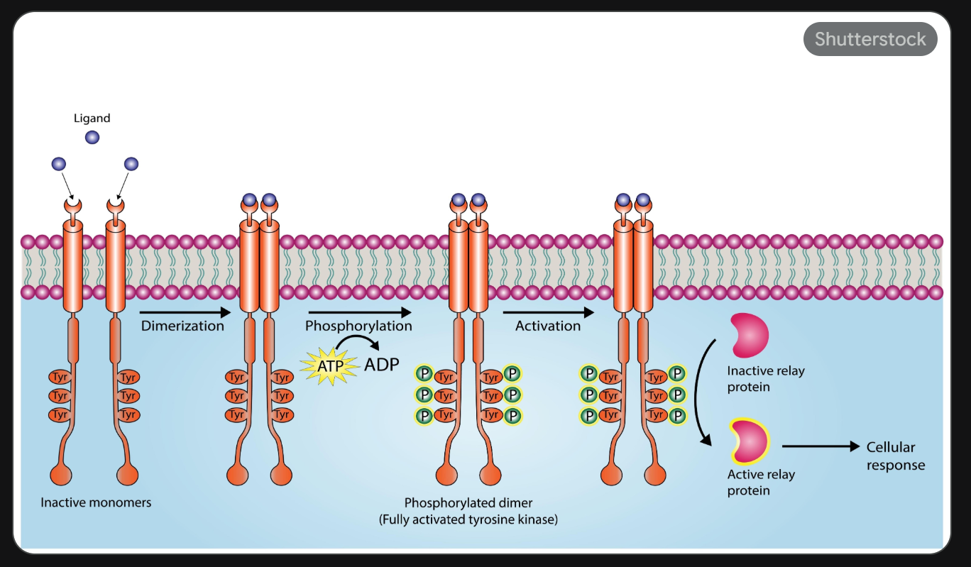
Protein Kinase Receptors
Ligand Binding: A signaling molecule (like insulin) binds to the extracellular portion of the receptor.
Dimerization/Activation: This binding causes the receptor subunits to come together or change shape.
Autophosphorylation: The "kinase" part of the receptor (on the inside of the cell) becomes active. It uses ATP to add phosphate groups to its own "tails."
Signal Propagation: These new phosphate groups act as "docking stations" for other signaling proteins inside the cell, starting the transduction cascade.
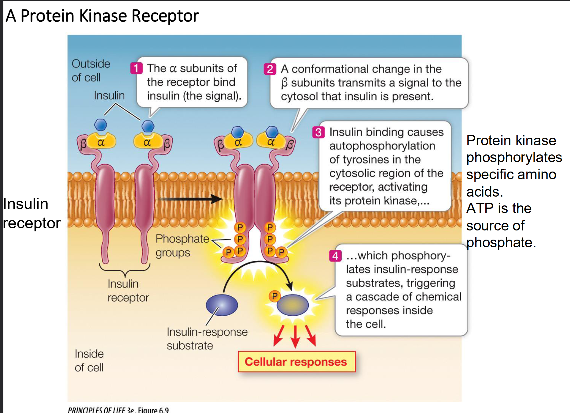
Ex. Mammalian insulin receptor
Phosphorylates itself and other insulin response substrates
Initiates insertion of glucose transporters into the plasma membrane
Example: The Insulin Receptor
The insulin receptor is a dimer, meaning it consists of two identical halves.
Reception: Insulin binds to the alpha subunits (the parts outside the cell).
Activation: This causes a conformational change that is transmitted to the beta subunits (the parts inside the cytoplasm).
Transduction: The beta subunits perform autophosphorylation. This "turns on" the receptor, allowing it to phosphorylate other proteins called Insulin Response Substrates (IRS).
Response: These IRS proteins trigger multiple pathways, such as:
Moving glucose transporters to the cell membrane to let sugar in.
Starting glycogen synthesis (storing energy).
Altering gene expression for cell growth.
The G Protein-Coupled Receptor (GPCR) Pathway
Reception: A ligand binds to the GPCR, causing a shape change in the receptor.
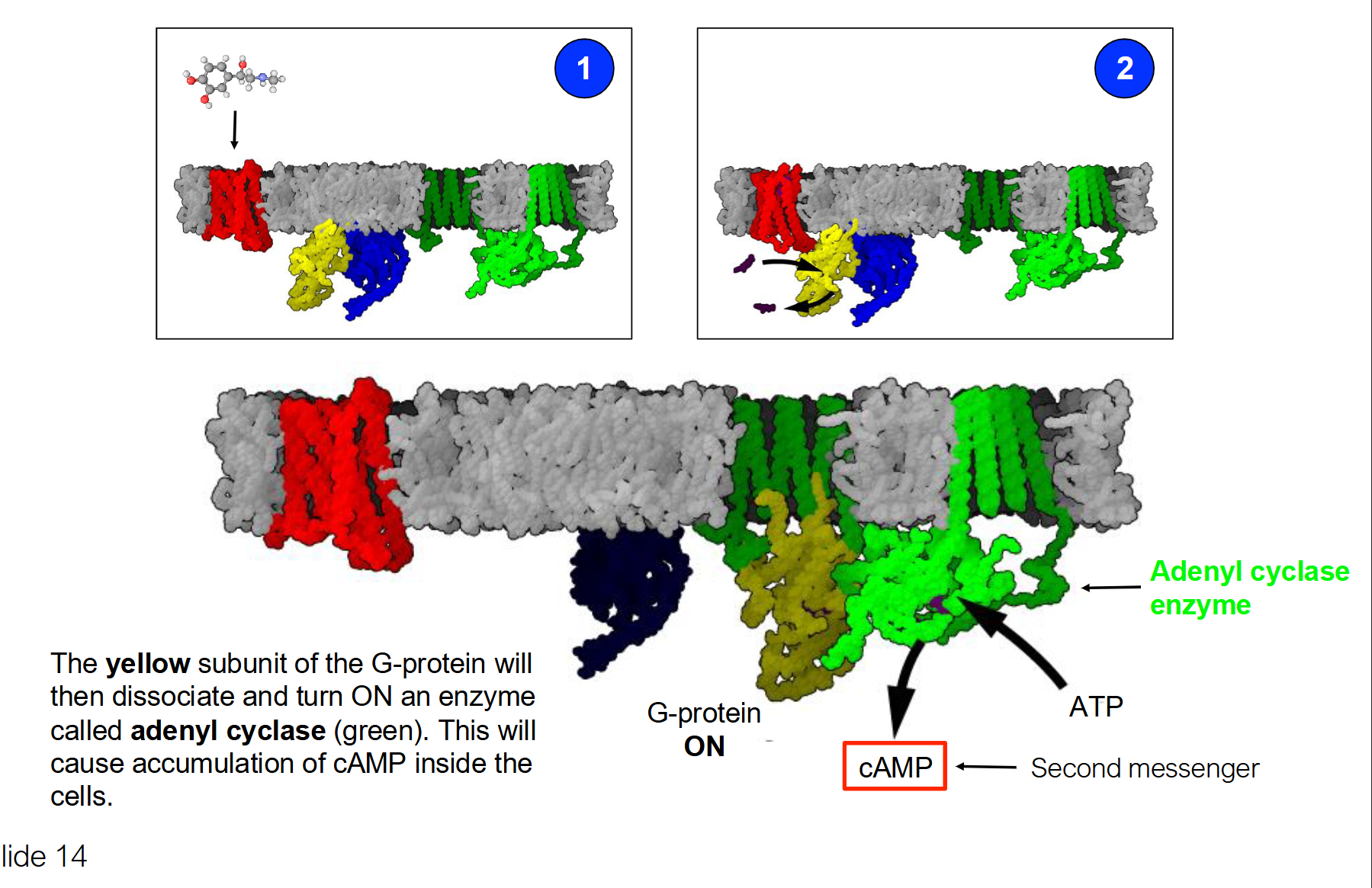
Activation: The receptor binds to an inactive G protein (alpha, beta, gamma subunits), causing the alpha subunit to swap GDP for GTP.
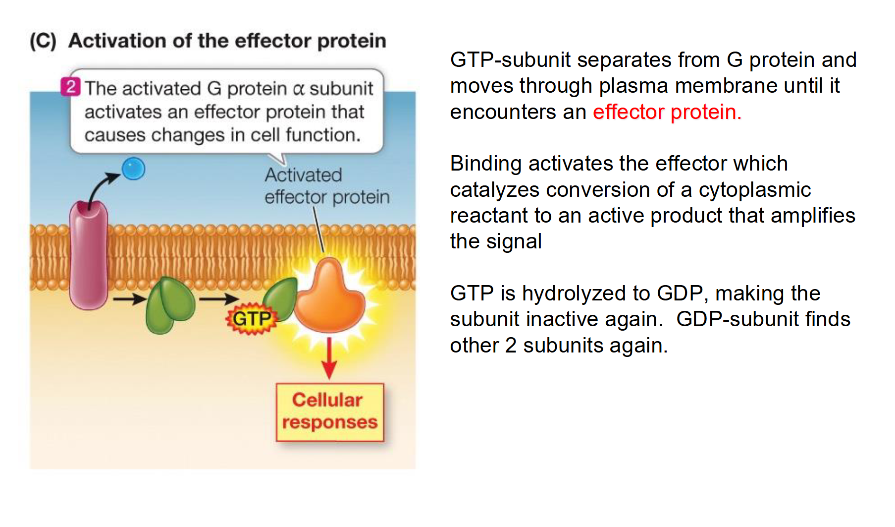
Dissociation: The active alpha subunit (with GTP) breaks away and moves along the membrane to activate an effector protein (like Adenyl Cyclase).
Second Messenger: The effector produces a second messenger (like cAMP), which triggers a downstream phosphorylation cascade and cellular response.
Termination: The alpha subunit eventually hydrolyzes GTP back to GDP, becomes inactive, and reunites with the beta and gamma subunits to reset the cycle.
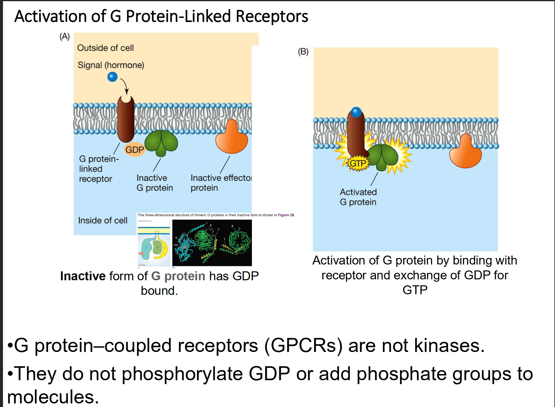
What is a G protein, and how does it act as a molecular switch in cell signaling?
Structure: A heterotrimeric protein consisting of three subunits: alpha, beta, and gamma (gamma).
The "OFF" State: The G protein is inactive when the alpha subunit is bound to GDP.
The "ON" State: When a ligand binds to the receptor, the alpha subunit releases GDP and binds GTP, causing it to dissociate from the other subunits.
Function: Once active, the alpha subunit (bound to GTP) moves along the membrane to activate an effector (like the enzyme Adenyl Cyclase).
Self-Regulation: It has built-in GTPase activity, meaning it eventually hydrolyzes GTP back into GDP to turn itself "OFF."
How does the G protein act as the essential link between an external hormone and the internal breakdown of glycogen?
The Activation Step: The hormone Epinephrine (ligand) binds to a GPCR, causing the G protein to swap GDP for GTP.
The Relay: The active alpha subunit dissociates and activates the enzyme Adenyl Cyclase.
The Second Messenger: Adenyl Cyclase produces cAMP, which carries the "breakdown" signal into the cytoplasm.
The Result: cAMP triggers the kinase cascade (PKA → Phosphorylase Kinase → Glycogen Phosphorylase) that ultimately chops glucose units off the glycogen chain.
Key Concept: The G protein is the molecular switch that translates an extracellular signal into an intracellular chemical message.
Compare and contrast the G Protein-Coupled Receptor (GPCR) pathway and the Protein Kinase Receptor pathway (e.g., Insulin receptor)
Mechanism of Activation:
GPCR: Relies on a "relay" system. The receptor recruits a separate G protein, which swaps GDP for GTP to become active.
Protein Kinase: Relies on autophosphorylation. The receptor subunits come together (dimerize) and add phosphate groups to themselves.
The "Switch":
GPCR: The switch is the exchange of nucleotides (GDP/GTP).
Protein Kinase: The switch is the covalent addition of a phosphate group from ATP.
Intermediate Steps:
GPCR: Usually activates an effector enzyme (like Adenyl Cyclase) to produce a second messenger (like cAMP).
Protein Kinase: The phosphorylated receptor provides docking sites for internal signaling proteins (like IRS) to bind directly.
Example from Notes:
GPCR: Epinephrine triggering glycogen breakdown.
Protein Kinase: Insulin triggering glucose uptake.
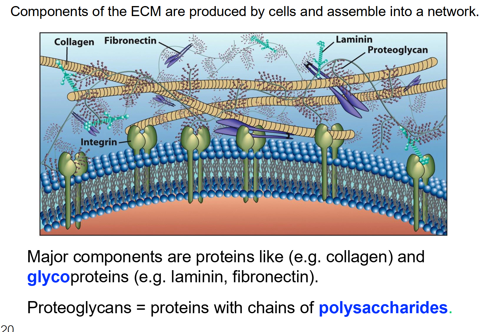
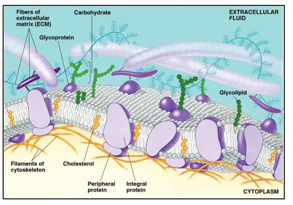
What is the ECM?
The ECM is an organized network of molecules (like proteins and carbohydrates) that cells produce and then "spit out" into the space around them.
It is a complex meshwork that connects cells into communities to form tissues like skin, bone, or muscle.
Key Components:
Fibers (like Collagen): These provide strength. Think of them as the steel cables in a bridge.
Glycoproteins & Proteoglycans: These are proteins with sugar chains attached. They help with hydration and trapping signaling molecules.
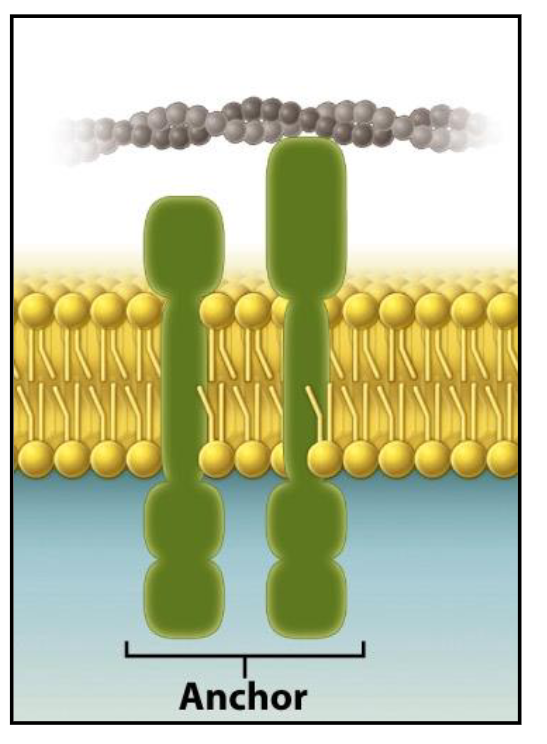
Anchor Proteins (Integrins): These are membrane proteins that physically "anchor" the inside of the cell (the cytoskeleton) to the ECM outside. This creates a continuous physical link between the cell and its environment
Anchor Proteins and the Extracellular Matrix (ECM)
They play an important role by interacting with and physically linking the cell to the components of the extracellular matrix.
Multicellular organisms are composed of tissues and organs consisting of communities of cells.
These cells work together to perform a function (e.g., skin, liver, leaves).
The ECM is an organized network of material produced and secreted by cells.
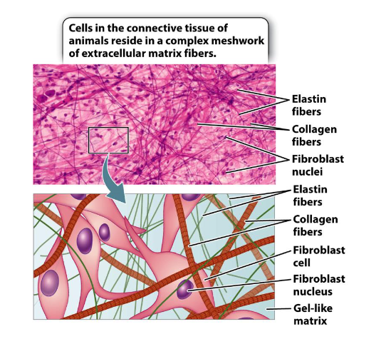
Example: The ECM is abundant in connective tissues of animals (e.g. tendons, ligaments, dermis)
List the four main functions of the ECM
Cell adherence: It provides a surface for cells to grip onto so they don't just float away.
Communication between cells: When the ECM is stretched or chemically changed, it sends signals through those anchor proteins to tell the cell how to behave or grow.
Cell shape, mechanical support, and structural integrity: It gives tissues their shape and mechanical strength (e.g., the hardness of bone or the elasticity of skin).
Serving as a barrier/filtering out particles: acts as a physical sieve, controlling which nutrients or particles can reach the cell.
According to the Misrepair-accumulation theory, how does the Extracellular Matrix (ECM) contribute to the development of wrinkles?
The ECM of the skin is packed with two primary types of protein fibers:
Collagen: Provides the skin with strength and structural integrity.
Elastin: Provides elasticity, allowing the skin to "snap back" after being stretched.
Wrinkles are caused by the accumulation of incorrectly repaired (altered) collagen and elastin fibers.
Instead of healthy regeneration, the tissue undergoes fibrosis (thickening and scarring), which disrupts the skin's structural integrity and leads to visible folding.
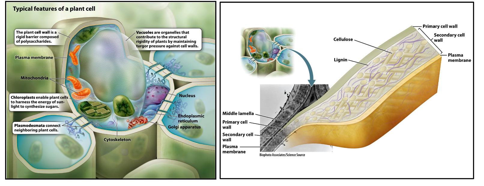
Compare and contrast the Extracellular Matrix (ECM) in animal cells versus plant cells. Focus on composition and primary function.
Identity: In plants, the ECM is the Cell Wall.
Composition:
Animals: Primarily protein-based (collagen, elastin).
Plants: Primarily carbohydrate-based (cellulose, hemicellulose, pectin, proteins).
Function:
Both: Provide adherence, signaling, and structural integrity.
Plants (Unique): Acts as a skeletal equivalent for the whole organism and provides a rigorous physical defense against pathogens and mechanical stress.
The Endosymbiotic Theory
Definition: An evolutionary theory stating that eukaryotic cells originated from a symbiotic relationship between different types of prokaryotes.
The Process: A large host cell engulfed smaller, specialized bacteria (aerobic bacteria and cyanobacteria) which eventually evolved into organelles.
Key Figures:
Konstantin Mereschkowski: First proposed the idea (1905).
Lynn Margulis: Advanced the theory with microbiological evidence (1967).
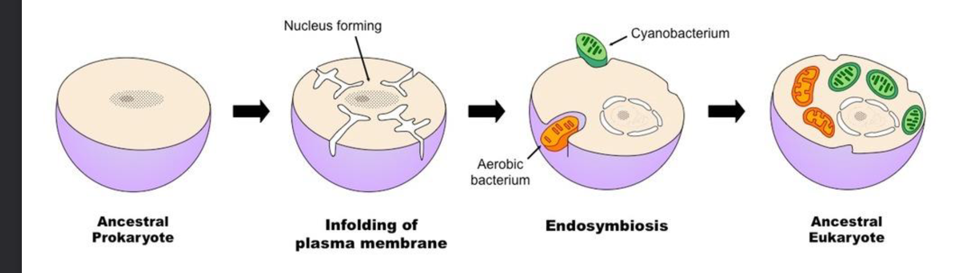
List two major pieces of evidence that support the Endosymbiotic Theory.
Genetic Evidence: Mitochondria and chloroplasts (plastids) contain their own circular DNA, which is structurally similar to bacterial DNA.
Reproduction: These organelles reproduce independently of the cell through binary fission, the same method used by bacteria.
Bonus: They also possess a double membrane, suggesting the inner layer is the original bacterial membrane and the outer layer is the host's vesicle.
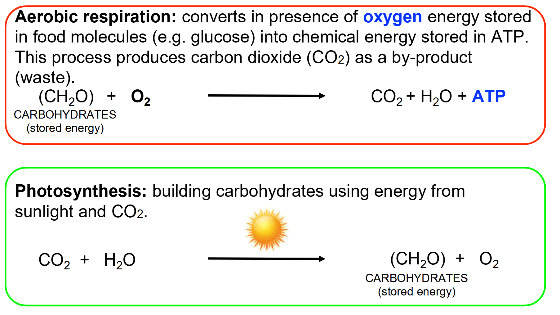
Summarize Aerobic Respiration and Photosynthesis
Aerobic Respiration:
Converts in presence of oxygen energy stored in food molecules (e.g. glucose) into chemical energy stored in ATP.
This process produces carbon dioxide (CO2) as a by-product
Occurs in the Mitochondria
Photosynthesis:
Building carbohydrates using energy from sunlight and CO2
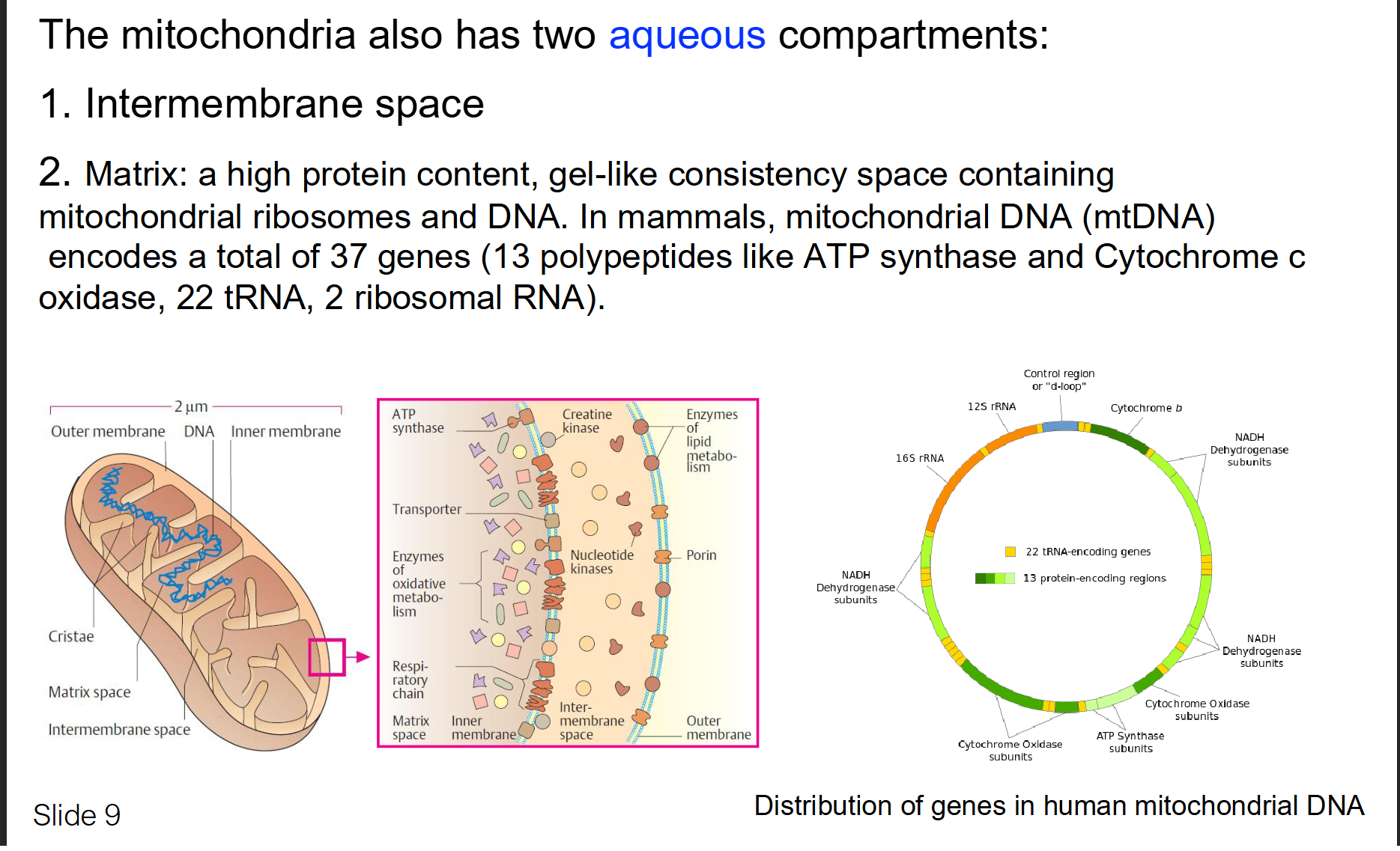
Describe the two membranes of the mitochondria
Outer Mitochondrial Membrane (OMM):
Contains porins (large channels) that make it permeable to molecules like ATP and sucrose.
Contains many enzymes with diverse metabolic functions. An example is monoamine oxidases that breaks down monoamines ingested from food, as well as monoamine neurotransmitters (dopamine and serotonin)
Inner Mitochondrial Membrane (IMM):
High protein to lipid ration (3:1)
Folded into cristae to increase surface area for energy production and contain machinery for aerobic respiration and ATP formation
Phospholipid called cardiolipin → a characteristic of bacterial membranes and needed for optimal function of many enzymes
The Matrix: The innermost gel-like space containing mtDNA and ribosomes.
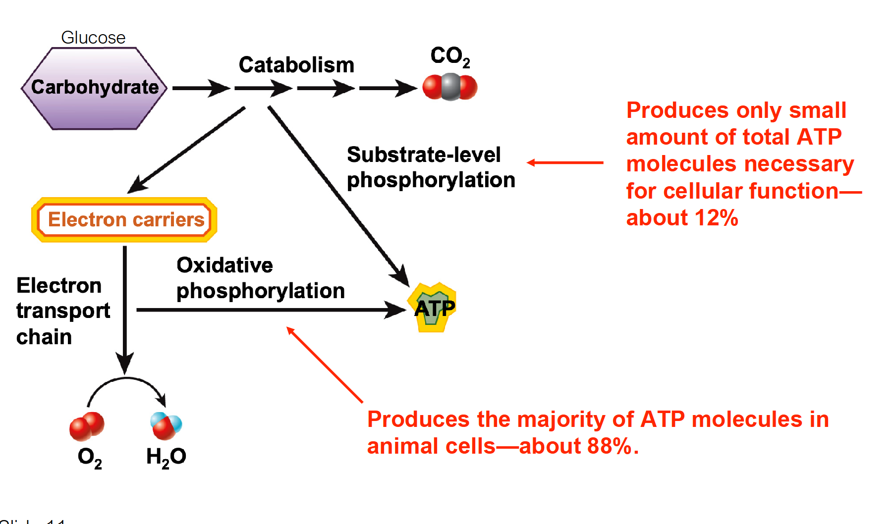
What is the difference between Substrate-level phosphorylation and Oxidative phosphorylation?
Cellular Respiration: Involves a series of catabolic reactions → Breaking down carbohydrates (glucose) to form ATP
Substrate-level phosphorylation: A direct chemical reaction (e.g., in glycolysis) that transfers a phosphate group to ADP to form ATP.
Oxidative phosphorylation: Uses electron carriers to create an electrochemical gradient across the inner membrane. This gradient powers ATP synthase to produce the majority of the cell's ATP.
Requirement: Oxidative phosphorylation requires oxygen (aerobic), whereas substrate-level can occur without it.
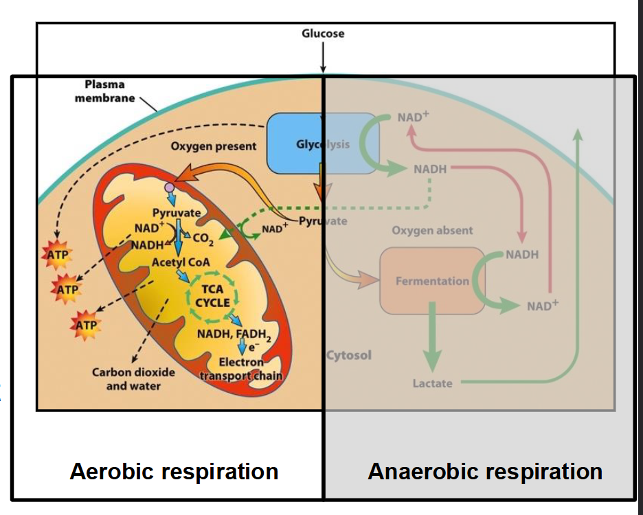
Does SLP or OP produce more ATP?
Oxidative Phosphorylation produces a lot more!
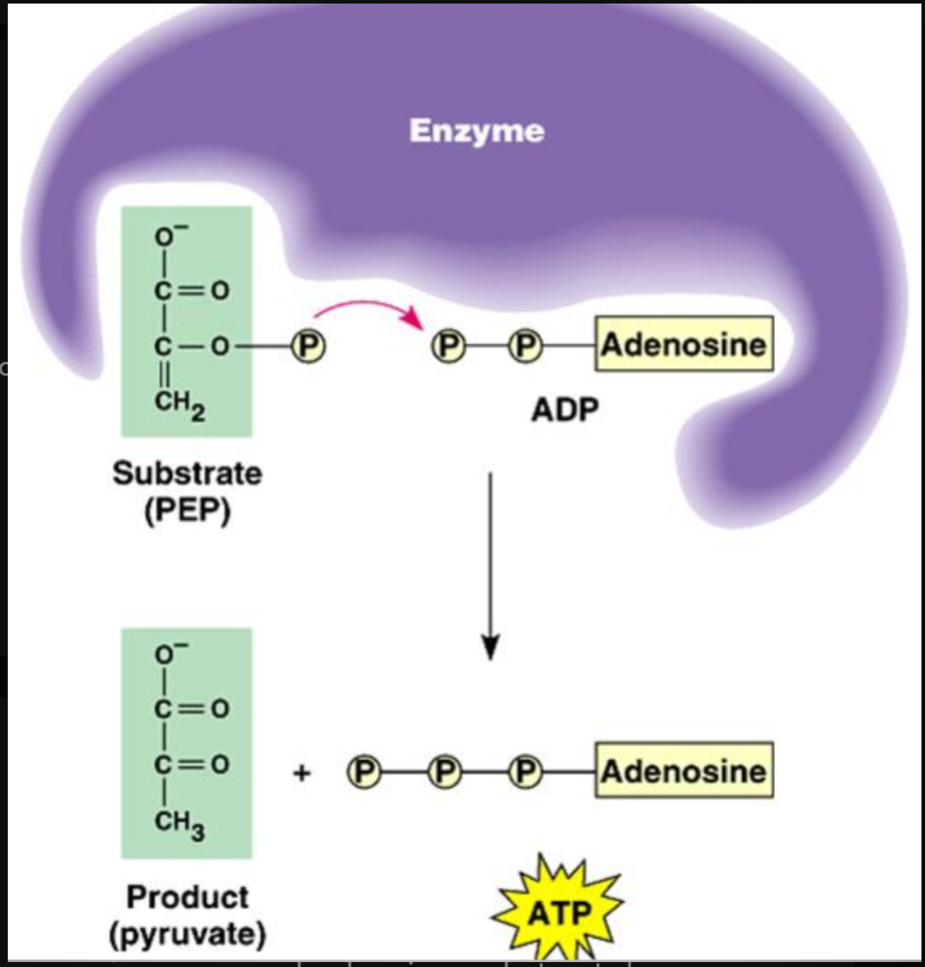
Substrate Level Phosphorylation Overview
In this process, a phosphate group is physically moved from a high-energy "donor" molecule (the substrate) directly onto ADP (adenosine diphosphate) to create ATP.
Components:
ADP: The "empty battery" waiting to be charged.
Phosphorylated Substrate: A metabolic intermediate molecule that is holding onto a phosphate group.
NAD+ and FAD: Electron carriers that are reduced
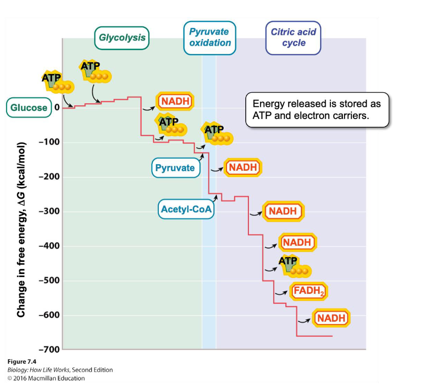
Glycolysis: Happens in the cytosol. For every molecule of glucose, the cell nets 2 ATP through substrate-level phosphorylation.
The TCA (Krebs) Cycle: Happens in the mitochondrial matrix. This cycle produces 1 ATP (or GTP) per turn via this method.
Glycolysis
Starts with glucose (6C sugar) and produces two 3C pyruvate (pyruvic acid) molecules
Occurs in cytoplasm
Anaerobic process: Does not require oxygen
2 NAD+ → 2 NADH that enter ETC
Net Profit of 2 ATP (4 Produced in total, but 2 were invested to break glucose down)
The overall goal of glycolysis is to break down glucose into smaller pyruvate molecules, producing a small amount of ATP and NADH for energy use.
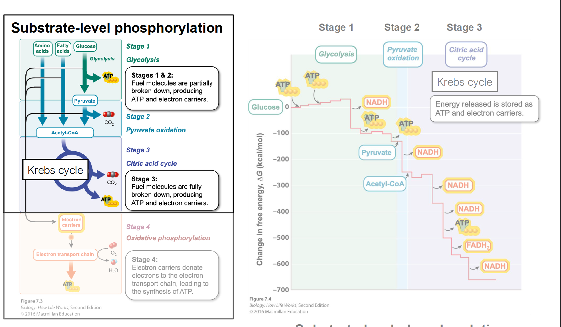
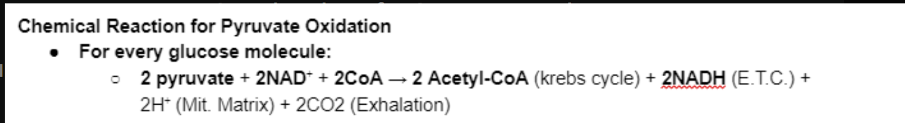
Pyruvate Oxidation
Following glycolysis, two pyruvate molecules are transported through the two mitochondrial membranes into the matrix
The main purpose of pyruvate oxidation is to convert pyruvate (from glycolysis) into acetyl-CoA, which can then enter the Krebs cycle (Citric Acid Cycle) for further energy extraction.
This process also generates NADH and releases carbon dioxide (CO₂).
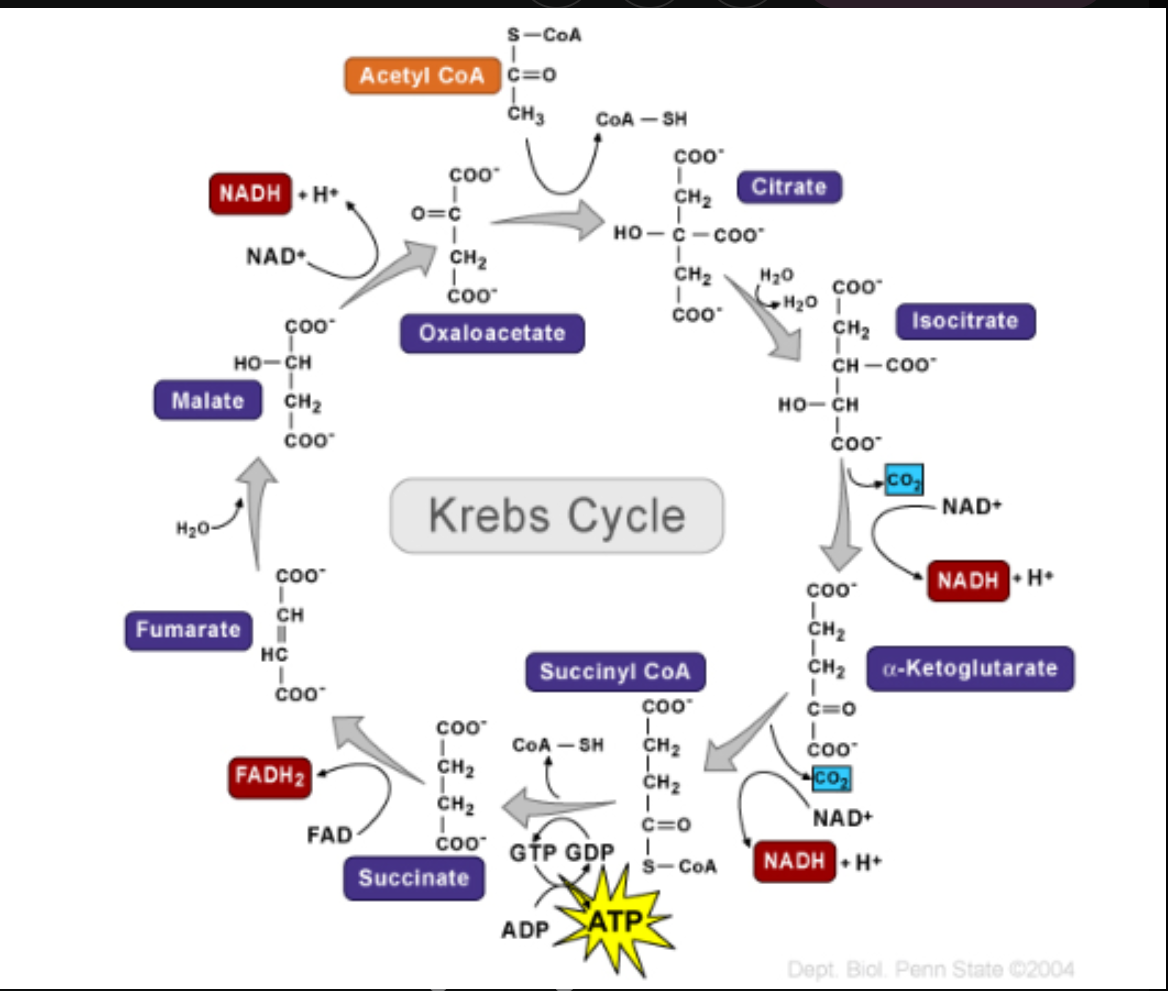
The Citric Acid Cycle
A.K.A Kreb’s Cycle
Result in oxidation of acetyl group to CO2
Completes the conversion of all carbon atoms originally in glucose to CO2
Synthesizes ATP, NADH and FADH2
Its main purpose is to fully oxidize the acetyl group from acetyl-CoA into carbon dioxide (CO₂) and capture high-energy electrons in the form of NADH and FADH₂ for use in the electron transport chain.
The cycle also generates a small amount of ATP (or GTP).
The Electron Carriers
Coenzymes acting as electron carriers can exist either as:
Oxidized—can accept electrons
Reduced—can donate electrons when returning to their oxidized state
Nicotinamide adenine dinucleotide
Oxidized = NAD+
Reduced = NADH
Flavin adenine dinucleotide
Oxidized = FAD
Reduced = FADH2
Oxidation of NADH and FADH2 allows electrons and energy to be transferred
The ETC
Delivery: Electron carriers (NADH and FADH₂) from Glycolysis and the TCA cycle arrive and "dump" their high-energy electrons into the chain.
The Chain Reaction: As these electrons move from one complex to the next, they lose energy.
The Proton Pump: This lost energy isn't wasted. Complexes I, III, and IV use it as fuel to pump Hydrogen ions (H+) from the Matrix into the Intermembrane Space.
The Final Destination: At the very end of the chain, Oxygen acts as the final electron acceptor. It grabs the "spent" electrons and some H+ ions to form Water (H2O). This is why you need to breathe!
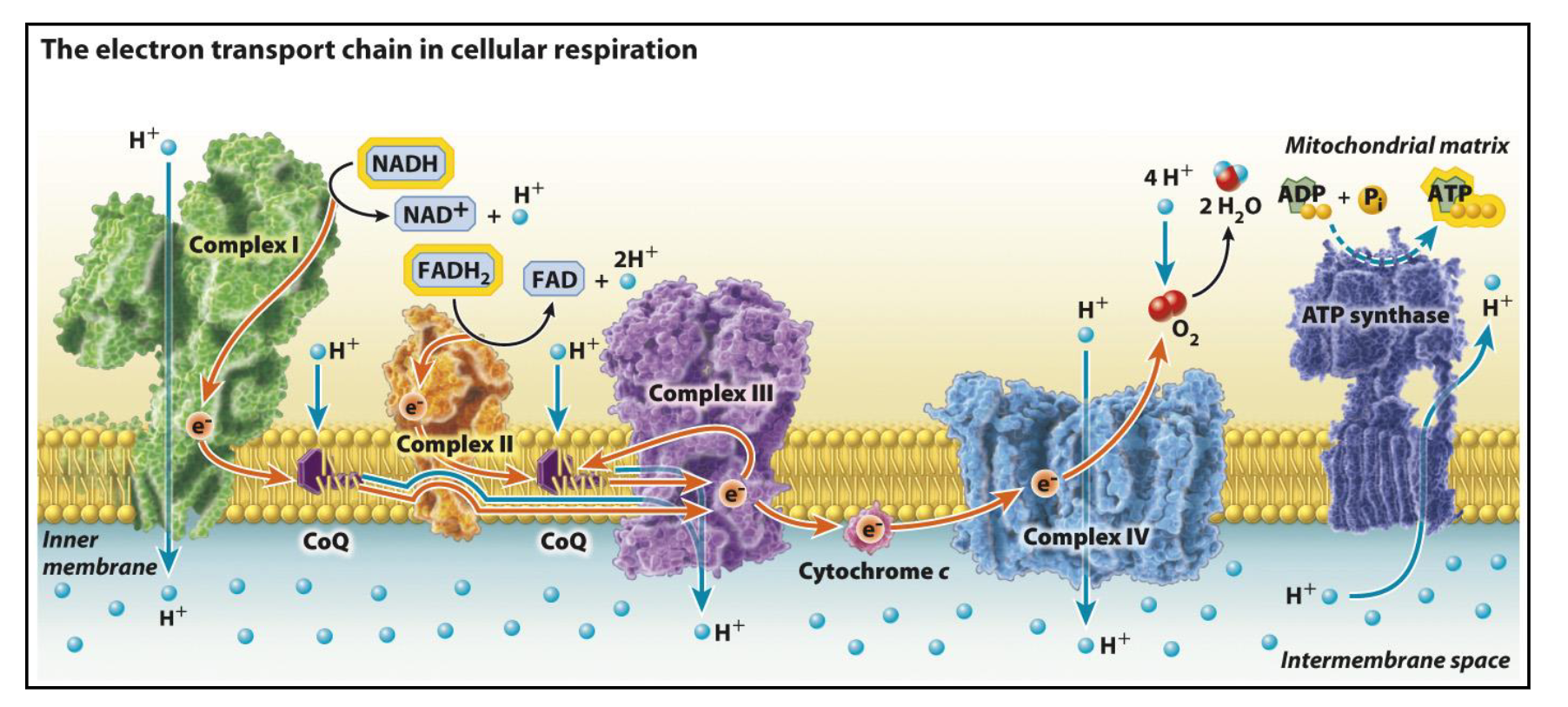
The Electrochemical Gradient and Chemiosmosis: The ATP Factory
Because the ETC is constantly pumping H+ ions into the narrow intermembrane space, that area becomes highly concentrated with positive charges.
This creates a proton-motive force.
The ions desperately want to diffuse back into the matrix (where the concentration is lower), but they cannot pass through the phospholipid bilayer because they are charged.
Since the H+ ions are trapped, they have only one "doorway" back into the matrix: a specialized protein called ATP Synthase.
The Turbine: As H+ ions flow through ATP synthase, they cause the protein to spin like a water wheel or a turbine.
The Payoff: This mechanical spinning energy is used to capture an inorganic phosphate and attach it to ADP, creating ATP.
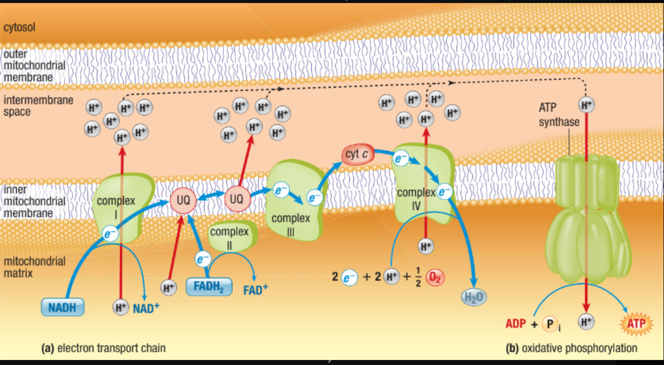
What is the role of Oxygen and the Proton Gradient in oxidative phosphorylation?
Oxygen: Acts as the final electron acceptor at the end of the ETC. It combines with electrons and H+ to form water (H2O). Without it, the chain backs up and stops.
One of the most electronegative elements
Proton Gradient: Energy from passing electrons is used to pump H+ ions into the intermembrane space.
ATP Synthesis: The high concentration of H+ creates a "pressure" (proton-motive force) that flows through ATP Synthase, spinning it like a turbine to manufacture ATP.
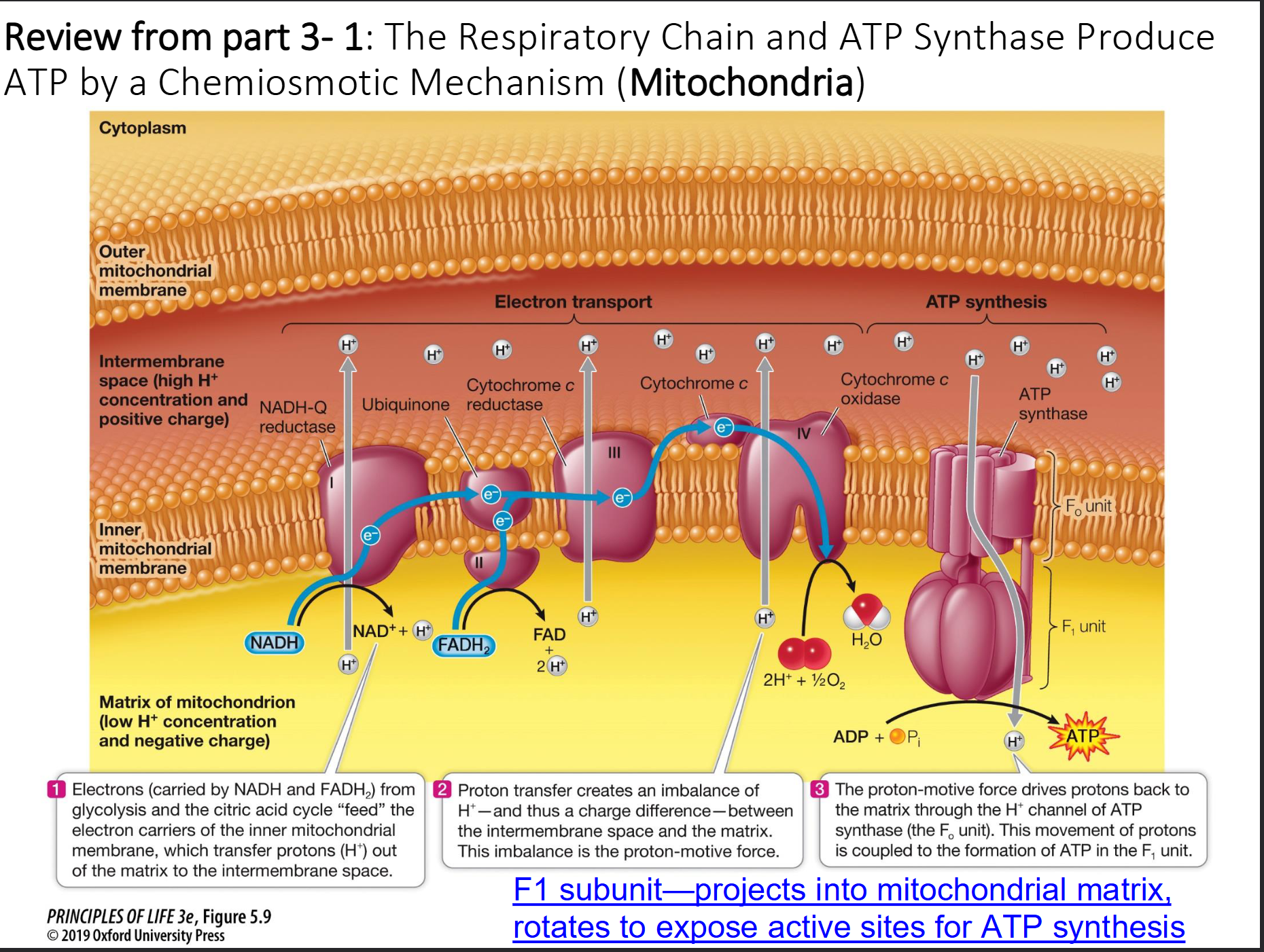
NADH and FADH2
NADH
Passes electrons to NADH dehydrogenase (1st protein complex)
NADH oxidation pumps 3 protons and are responsible for producing 3ATP
FADH2
Passes electrons to complex II
FADH2 oxidation pumps 2 protons and are responsible for producing 2ATP
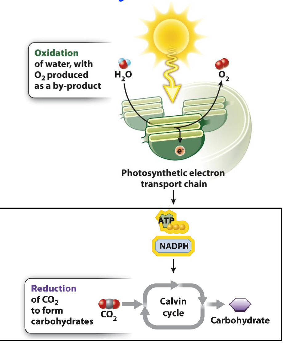
Photosynthesis and its two stages
Photosynthesis: Building carbohydrates (CH2O) using energy from sunlight and CO2.
CH2O is then used as a starting point for making other molecules or synthesizing ATP through cellular respiration (mitochondria)
#1 Light Dependent Reactions:
Directly associated with the absorption of light
#2 Light Independent Reactions:
A.K.A Calvin Cycle
DO NOT require light but need products of light dependent reactions
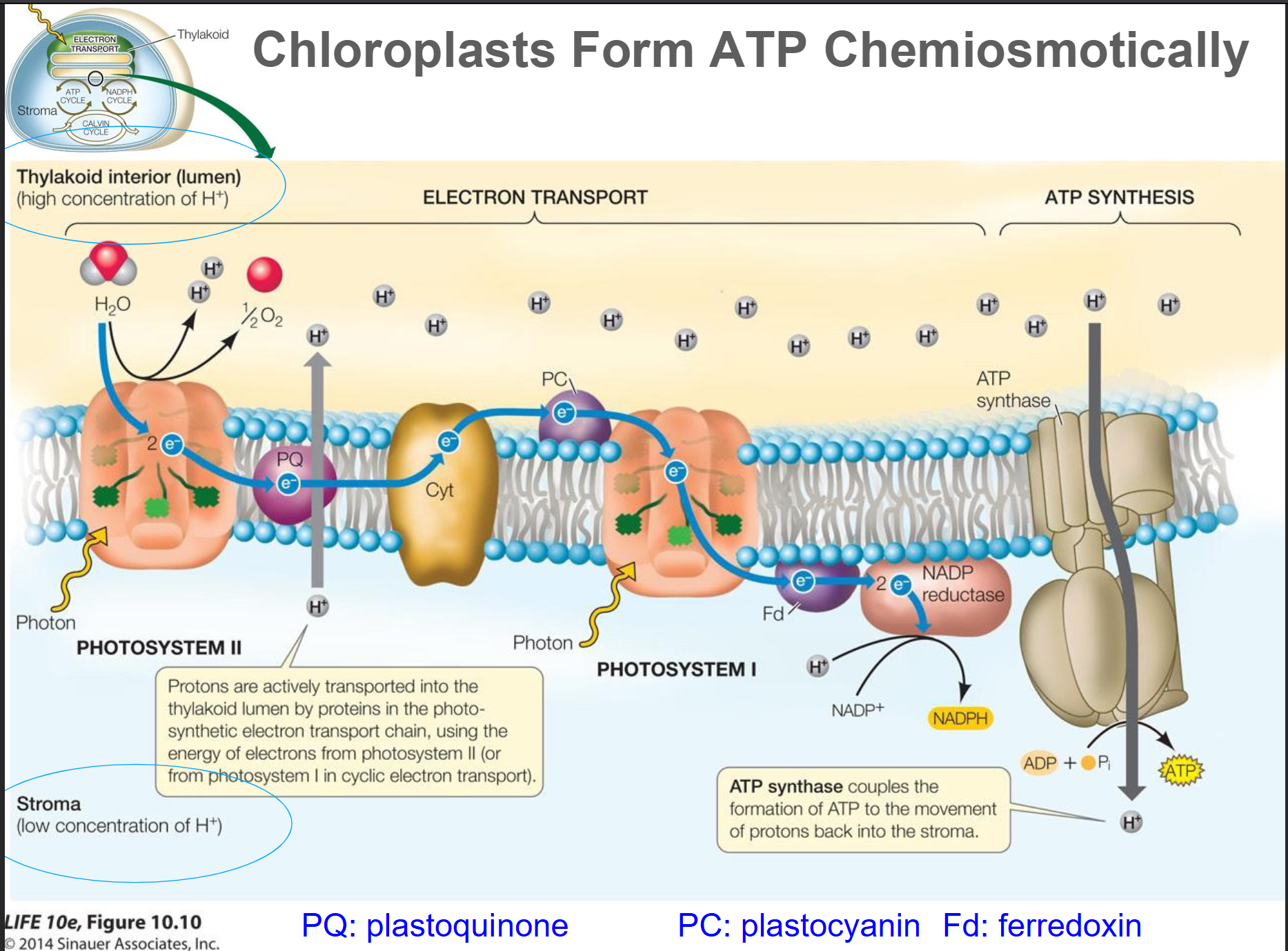
Light Dependent Reactions
Light energy is captured by pigment molecules (chlorophyll)
Used to make ATP and NADPH (Nicotinamide adenine dinucleotide phosphate)
A water molecule is split and an ETC transfers electrons to NADP+, establishing a proton gradient to make ATP
These products are used in the Calvin cycle to synthesize glucose
Oxygen is produced as a waste product and is released
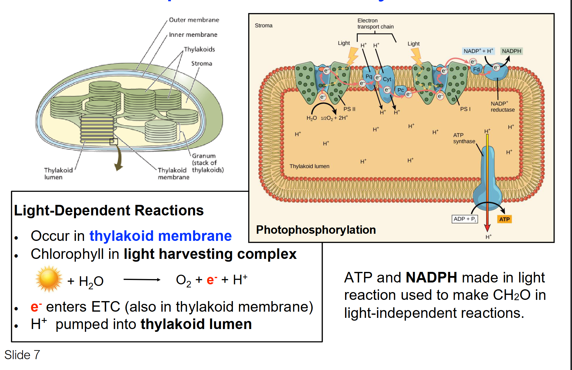
What are the key steps in the light dependent reaction?
Absorption of Light: Chlorophyll and other pigments in the photosystem II absorb light energy, which excites electrons to a higher energy level.
Splitting of Water: This excited state triggers the splitting of water molecules into oxygen, protons (H⁺), and electrons. Oxygen is released as a byproduct.
Electron Transport Chain (ETC): The excited electrons travel through the ETC, losing energy as they go, which is used to pump protons across the thylakoid membrane, creating a proton gradient.
ATP Synthesis: As protons flow back across the membrane through ATP synthase (due to the gradient), ATP is generated from ADP and inorganic phosphate.
Formation of NADPH: Electrons reach photosystem I, get re-energized by light, and are then transferred to NADP⁺ to form NADPH.
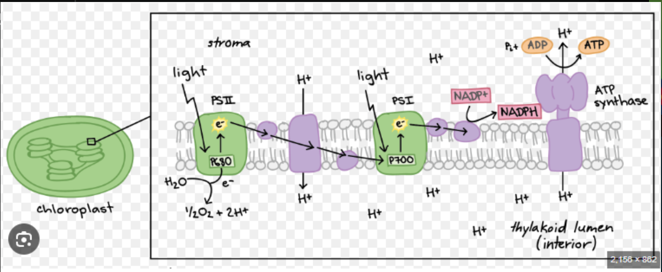
Summary of Light Dependent Reaction
The Path: Electrons start at Photosystem II (PSII), move through an electron transport chain to Photosystem I (PSI), and finally end up at NADP⁺.
Photolysis: To replace the electrons leaving PSII, water molecules are split (H2O → 2H+ + ½ O2 + 2e-. This is why plants release oxygen.
Products: It produces both ATP (via a proton gradient) and NADPH (the final electron catcher).
Purpose: Provides the exact "ingredients" (ATP and NADPH) needed for the Calvin Cycle to build sugar.
Summarize the light independent reaction (Calvin Cycle)
CO2 Fixation
Electrons in NADPH and the ATP’s used to convert CO2 into organic compounds/carbohydrates (CH2O)
First into 3-carbon compounds which are further processed to make glucose
Take place in the stroma of chloroplasts and do not require light directly.
Compare the location, energy source, and final electron acceptor for the ETC in plants (Photosynthesis) vs. animals (Cellular Respiration).
Feature | Animal ETC (Mitochondria) | Plant ETC (Chloroplast) |
Organelle Location | Inner Mitochondrial Membrane (IMM) | Thylakoid Membrane |
Primary Energy Source | Chemical energy from Glucose (via NADH/FADH₂) | Light energy from Photons |
Final Electron Acceptor | Oxygen (O2) (forming water) | NADP⁺ (forming NADPH) |
Proton Pumping Direction | Matrix → Intermembrane Space | Stroma → Thylakoid Lumen |
By-product | Water (H2O) | Oxygen (O2) |
Where do the electrons originally come from for the Mitochondrial ETC versus the Chloroplast ETC?
Animals (Mitochondria): Electrons are stripped from organic food molecules (like Glucose) during Glycolysis and the TCA cycle.
Plants (Chloroplasts): Electrons are stripped from Water (H2O) through a process called photolysis in Photosystem II.
Key Distinction: Animals use oxygen to take electrons away at the end; plants create oxygen by taking electrons from water at the beginning.