2026 RHS Exam Prep (DANB) – High Yield
1/161
There's no tags or description
Looks like no tags are added yet.
Name | Mastery | Learn | Test | Matching | Spaced | Call with Kai |
|---|
No analytics yet
Send a link to your students to track their progress
162 Terms
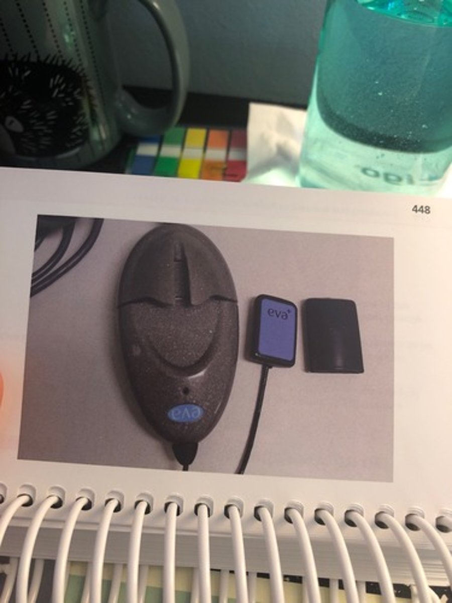
Image receptor
records the x-ray image (sensor, PSP, or film)
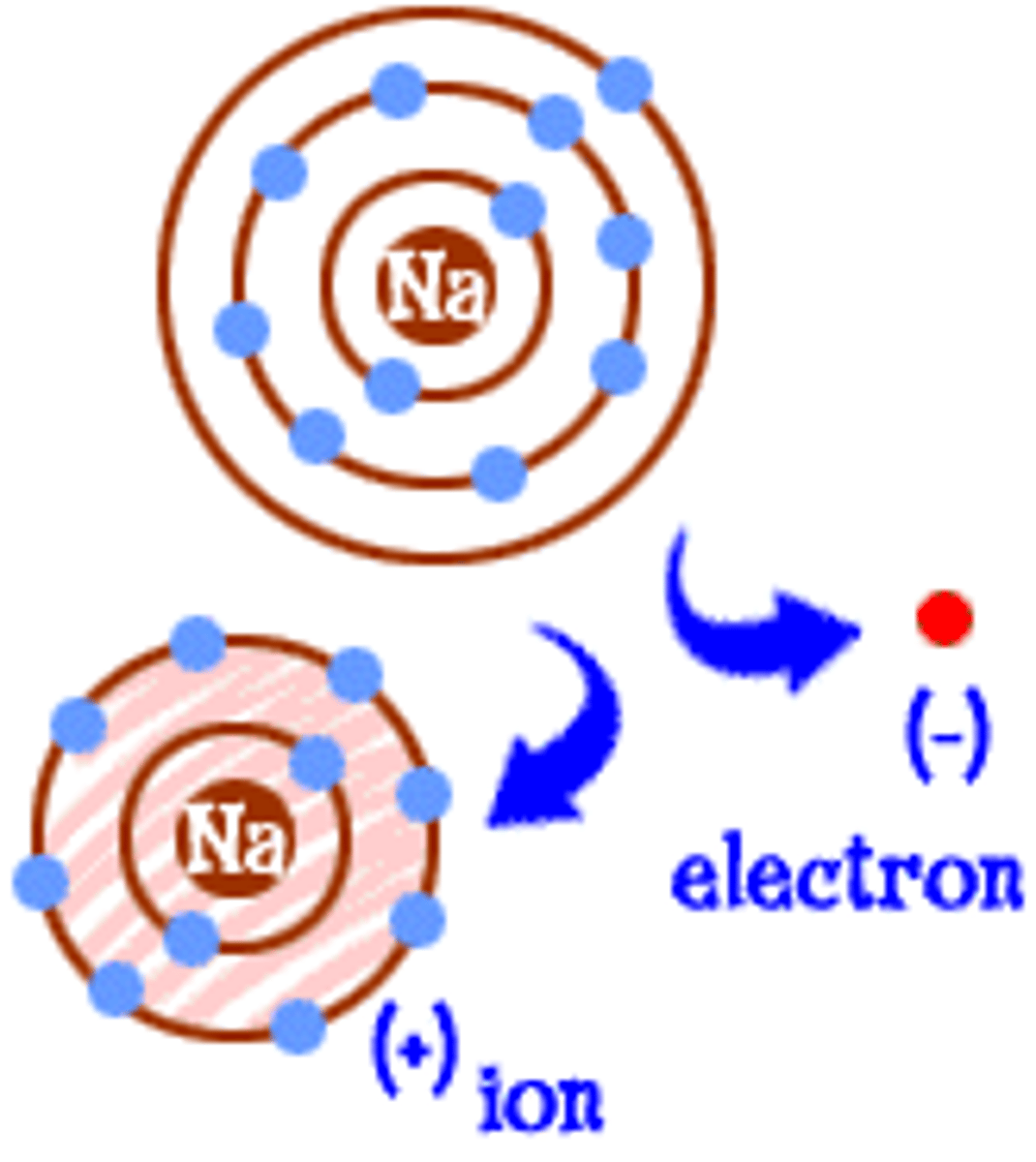
Ionization
Process where atoms gain or lose electrons and become charged
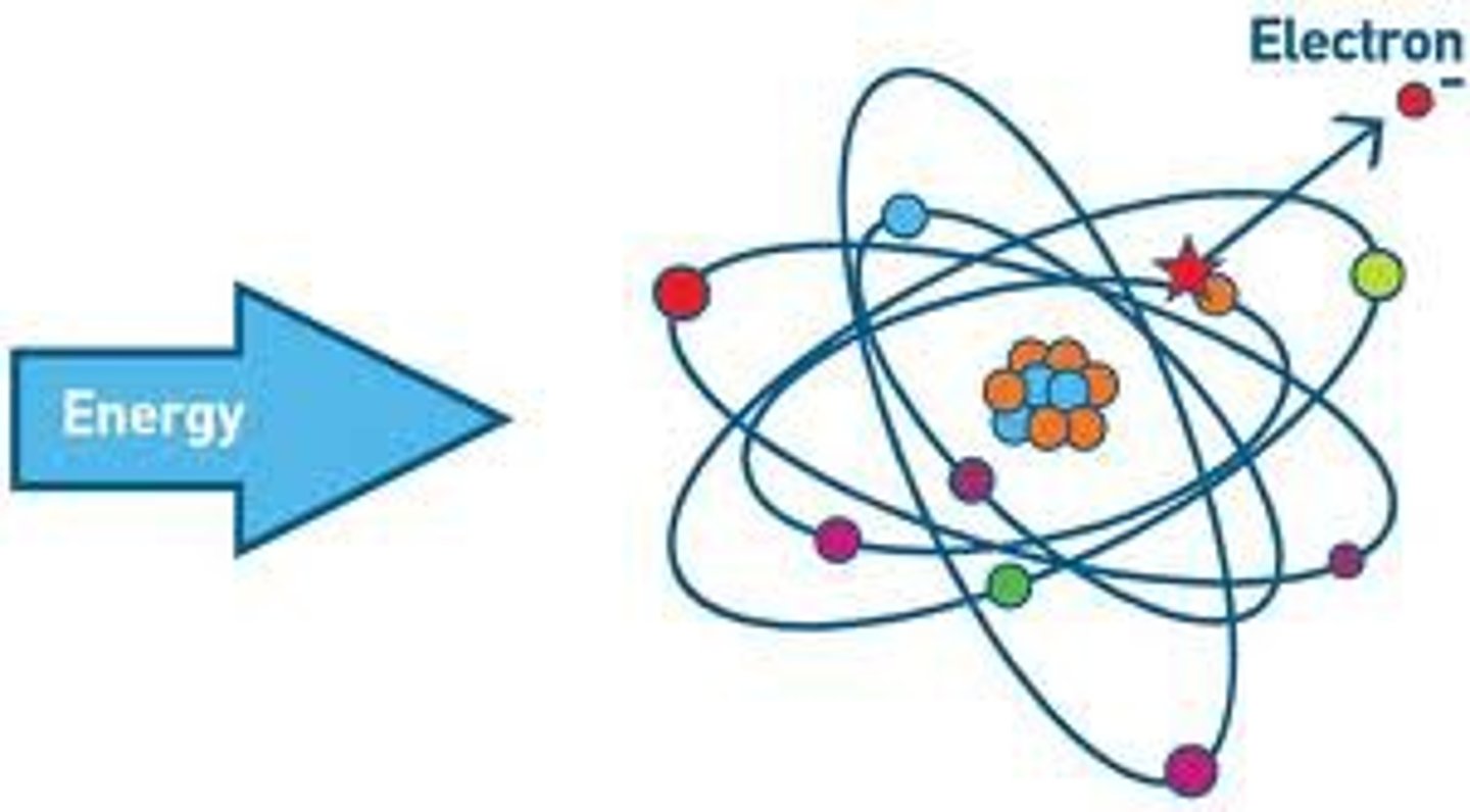
Ions
Atoms with a positive or negative charge
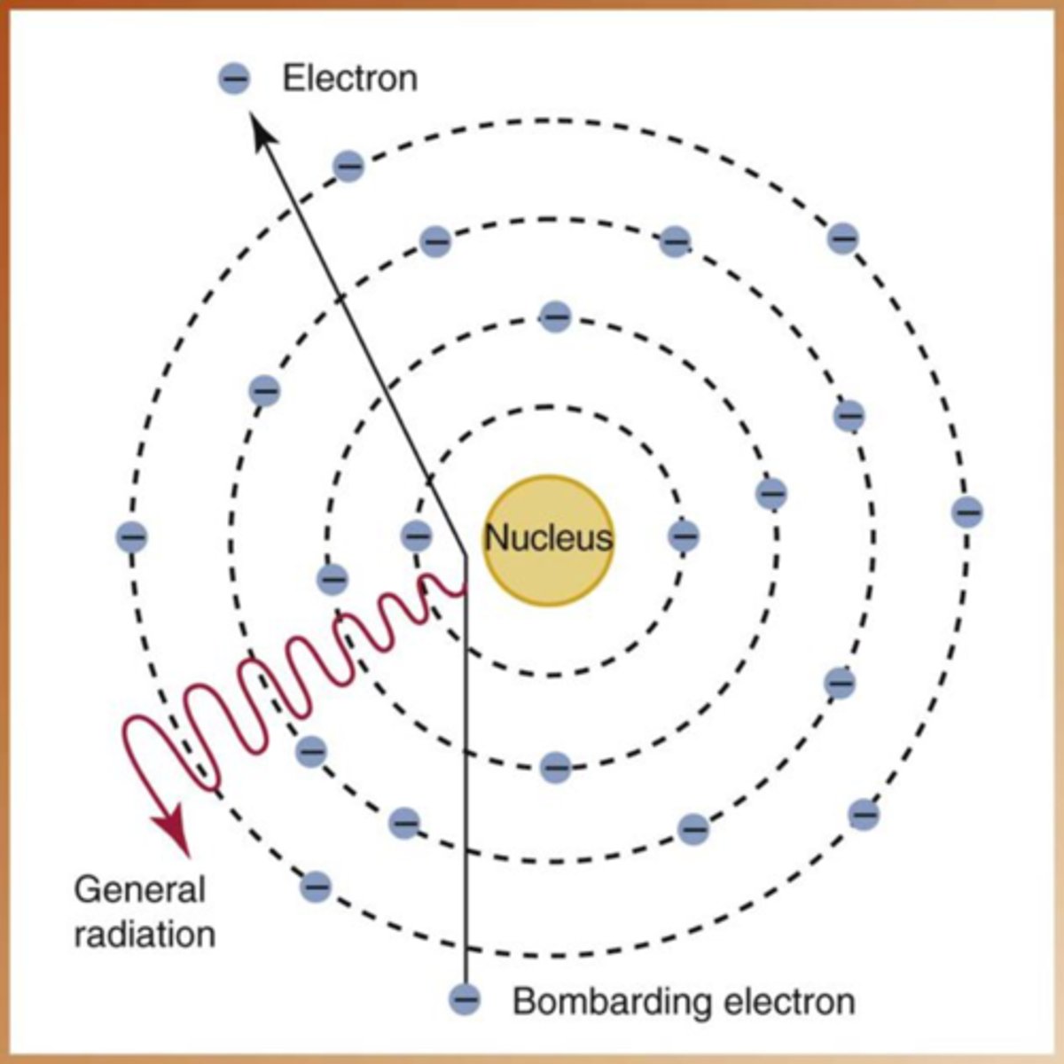
Bremsstrahlung radiation
X-rays produced when electrons are slowed near the nucleus
Tubehead components
X-ray tube head , extension arm, and control panel
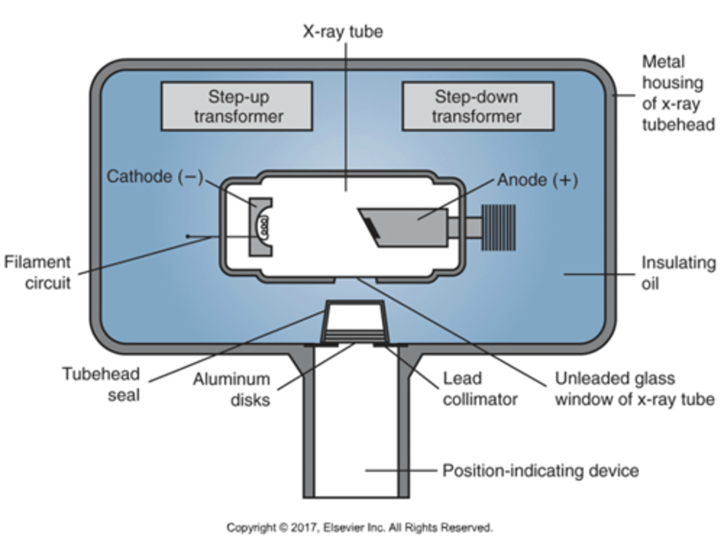
Insulating oil
Absorbs heat and prevents overheating in the tubehead
Aluminum filter
Removes low-energy, non-penetrating x-rays
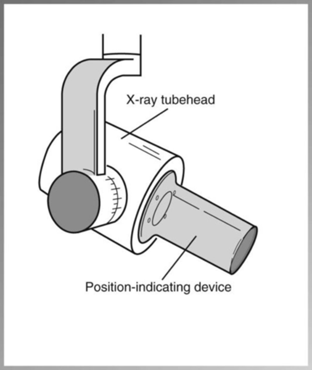
Position Indicating Device (PID)
Lead-lined, open-ended cylinder that directs the x-ray beam
Short wavelength x-rays
High energy and greater penetration
Long wavelength x-rays
Low energy and less penetration
Photons
Packets of x-ray energy no weight or mass
Cathode
Negative electrode that produces electrons
Anode
Positive electrode that converts electrons into x-rays
Energy converted to heat
99% of electron energy becomes heat
Energy converted to x-rays
1% of electron energy becomes x-rays
Kilovoltage peak (kVp)
Controls beam quality and penetrating power
Milliamperage (mA)
Controls quantity of x-rays produced
Exposure time
Controls length of exposure and image density
Impulses
Units used to measure exposure time (60 impulses = 1 second)
Secondary radiation
Radiation produced when the primary beam interacts with matter
Primary radiation
Radiation emitted directly from the x-ray tube
X-ray interaction with atoms
Can disturb electrons and cause ionization
Electrons
small, negatively charged particles
X-radiation
High energy ionizing electromagnetic radiation
X-ray tube head
equipment consisting of the x-ray tube, collimator, and operator controls; permits manipulation of x-ray tube in many directions for proper positioning
X-Ray Extension Arm
wires located inside attached to tube head and control panel

X ray control panel
Master Switch ; indicator light ; selector buttons and exposure buttons
X-ray image
film based or digitally produced recording of anatomic structures
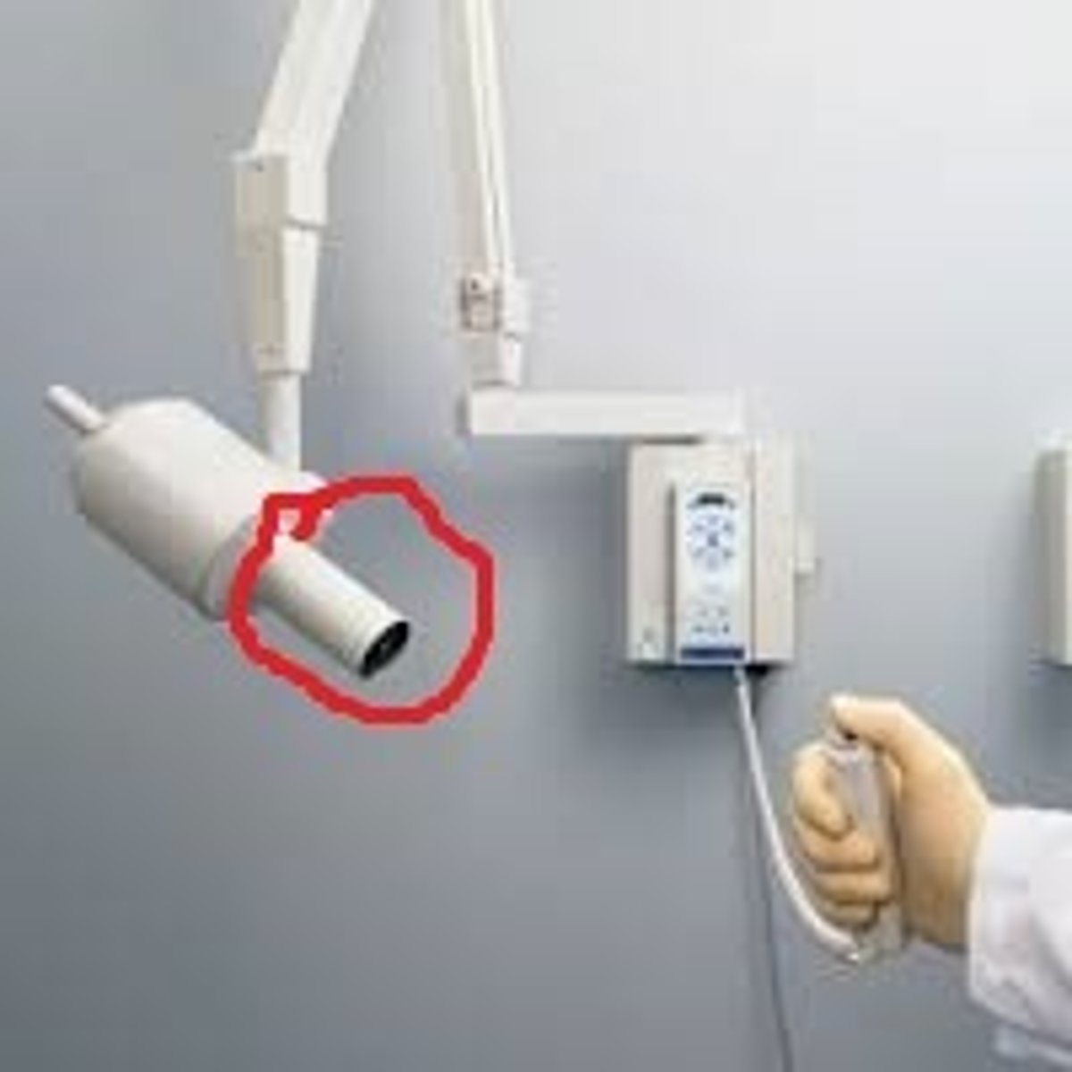
Position indicating device (PID)
Aims and shapes the x-ray beam; open ended, lead lined cylinder that extends from the opening of the metal housing of the tubehead. Sometimes referred to as the cone.
XCP
To position and hold an Xray

Scatter radiation
Radiation that is deflected from its original path after interacting with matter
Radiolucent
Structures that allow x-rays to pass through and appear dark on the image
Examples of radiolucent structures
Pulp, air spaces, soft tissue
Radiopaque
Structures that absorb x-rays and appear light or white on the image
Examples of radiopaque structures
Enamel, bone, metal restorations
Sharpness
The ability to see fine lines and detail in an image
Density
The overall darkness or blackness of a radiograph
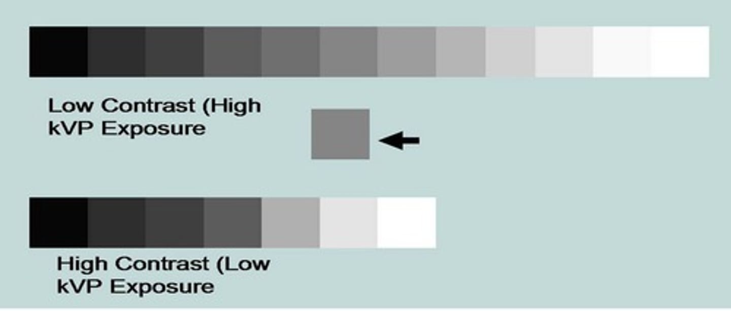
Contrast
The difference in shades between black, gray, and white on a radiograph
What does high kVp produce
Low contrast (long scale contrast) with many shades of gray
What does low kVp produce
High contrast (short scale contrast) with fewer shades
What does increasing kVp do
Increases density and decreases contrast
What does decreasing kVp do
Decreases density and increases contrast
What does increasing mA do
Increases density by increasing number of x-ray photons
What does decreasing mA do
Decreases density
What does increasing exposure time do
Increases density
What does decreasing exposure time do
Decreases density
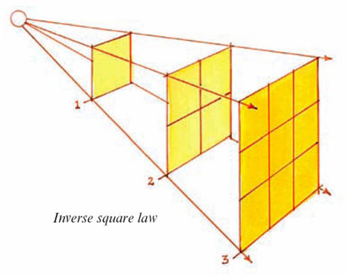
What is quantity in x-rays
The number of x-ray photons produced
What controls quantity of x-rays
mA and exposure time
What is amperage (mA)
A measurement of the number of electrons flowing
Penumbra
The blurred or fuzzy edge around an image
What causes magnification
Image receptor placed too far from the tooth or short PID
Latent period
Time between radiation exposure and appearance of effects
What is ionizing radiation
Radiation capable of removing electrons from atoms
Acute radiation dose
Large dose received in a short period of time
Chronic radiation dose
Small doses received over a long period of time
What is the tungsten target
Area of the anode where electrons strike to produce x-rays
Genetic cells
Reproductive cells (sperm and ova)
Genetic effects
Damage passed to future generations
Somatic cells
Body cells that are not reproductive
Somatic effects
Damage not passed to future generations
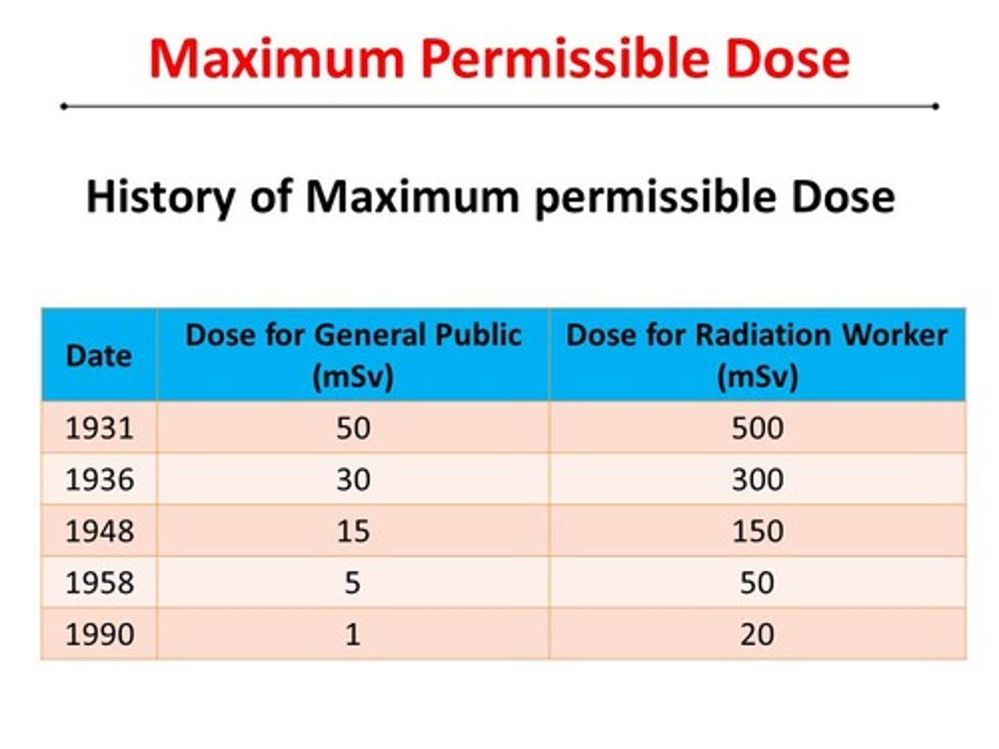
Maximum permissible dose (MPD)
5.0 rems (5000 millirems) per year for occupational exposure
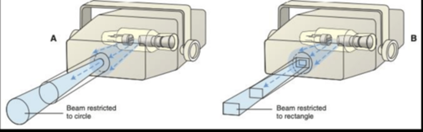
What does a collimator do
Restricts size and shape of x-ray beam to reduce exposure
Types of collimation
Round or rectangular
What is a PID
Position indicating device that directs the x-ray beam
PID lengths
8, 12, or 16 inches
Which PID reduces radiation exposure
Long PID reduces exposure more than short PID
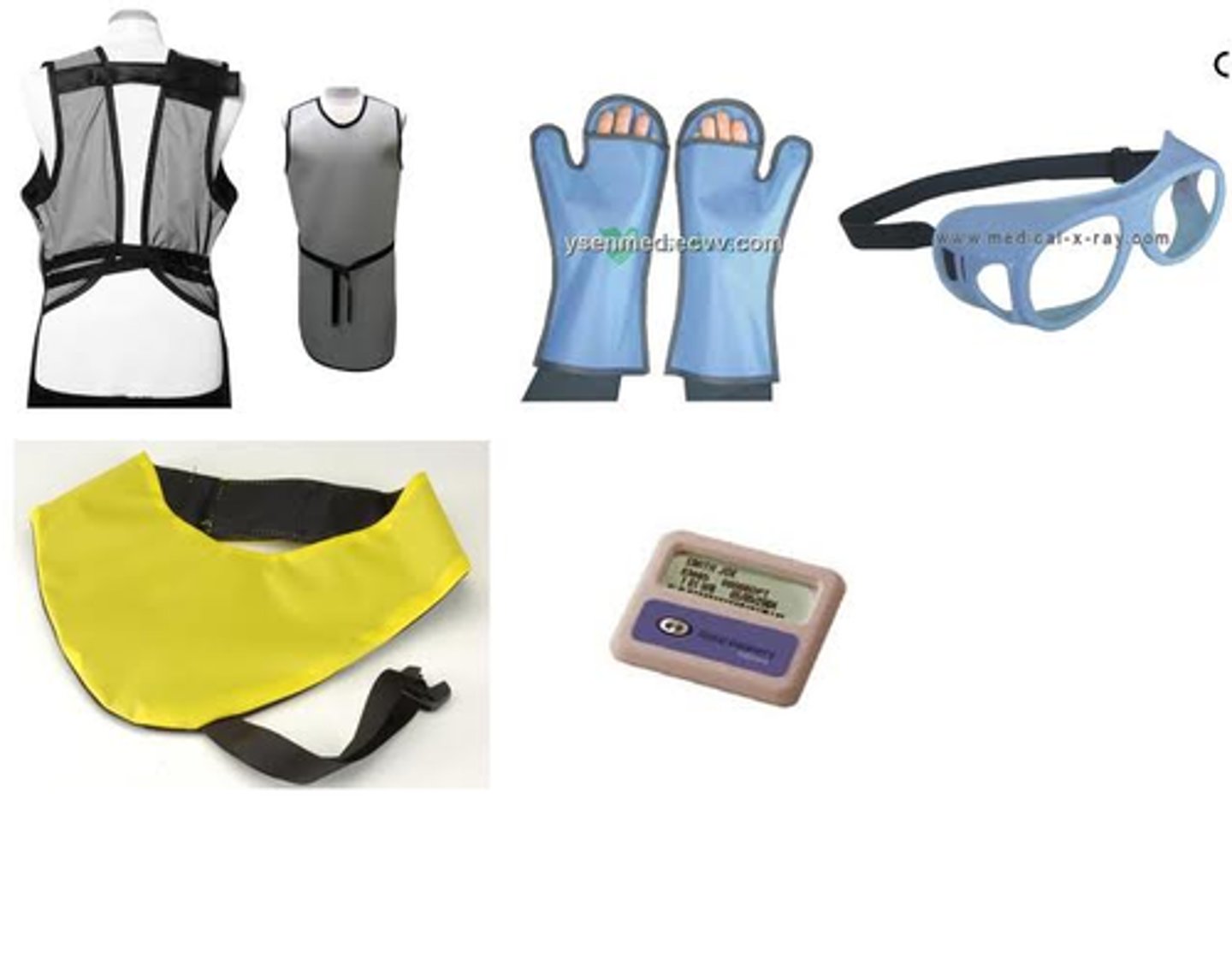
Why should lead aprons not be folded
Folding can cause cracks that reduce protection
What does the aluminum filter do
Removes low-energy, long wavelength x-rays
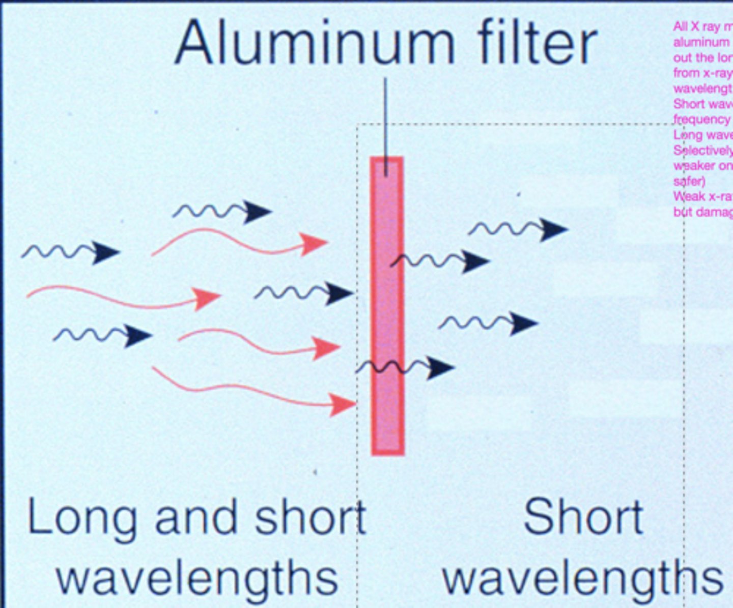
Required aluminum filtration
Minimum 2.5 mm aluminum for machines at 70 kVp or higher
What is a dosimeter
Device that measures occupational radiation exposure
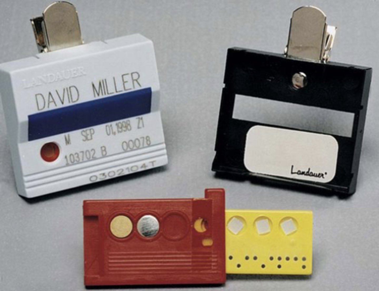
Another name for dosimeter
Personal radiation monitoring badge
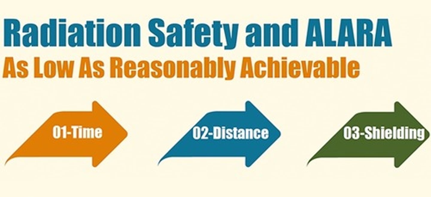
What does ALARA stand for
As Low As Reasonably Achievable
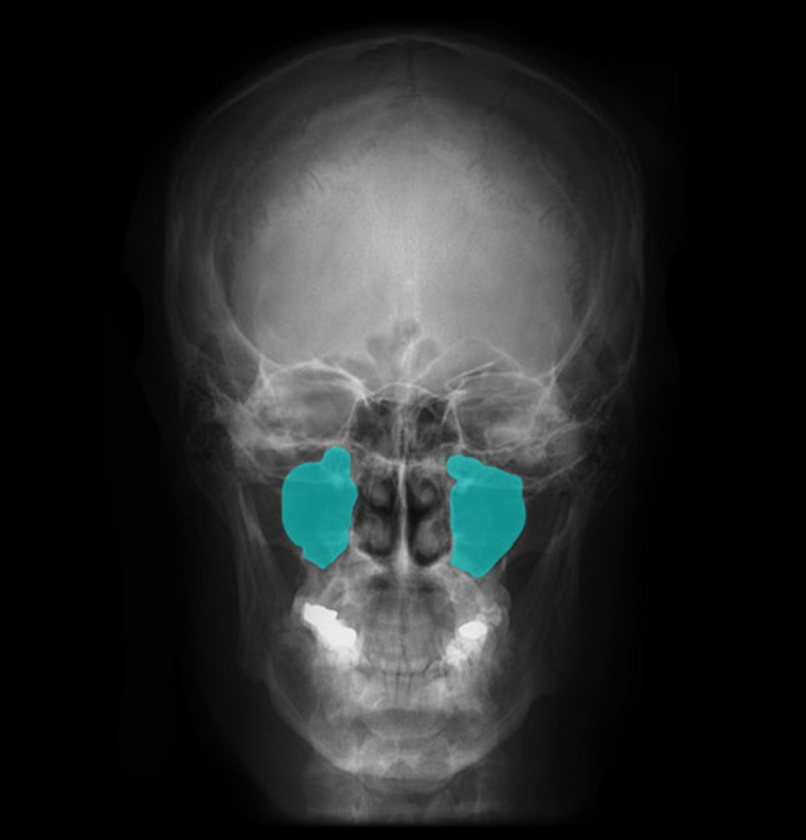
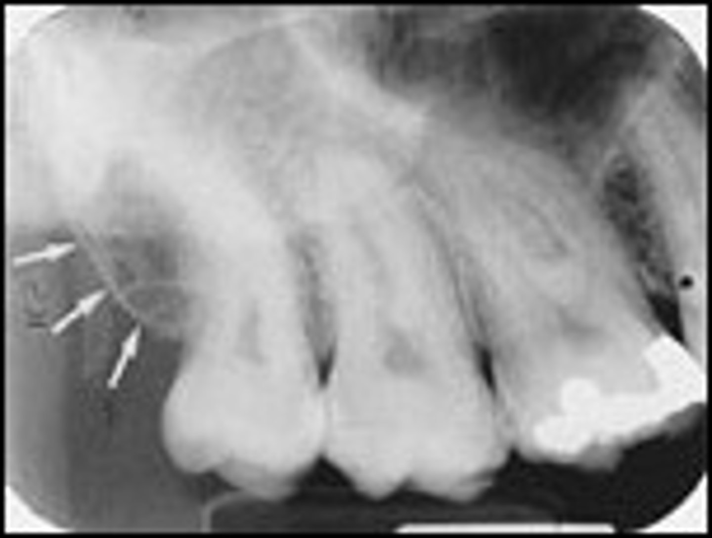
Maxillary sinus
Radiolucent space above posterior maxillary teeth
Floor of maxillary sinus
Radiopaque line outlining the maxillary sinus
Nasal cavity
Radiolucent area above maxillary incisors
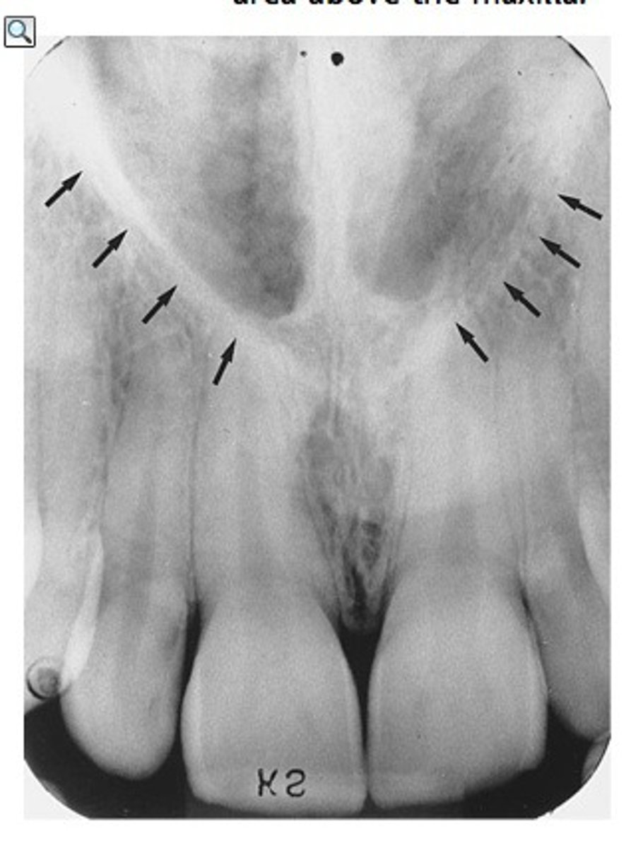
Nasal septum
Radiopaque vertical line dividing the nasal cavity
Anterior nasal spine
Radiopaque V-shaped structure above maxillary central incisors
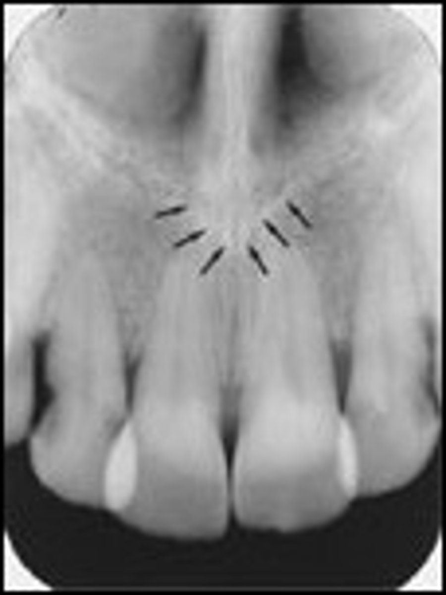
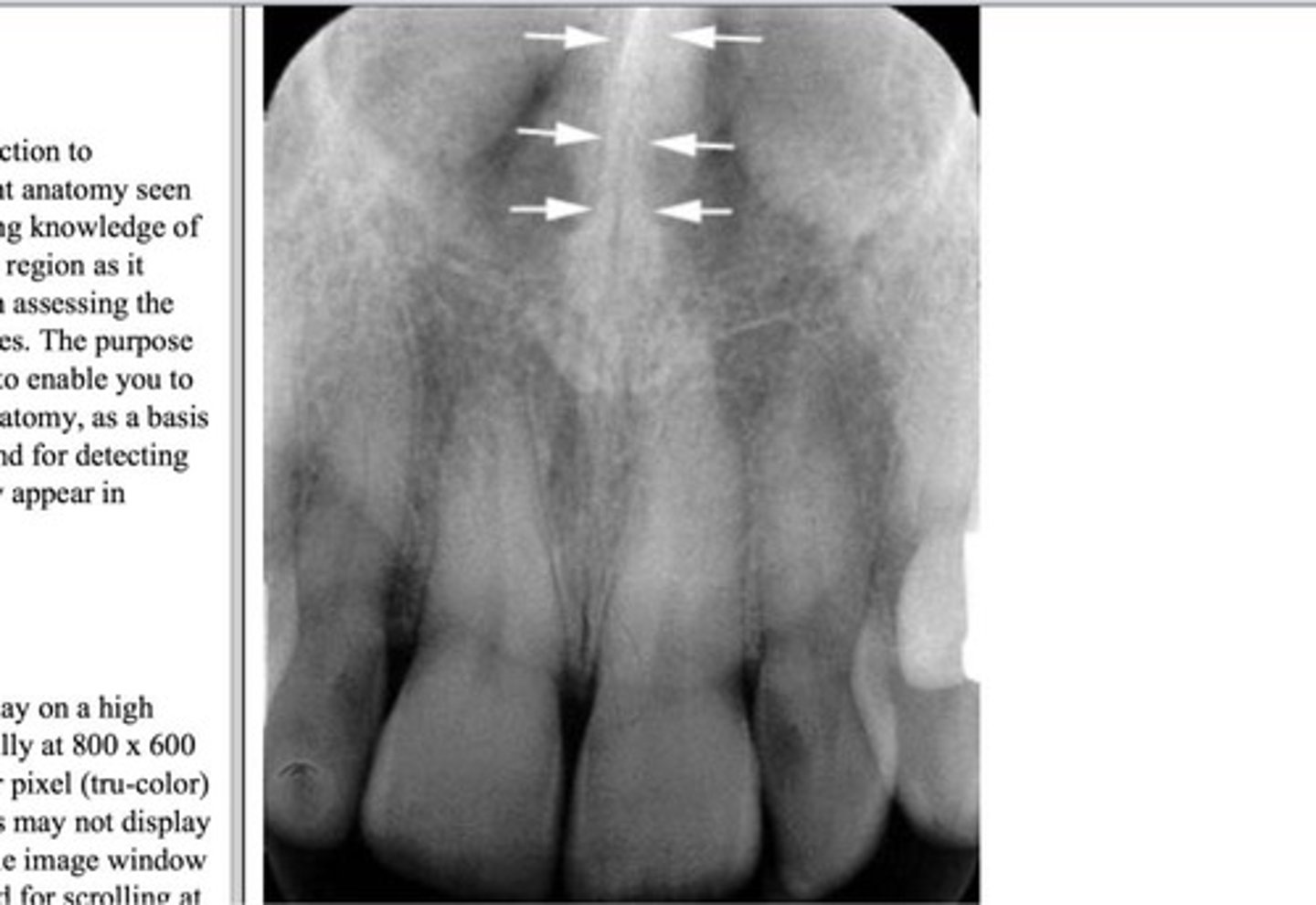
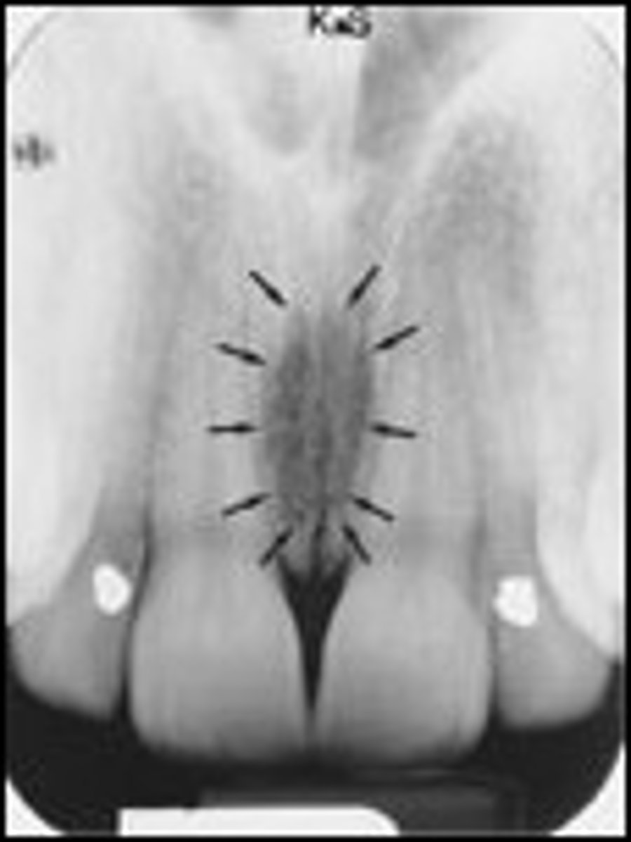
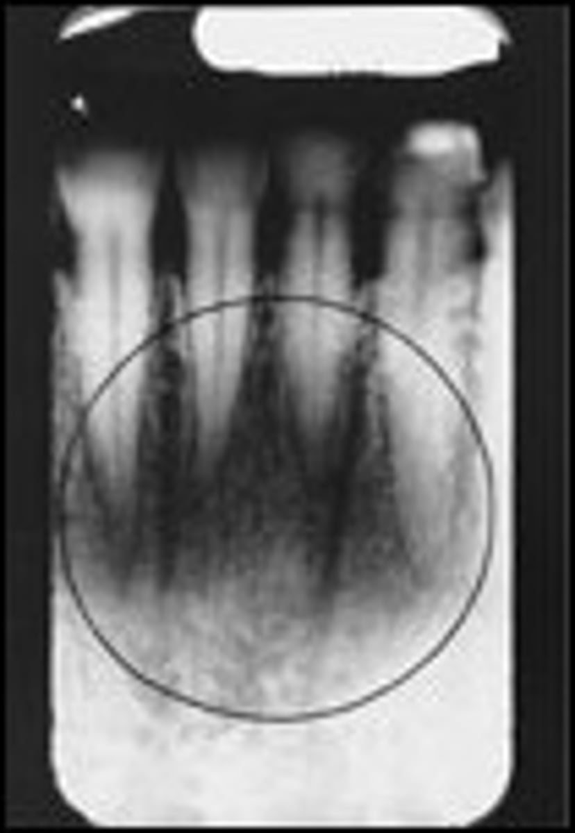
Incisive foramen
Radiolucent area between maxillary central incisors
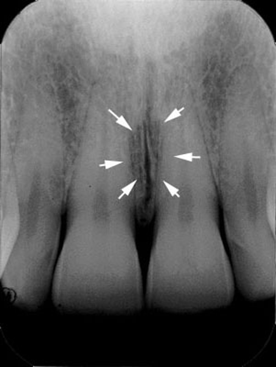
Median palatine suture
Radiolucent line between maxillary central incisors
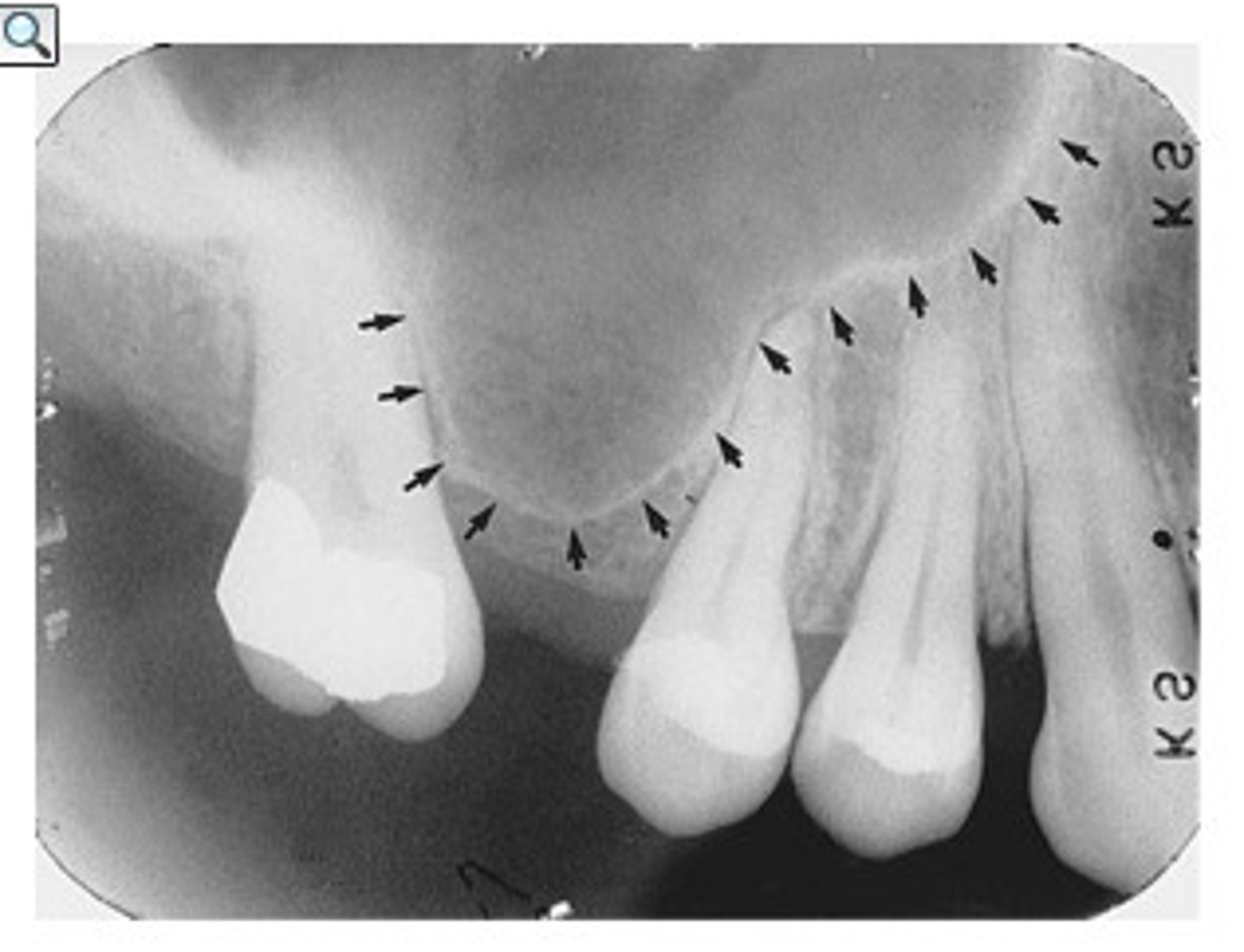
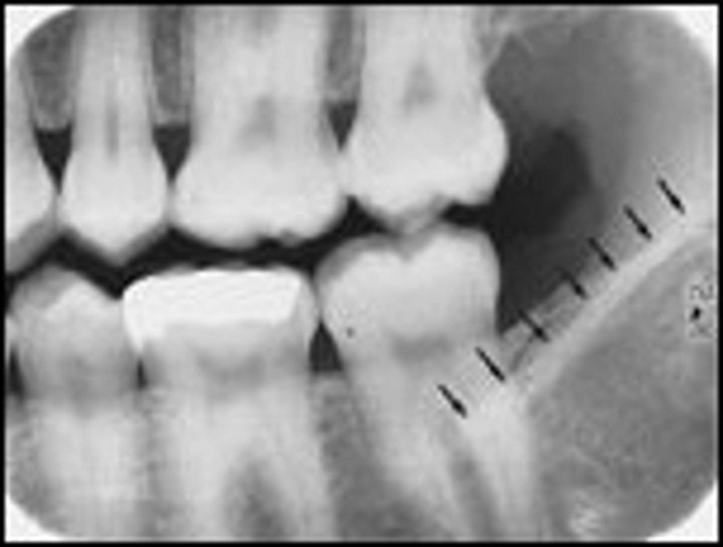
Zygomatic process
Radiopaque U- or J-shaped structure above maxillary molars
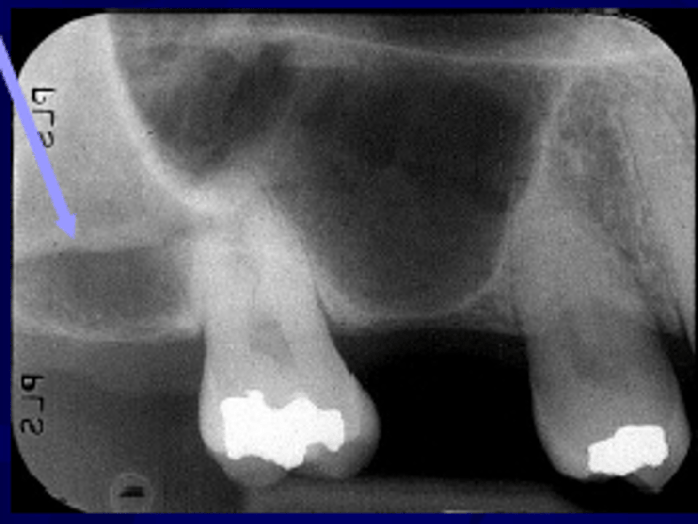
Mental foramen
Radiolucent round/oval area near mandibular premolars
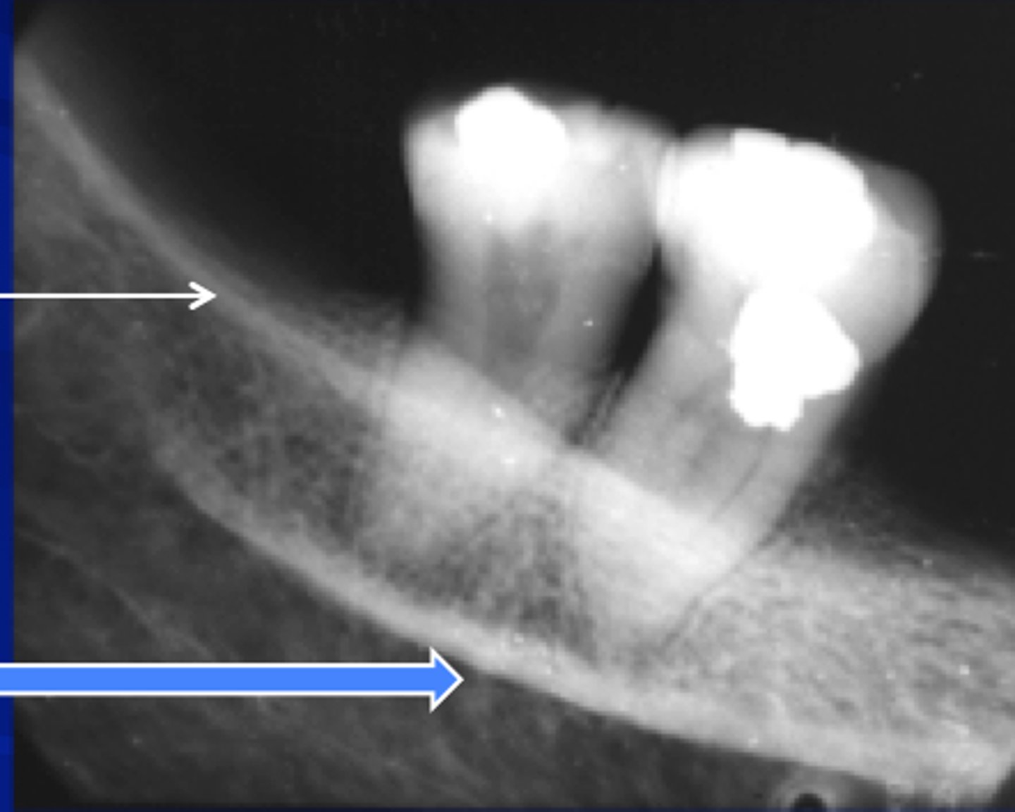
Mandibular canal
Radiolucent band with radiopaque borders below mandibular molars
Mental ridge
Radiopaque line in anterior mandible
Mental fossa
Radiolucent area above the mental ridge
Genial tubercles
Radiopaque ring below mandibular incisors

Lingual foramen
Radiolucent dot inside genial tubercles
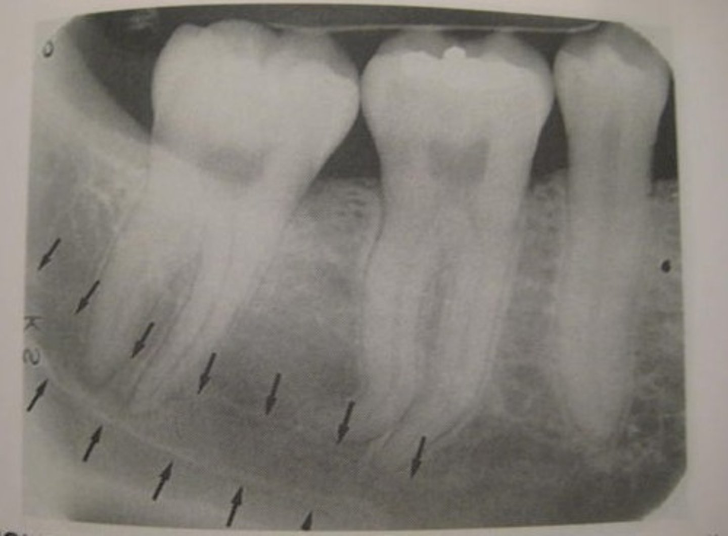
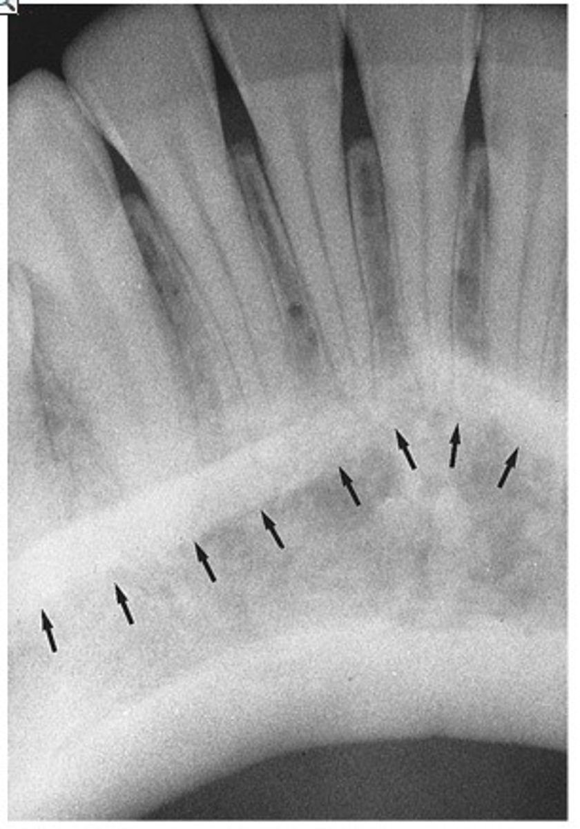
External oblique ridge
Radiopaque line above mandibular molars
Internal oblique ridge
Radiopaque line below external oblique ridge
Cancellous bone molar region identification
Maxillary tuberosity (posterior rounded area behind maxillary molars)
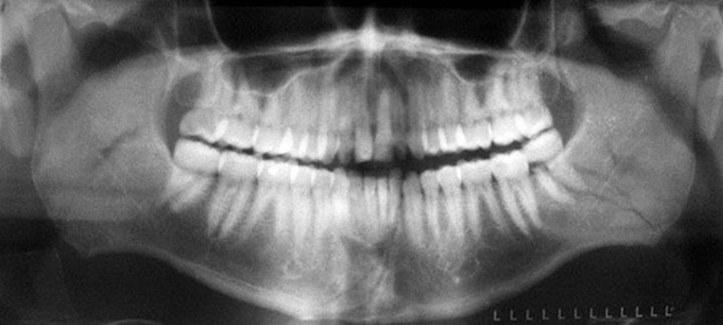
White shadow in center of panoramic image
Patient is slumped; correct by standing up straight / elongating spine
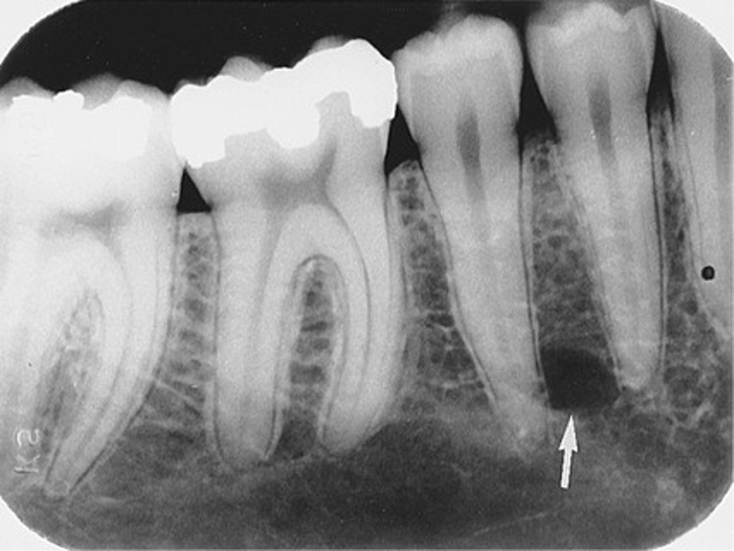
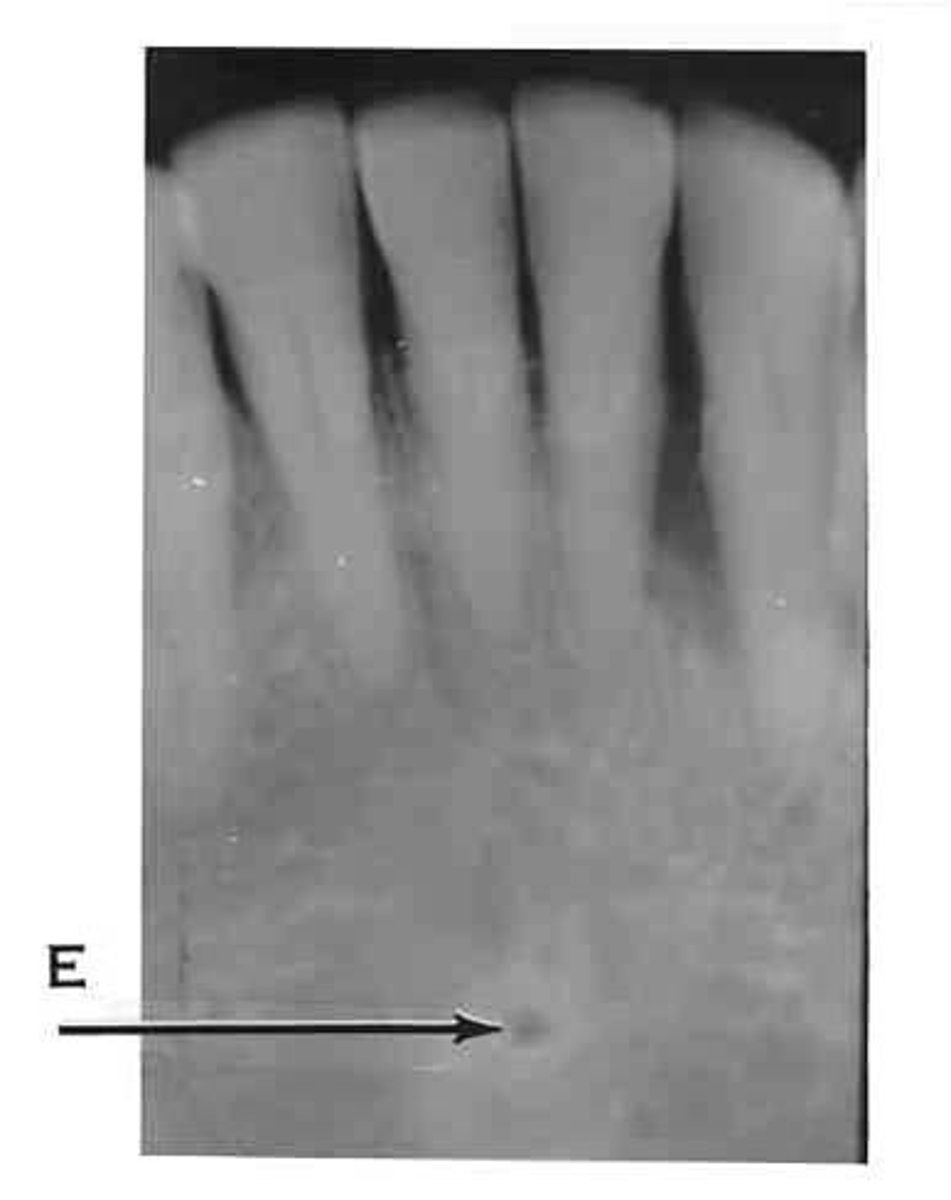
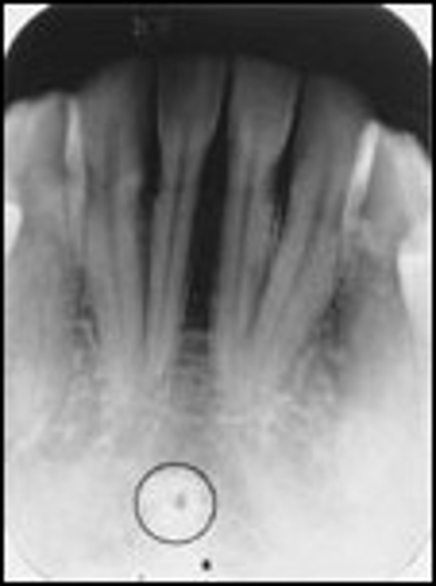
Radiolucent lines in mandibular anterior region
Nutrient canals
Radiolucent landmark in mandibular incisors
Lingual foramen
PID classification
Non-critical
Disinfectant for PID and contaminated surfaces
Intermediate-level disinfectant (EPA registered)

Gloves for cleaning operatory after radiographs
Utility gloves

Agency that protects workers from infection
OSHA
Radiograph to capture mandibular nerve near third molars
Panoramic radiograph
Digital imaging always requires
Computer monitor
Sensor holder classification
Semi-critical
First step when disinfecting sensor for first time
Read manufacturer instructions