CH 20: Enamel, Dentin, and Pulp: Development, Abnormalities, and Structure
1/102
There's no tags or description
Looks like no tags are added yet.
Name | Mastery | Learn | Test | Matching | Spaced | Call with Kai |
|---|
No analytics yet
Send a link to your students to track their progress
103 Terms
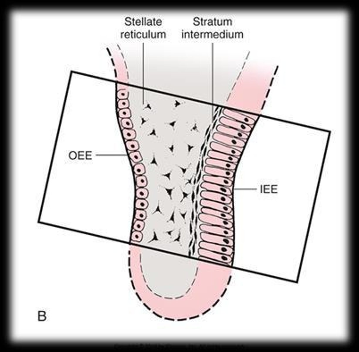
What are the cells of the inner enamel epithelium (IEE) responsible for?
They differentiate into enamel-forming cells known as ameloblasts.
enamel develops from the?
enamel organ, which was developed by the ectoderm
dentin and pulp develop from the?
dental papilla which is derived from mesoderm
What is the relationship between enamel formation and dentin formation?
Enamel formation is interrelated with dentin formation, as both processes occur simultaneously during tooth development.
What role does the dental papilla play in tooth development?
The dental papilla determines the shape of the developing crown of the tooth and has genetic control over it.
Dental papilla cells form when enamel organ is in
cap stage
the inner enamel epithelium (IEE) during the bell stage:
become taller and are known as preameloblasts
peripheral cells of the dental papilla adjacent to preameloblasts become?
low columnar/cuboidal cells called odontoblasts
Odontoblast move away from preameloblasts toward the center of the dental papilla laying down ______
a matrix of mucopolysaccharide ground substance and collagen fibers
why does the preameloblast change its polarity during enamel/dentin formation?
they need to reposition their secretory machinery toward the future dentinoenamel junction (DEJ)
what happens when the change in polarity occurs?
the cell becomes and ameloblast- then begins to secrete and enamel matrix
What is the composition of enamel?
Enamel is composed of approximately 96% inorganic material (hydroxyapatite crystals) and 4% water and organic material.

enamel matrix consists of?
mucopolysaccharide and organic fiber
stages in embryonic development:
1. initiation stage - thickening of lamina
2. bud- dental lamina grows into bud
3. cap- bud forms a cap & gives rise to enamel organ, dental papilla-dental sac
4. bell- changes in polarityof pre-ameloblasts gives the signal to begin producing enamel
what secretes the dentin matrix
odontoblats
2 parts of enamel
rod sheath and rod
rod sheath
Outlines the rod and contains most of the fibrous organic substance
rod
made up of hydroxyapatite
properties of enamel
- avascular
- no nerves
- non-renewable
-not a static tissue, may undergo mineralization changes
What is the keyhole shape of an enamel rod?
An enamel rod has a wide upper end and a narrowed bottom end, contributing to its unique structure.
why are enamel rods key shaped?
the crystals in those parts are oriented in different directions; the secreting ameloblasts bulge outward in the irection of DEJ
how many ameloblasts come together to form enamel rod
3-4
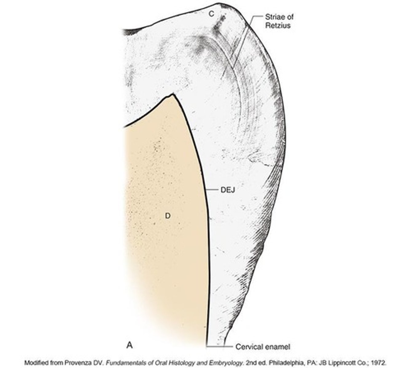
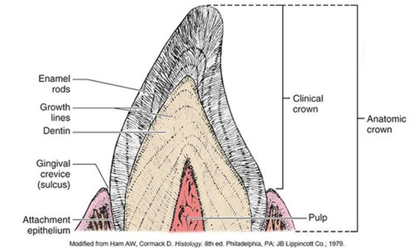
dentinoenamel junction
Line marking the junction of the dentin and the enamel.
ameloblast
Enamel forming cells
how do enamel rods fit tightly together
because of a cementing substance called interrod substances
Tomes's process of an ameloblast
responsible for the formation of enamel rods:
Secretes enamel matrix.
Forms enamel rods (prisms) from the tip of the process.
Forms interrod enamel from the sides of the process.
Creates the characteristic rod-and-interrod arrangement of enamel.
Hunter-Schreger bands
Light or dark bands oriented perpendicular to the DEJ, caused by the curvature of the rods
how often do the rods develop?
4 µm (micrometer) per day
What are striae of Retzius?
Striae of Retzius are brownish lines in enamel that mark changes in rod development, curving outwardly from the dentinoenamel junction (DEJ).
What happens during the mineralization stage of enamel development?
Ameloblasts lay down a matrix and deposit millions of hydroxyapatite crystals, establishing the initial crystal structure.
What occurs during the maturation stage of enamel development?
The hydroxyapatite crystals grow until they are tightly packed together. (4mm a day)
imbrication lines
surface manifestations of striae of retzius, horizontal, seen on labial surface
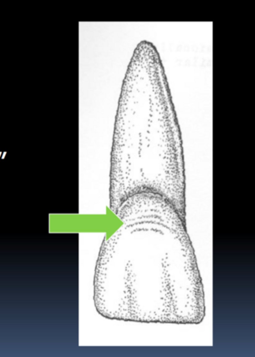
how can imbrication lines be affected?
nutritional deficiencies & unfavorable metabolic conditions
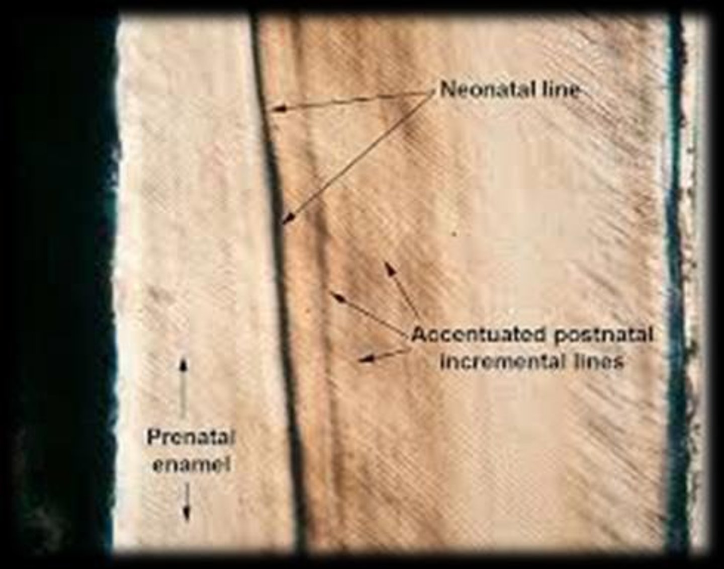
neonatal lines
created by nutritional disturbance at birth, on the surface of primary teeth
What are the two main components of enamel?
The two components are the enamel rod and the rod sheath.
What is hypoplastic enamel?
Hypoplastic enamel is a condition where enamel is underdeveloped, leading to thinner enamel than normal.
What is hypocalcified enamel?
Hypocalcified enamel is characterized by enamel that is poorly mineralized, leading to a softer structure.
What is the significance of the dental lamina in tooth development?
The dental lamina is crucial for the initiation stage of tooth development, where it thickens to form the basis for tooth buds.
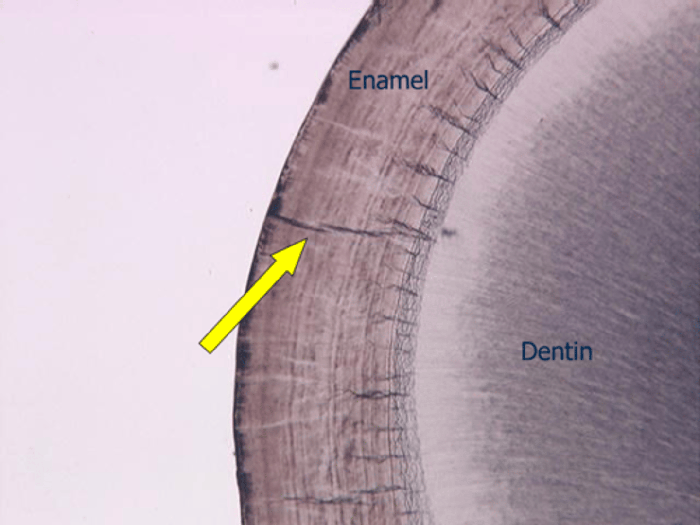
What are enamel tufts?
Enamel tufts are small, dark structures found at the DEJ, believed to be areas of less mineralization.
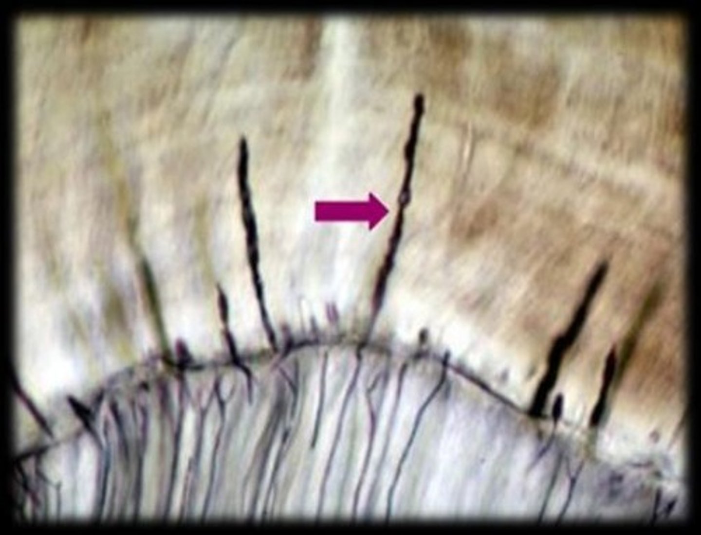
What are enamel spindles?
Enamel spindles are extensions of odontoblasts that extend into the enamel, possibly playing a role in sensation.
What is the role of the inner enamel epithelium (IEE) during the bell stage?
During the bell stage, IEE cells become taller and differentiate into preameloblasts.
What is the function of the outer enamel epithelium (OEE)?
The OEE serves as a protective layer for the enamel organ during tooth development.
How do ameloblasts change during enamel formation?
Ameloblasts change polarity and begin secreting enamel matrix as they move toward the OEE.
What is the primary unit of enamel's structure?
The primary unit of enamel's structure is the enamel rod, composed of hydroxyapatite crystals.
What is the significance of the bell stage in tooth development?
The bell stage is critical for the differentiation of cells and the initiation of enamel and dentin formation.
What is the process of condensation in tooth development?
Condensation refers to the rounding and densification of mesenchymal cells adjacent to the enamel organ during the cap stage.
What is the first stage of enamel development?
Mineralization stage, first seen at the tip of a cusp.
What are imbrication lines?
Surface manifestations of Striae of Retzius that are horizontal in direction, seen on the labial surface of anterior teeth.
What happens to ameloblasts during enamel organ fate?
Ameloblasts move away from the DEJ toward the OEE, compressing the middle layers and signaling the cessation of enamel formation.
What is Nasmyth's membrane?
Also known as primary cuticle, it covers the crown of the tooth and is worn away by toothbrushing or polishing.
reduced enamel epithelium
Layers of flattened cells overlying enamel surface from compressed enamel organ.
What does the REE produce to hold the gingiva to the tooth?
secondary enamel cuticle, or epithelial attachment, found at the base of the ginigval sulcus
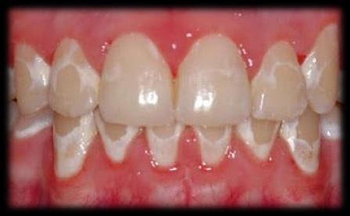
hypocalcified enamel
White spots on the teeth, insufficient growth of the enamel crystals deposited in the matrix
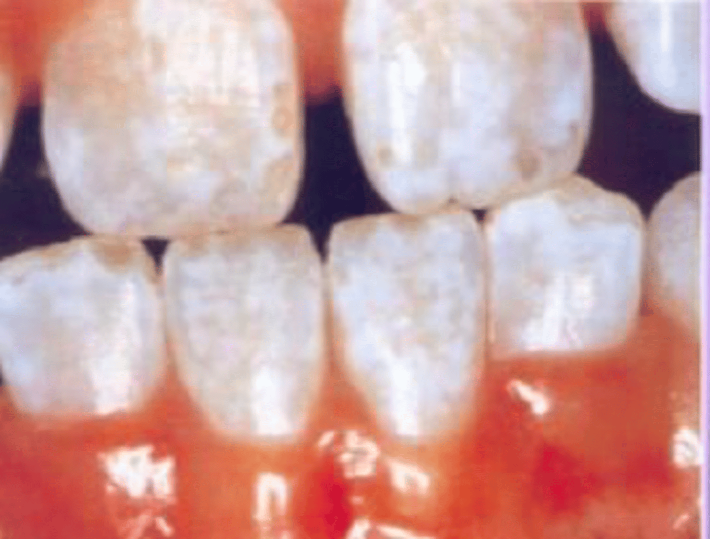
why does hypocalcification occur
if the crystals do not grow to full size, they wont be packed together, then the enamel is less than 96% inorganic material
What is enamel fluorosis?
A form of enamel hypocalcification caused by excessive fluoride, leading to discoloration ranging from white flecks to brownish spots.
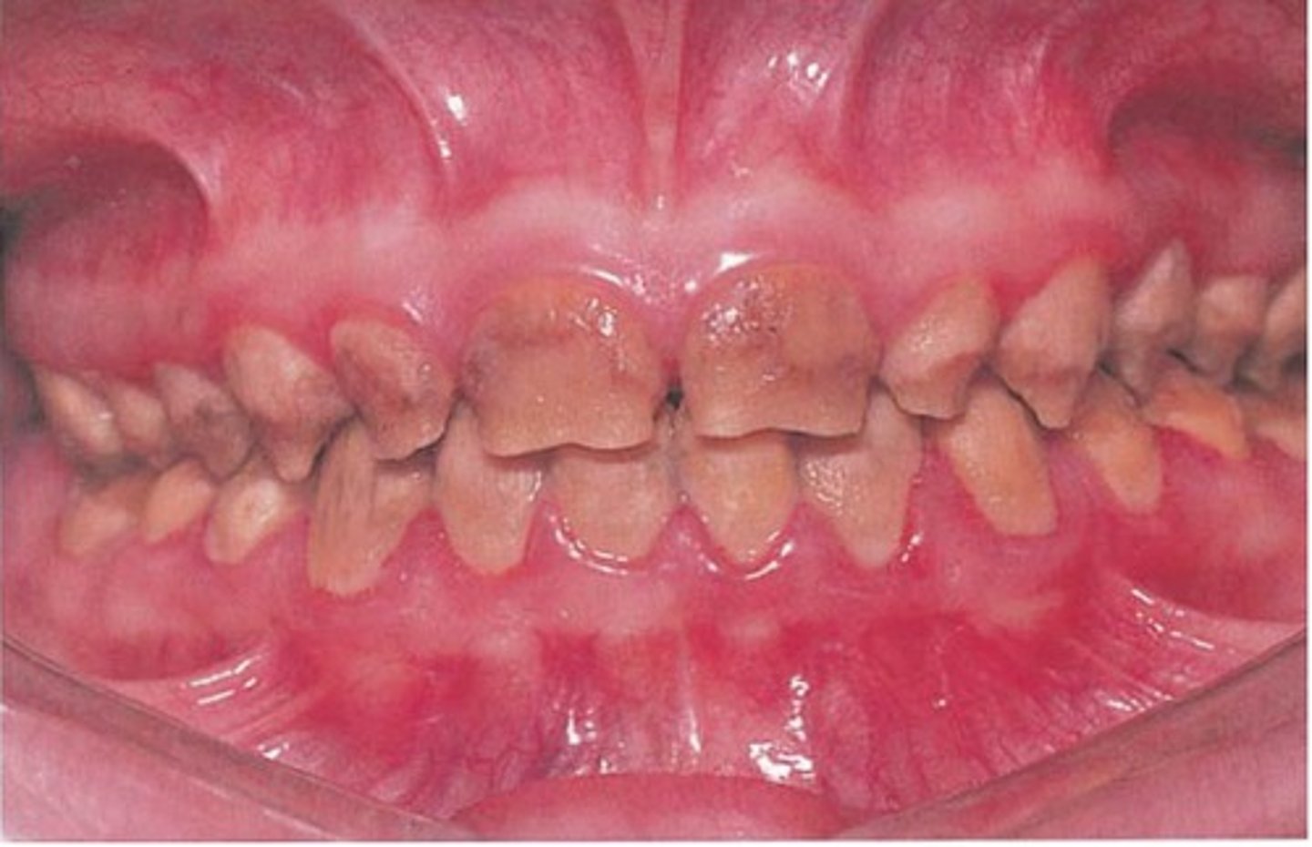
What is amelogenesis imperfecta?
A hereditary anomaly related to hypocalcification, characterized by thin, yellow enamel that fractures easily.
What are enamel lamellae? common
Hairline cracks in enamel caused by developmental problems or trauma, often due to rapid temperature changes.
enamel lamellae uncommon
developmental defect & result of one or more ameloblasts stopping enamel production, leaving a space between other enamel rods and providing a potential pathway for bacteria to enter
What is an enamel tuft?
A small area of hypocalcified enamel seen at the DEJ, extending about a third of the way through enamel, with no clinical significance. (odontoblastic process)
What is an enamel spindle?
A cellular extension of the odontoblast trapped between ameloblasts, seen histologically and may contribute to slight hypersensitivity.
What is attrition?
Loss of tooth structure through tooth-to-tooth contact, resulting in shiny facets and matching wear on occluding surfaces.
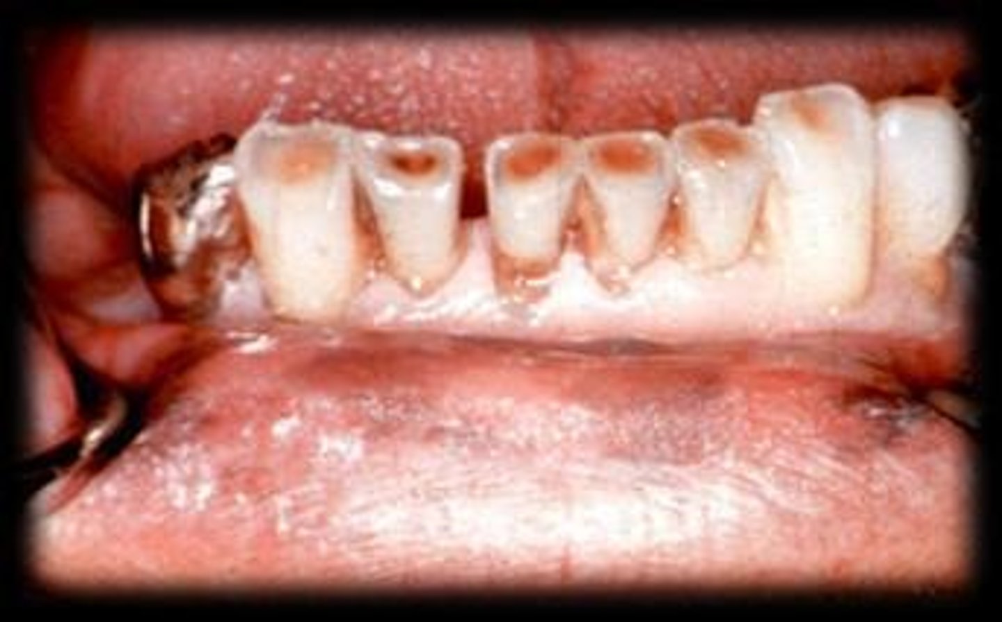
What is erosion in dental terms?
Loss of tooth structure through chemical means, leading to broad concavities within smooth surface enamel.
What is abrasion?
Loss of tooth structure through friction, often from tooth brushing or abrasive toothpaste, typically located at facial cervical areas.
What is abfraction?
Loss of tooth structure due to tensile and compressive forces during tooth flexure, resulting in deep, narrow V-shaped notches.
What are the components of pulp?
Pulp consists of connective tissue, blood vessels, and nerves, with age-related changes affecting its structure.
dentin compostion
70% inorganic hydroxyapatite and 20% organic, collagen, ground substance, !)% water
dentinal tubule
Long tube running from the DEJ
to the pulp; contains odontoblastic process
Peritubular dentin
-Wall of tubules
-Highly mineralized
Intertubular dentin
dentin found between the tubules (bulk)
formation of primary dentin
1. Odontoblasts differentiate from cells of the dental papilla.
2. Odontoblasts secrete an organic matrix called predentin (mainly collagen).
3. Predentin is deposited along the future dentinoenamel junction (DEJ) and dentinocemental junction (DCJ).
4.Calcium and phosphate are deposited, causing mineralization of predentin into dentin.
5.As dentin forms, odontoblasts move inward toward the pulp, leaving behind odontoblastic processes in dentinal tubules.
6.Dentin formation continues until the tooth root is completed.
peritubular dentin
Creates the wall of the dentinal tubule
when the tooth erupts into the oral cavity, the dentin that has formed is?
primary dentin
What is interglobular dentin?
A type of dentin characterized by areas of unmineralized matrix, typically seen in developing teeth.
formation of reparative dentin
1.Forms in response to injury or irritation (e.g., caries, wear, trauma, dental procedures).
2.Produced by surviving odontoblasts or newly differentiated odontoblast-like cells.
3.Deposited only at the site of injury to protect the pulp.
4. Often has a more irregular structure than primary or secondary dentin.
secondary dentin formation
1.Forms after root completion and continues slowly throughout life.
2. Produced by existing odontoblasts.
3. Deposited on the pulp side of dentin.
4. Causes gradual reduction in pulp chamber siz
amelogenesis imperfecta
-Hereditary defect of enamel formation
-Enamel hypoplasia, pits and/or grooves in teeth
-Teeth often discolored
-Different subtypes
What are dead tracts in dentin?
Spaces in dentin that occur when odontoblasts die, leaving behind empty spaces.
What is sclerotic dentin?
Dentin that becomes more mineralized and less permeable due to aging or trauma.
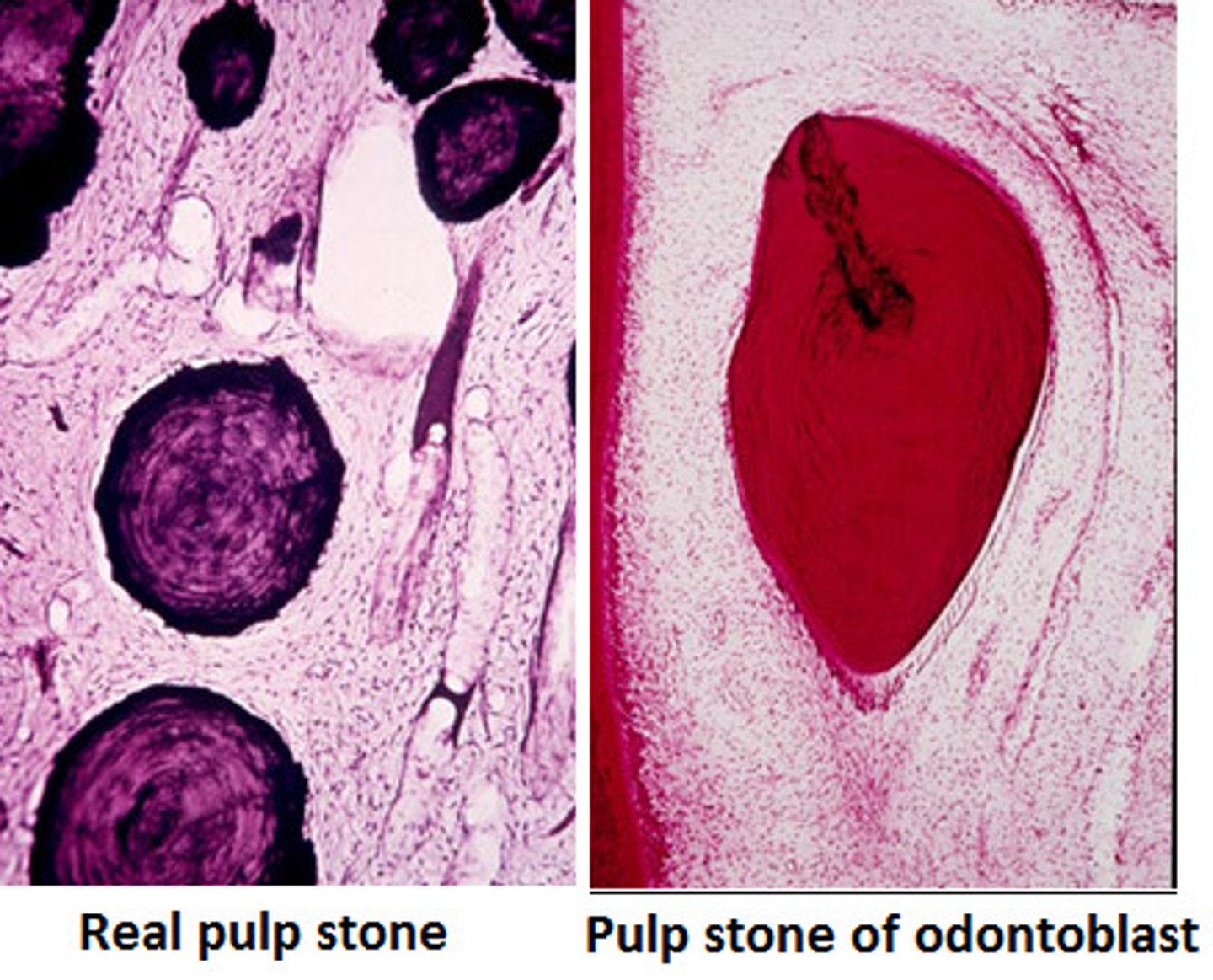
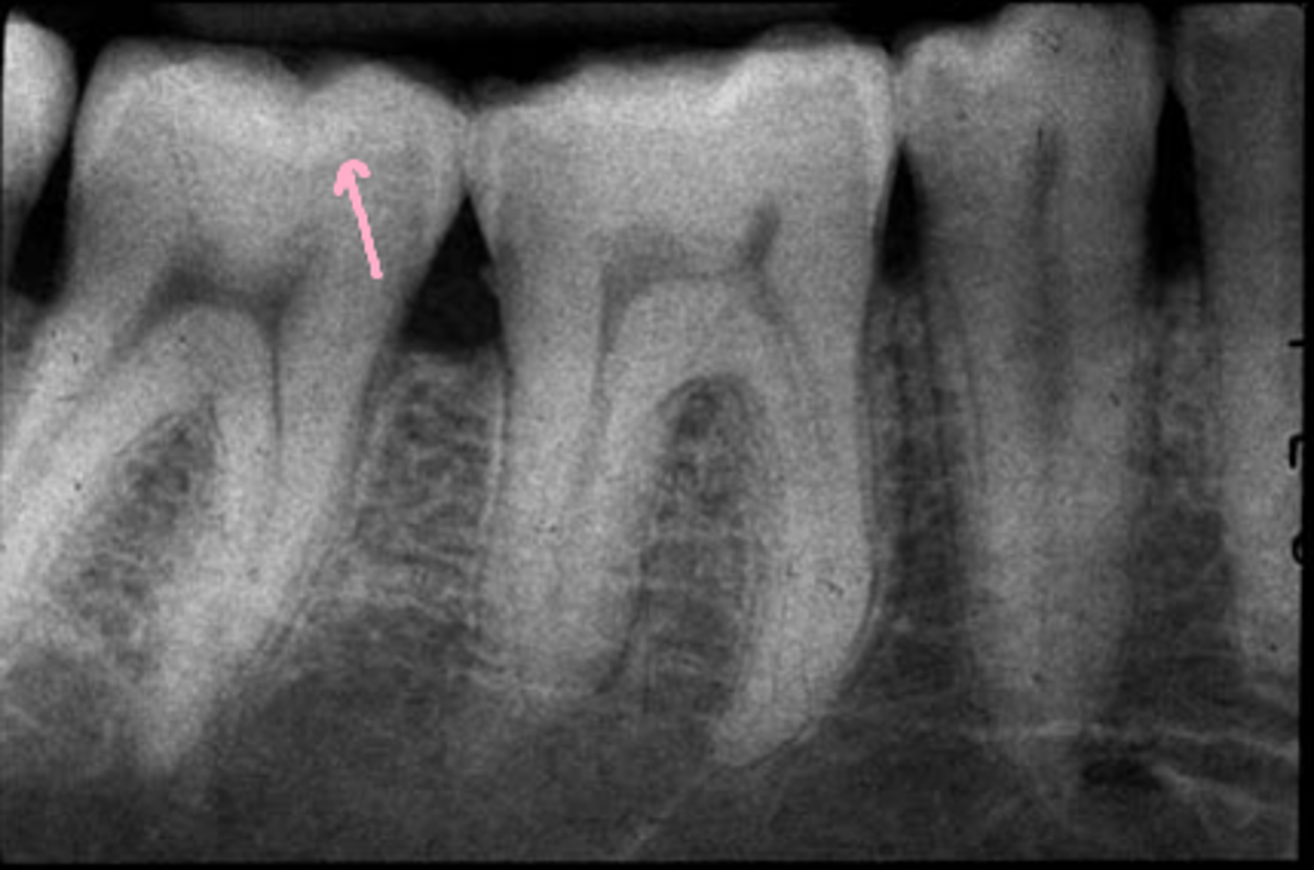
What are pulp stones?
Calcified structures found within the pulp chamber, which can be classified as true or false based on their origin.
What is the composition of dentin?
Dentin is about 70% inorganic and 30% organic.
What is the primary inorganic component of dentin?
Hydroxyapatite crystal.
What are the organic components of dentin?
Collagen, mucopolysaccharide ground substance, and water.
What are the three distinct areas of dentin?
Dentinal tubule, intertubular dentin, and peritubular dentin.
What does the dentinal tubule contain?
An odontoblastic process.
What is the bulk of dentinal material called?
Intertubular dentin.
What surrounds the dentinal tubules?
Peritubular dentin, which has a higher crystalline content.
What is primary dentin?
Dentin formed by odontoblasts secreting a matrix that calcifies to form intertubular dentin.
When does secondary dentin start forming?
When the tooth erupts and contacts the opposing tooth.
What happens to the pulp chamber as secondary dentin forms?
The size of the pulp chamber decreases.
What is reparative dentin?
Dentin formed in response to local trauma, located immediately beneath the area of trauma.
What types of trauma can lead to the formation of reparative dentin?
Occlusal, mechanical, or chemical trauma.
occlusal trauma
Abnormal occlusal relationships of the teeth, causing injury to the periodontium
mechanical truama
usually the result of the cavity preparations in the tooth, leads to death of odontoblasts
Chemical Trauma
results from acids produced by bacteria that causes dental caries, also if cavity is not lined when substances are used to fill teeth
What is dentinogenesis imperfecta?
A hereditary condition where dentin is gray, brown, or yellow, and pulp chambers are filled with dentin.
What develops from mesodermal tissue of the dental papilla?
Pulp.
What are the primary components of pulp?
Blood vessels, lymphatic vessels, nerves, fibroblasts, collagen fibers, and other connective tissue cells.
What type of nerves are primarily found in the pulp?
Sensory nerves that transmit pain sensation.
What happens to pulp as it ages?
Pulp becomes smaller and more susceptible to permanent damage.
What are true pulp stones?
Pulp stones that originate from odontoblasts.