WBC Identification
1/40
There's no tags or description
Looks like no tags are added yet.
Name | Mastery | Learn | Test | Matching | Spaced | Call with Kai |
|---|
No analytics yet
Send a link to your students to track their progress
41 Terms
neutrophils
55-70% of WBCs
lymphocytes
20-40% of WBCs
monocytes
2-8% of WBCs
eosinophil
1-4% of WBCs
basophils
0.5-1% of WBCs
bone marrow
WBCs are produced in the ?
granulocytes
neutrophils - basophils - eosinophils
agranulocytes
lymphocytes - monocytes
key to WBC identification
granules + nucleus shape = ?
myeloblast - promyelocyte - myelocyte - metamyelocyte - band - neutrophil
order of leukocyte development
plasma cells
in addition to the five types of mature WBCs that normamly appear in the peripheral blood, the ? are rarely seen
plasma cells
megakaryoblast - promegakaryocyte - immature meakaryocyte - mature megakaryocyte
order of platelet maturation
neutrophils
first responders
phagocytosis of bacteria
lymphocytes
B cells, antibodies, and T cells —> regulation
NK cells —> kill infected cells
monocytes
become macrophages
phagocytosis + antigen presentation
eosinophils
kill parasites, allergic responses
basophils
release histamine, inflammation and allergy
leukocyte examination order
nuclear chromatin pattern
nuclear shape
size and number of nucleoli, when present
cytoplasmic inclusions
nuclear/cytoplasmic (N/C) ratio
leukocytosis
white blood cell count that is above normal
leukopenia
white blood cell count below normal
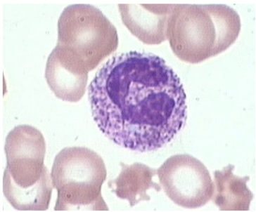
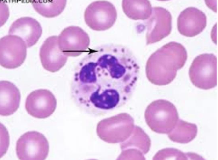
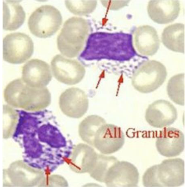
toxic granulation
deeply staining basophilic or blue-black, larger -than-normal granules found in the cytoplasm of neutrophils, bands, and metamyelocytes
toxic granulation
Dohle bodies
round or oval, small, clear, light-blue staining areas found in the neutrophil cytoplasm
Dohle Bodies
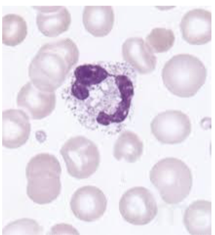
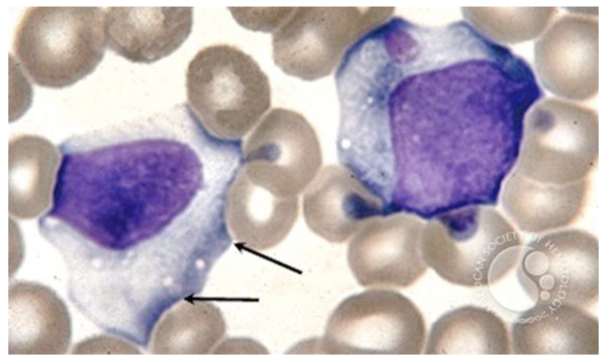
toxic vacuolization
vacuoles in the cytoplasm of neutrophils and bands
toxic vacuolization
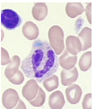
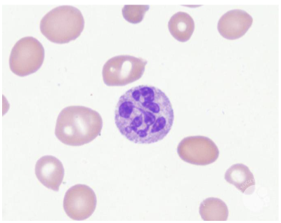
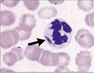
hyper segmentation
multiple lobes of nuclei
hyper segmentation
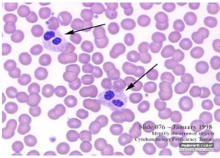
Pelger-Heut Anomaly
failure of neutrophils to segment
Pelger-Huet Anomaly
Chediak Higashi Anomaly
large granules in cytoplasm of neutrophils
Chediak-Higashi anomaly
May-Hegglin Anomaly
blue inclusions bodies seen in neutrophils
May-Hegglin Anomaly
Alder-Reilly anomaly
heavy, dark azurophilic granules
Alder-Reilly anomaly
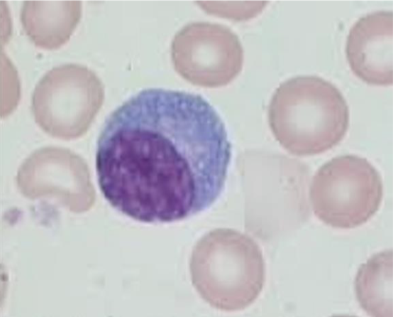
lymphocyte alterations
varient lymphocytes (reactive or atypical lymphocytes)
lymphocyte alterations
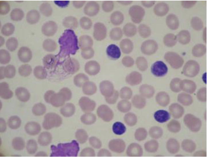
smudge/basket cells
damaged white blood cells
smudge/basket cells