Thigh Muscles
1/18
Earn XP
Description and Tags
thick thighs save lives
Name | Mastery | Learn | Test | Matching | Spaced | Call with Kai |
|---|
No analytics yet
Send a link to your students to track their progress
19 Terms
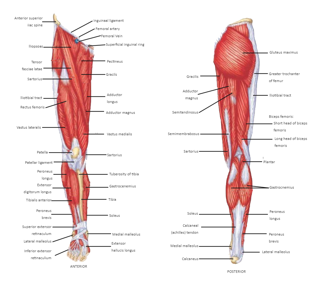
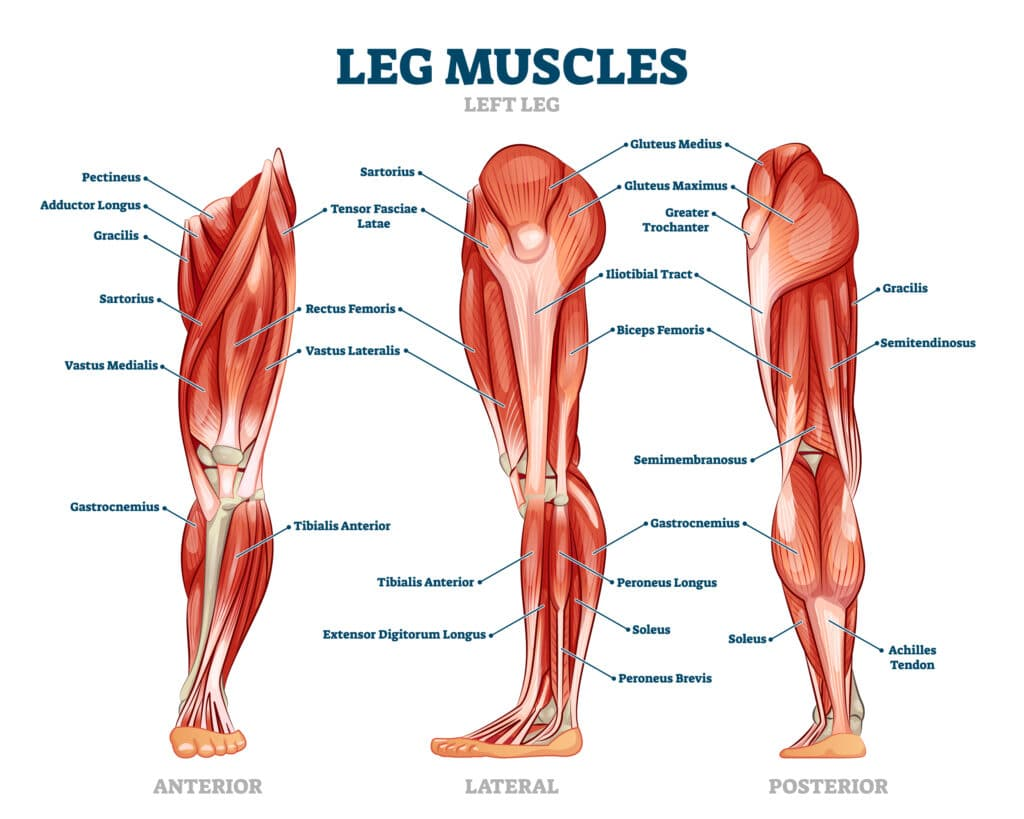
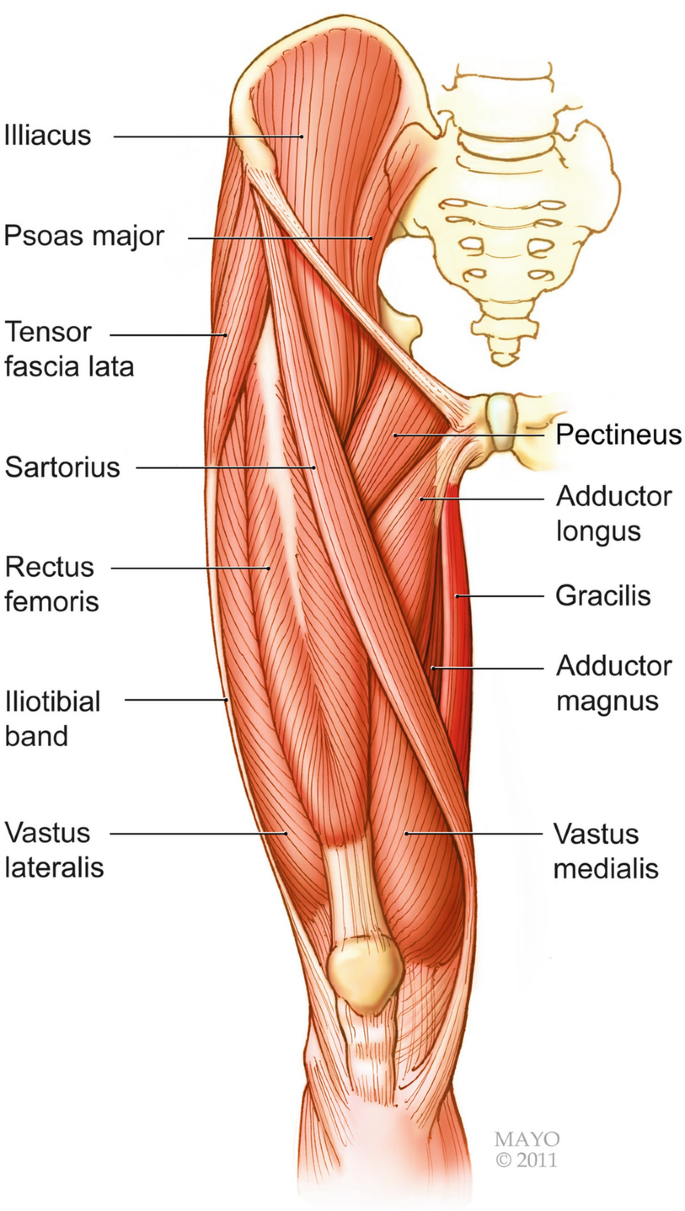
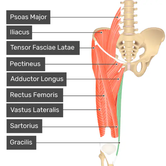
Overview of the Thigh
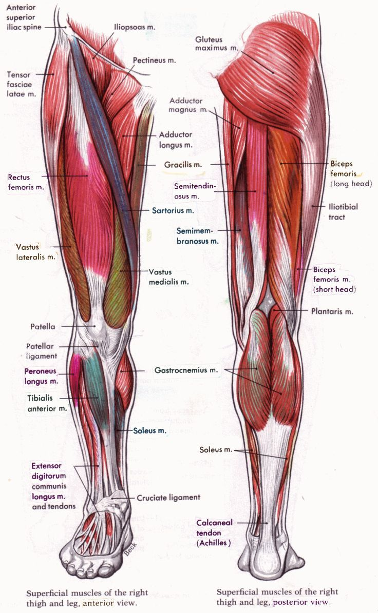
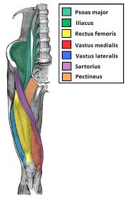
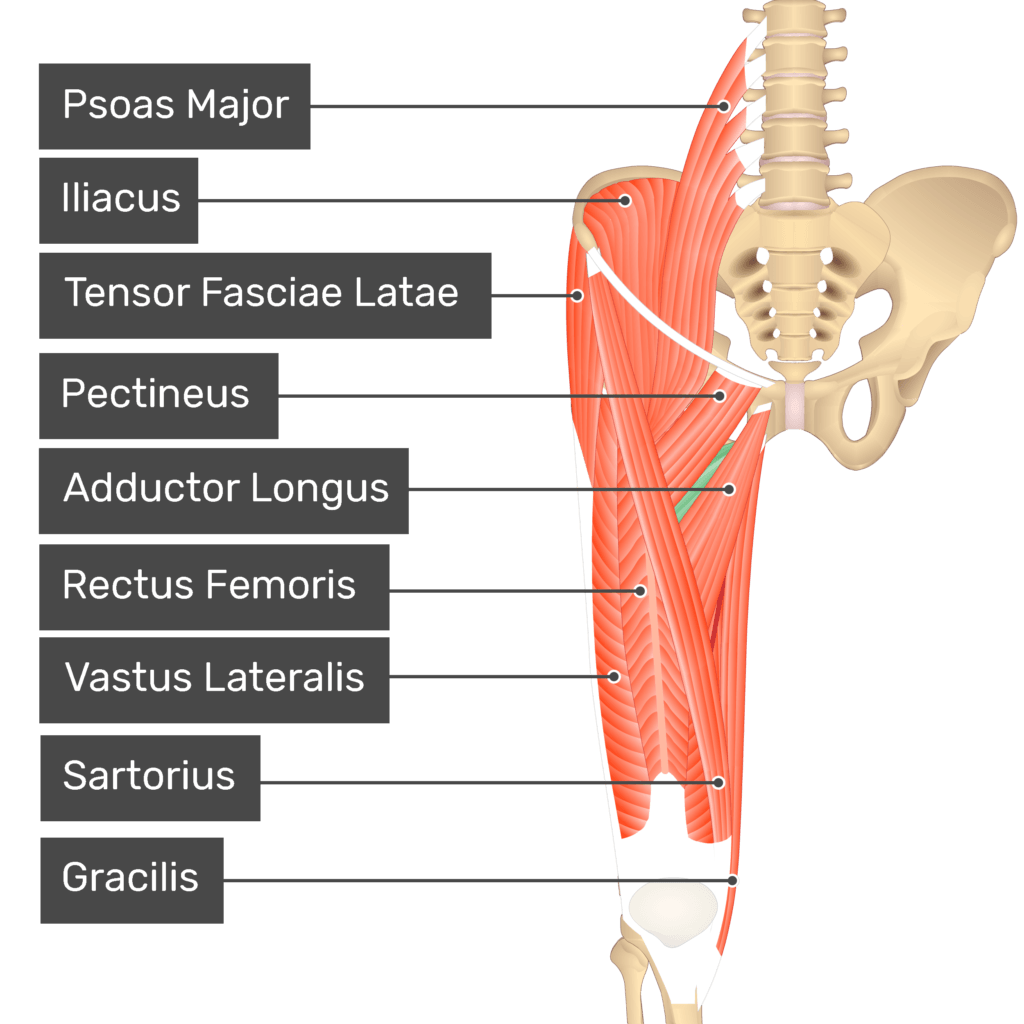
Anterior (flexor) Compartment
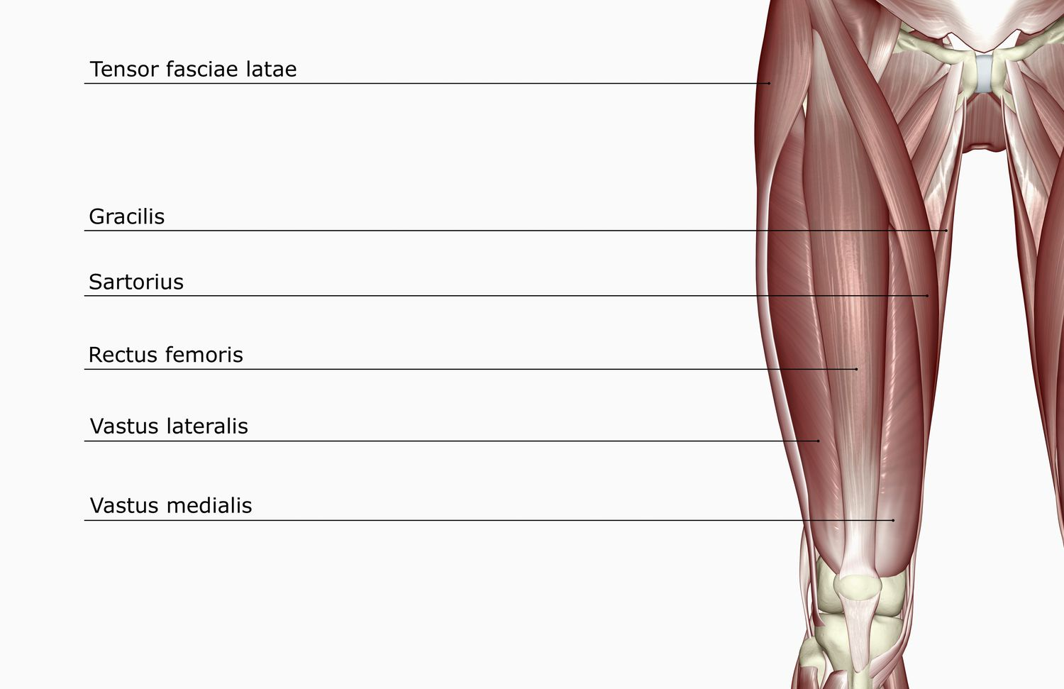
sartorius
quadriceps femoris
rectus femoris
vastus lateralis
vastus intermedius
vastus medialis
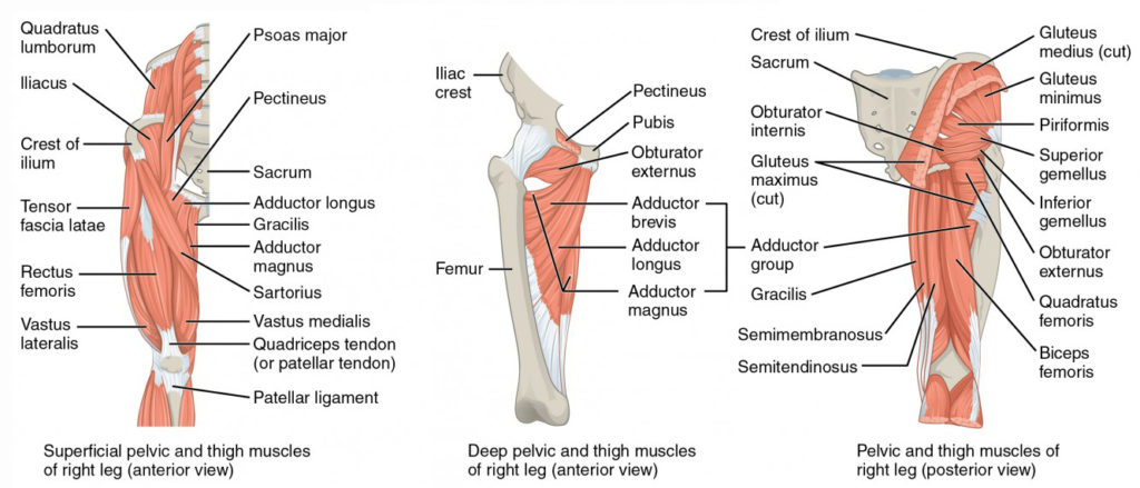
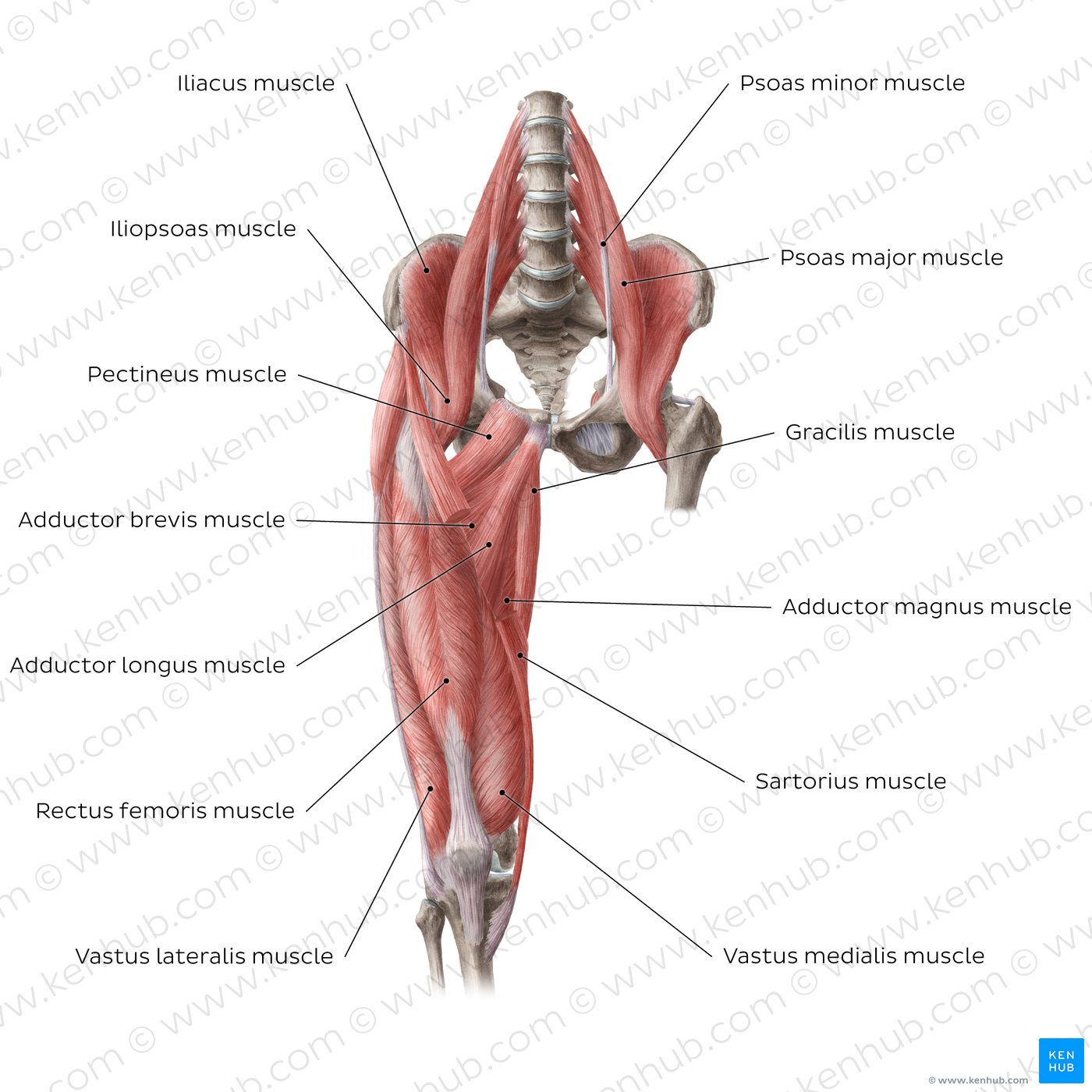
Medial (Adductor) Compartment
pectineus
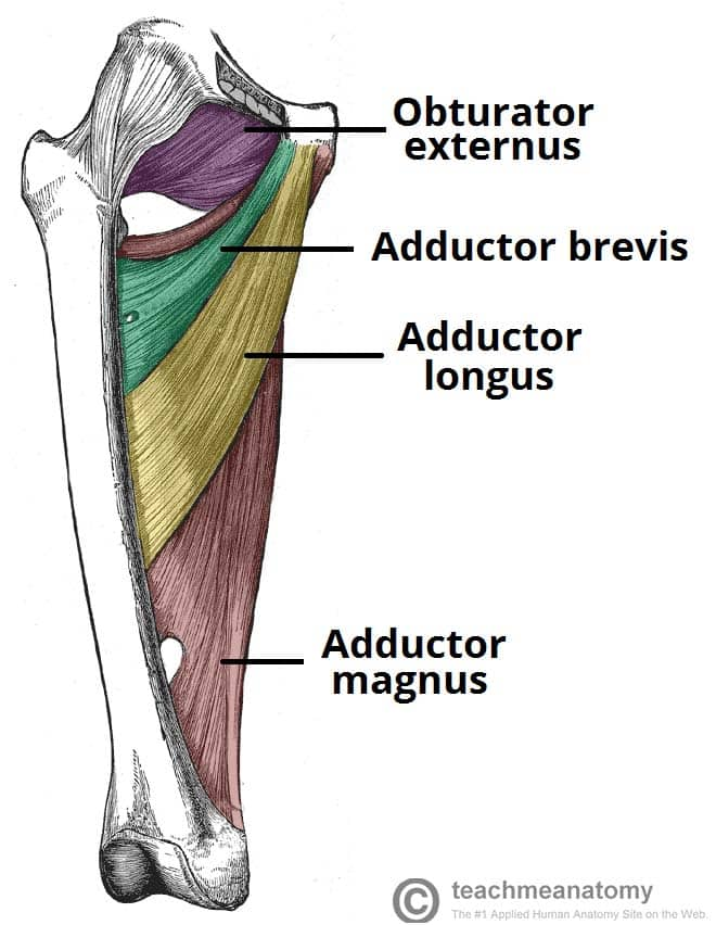
adductor longus
adductor brevis
adductor magnus
adductor minimus
gracilis
obturator externis
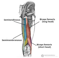
Posterior (Extensor) Compartment
biceps femoris
semimembranosus
semitendinosus
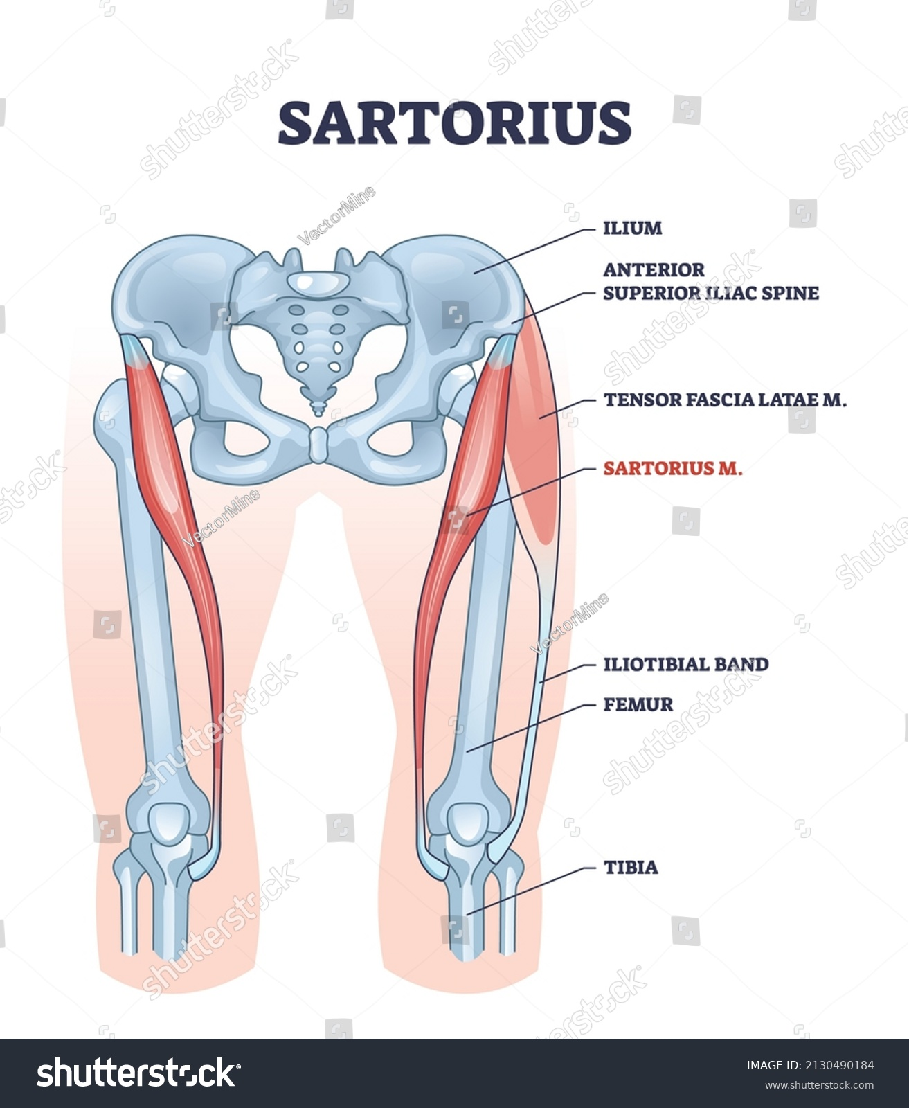
sartorius
the longest muscle in the human body
part of the anterior compartment along with quadriceps femoris
has an oblique course; arising from the anterior superior iliac spine, crossing to the medial side of the thigh, and inserting on the medial side of the proximal tibia
flexes, abducts, and externally rotates the thigh, internally rotates the leg
origin → anterior superior iliac spine (ASIS)
insertion → proximal end of tibia below medial condyle (via pes anserinus)
action → thigh flexion, abduction, and external rotation at the hip joint; leg flexion and internal rotation at the knee joint
innervation → femoral nerve (L2-L3)
blood supply → proximal third: branches of femoral artery, deep femoral artery, artery of quadriceps, lateral circumflex femoral artery
→ middle third: branches of femoral artery
→ distal third: branches of femoral and descending genicular arteries
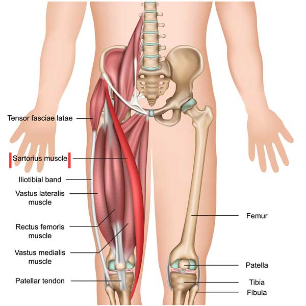
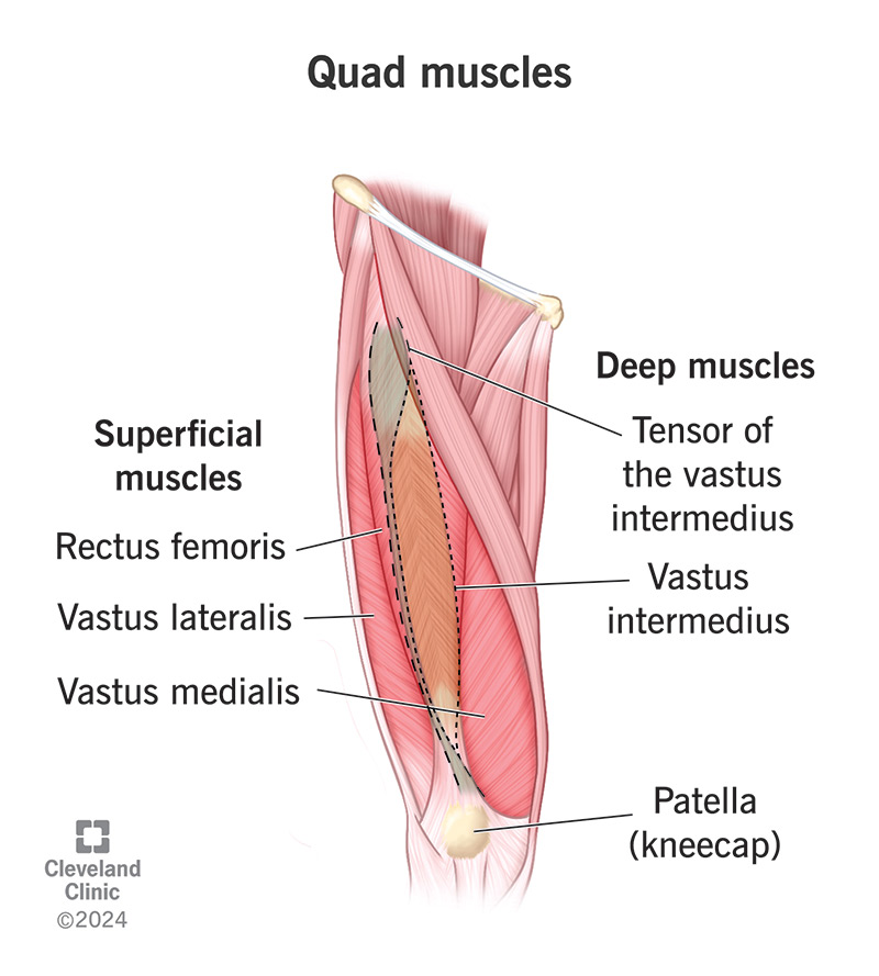
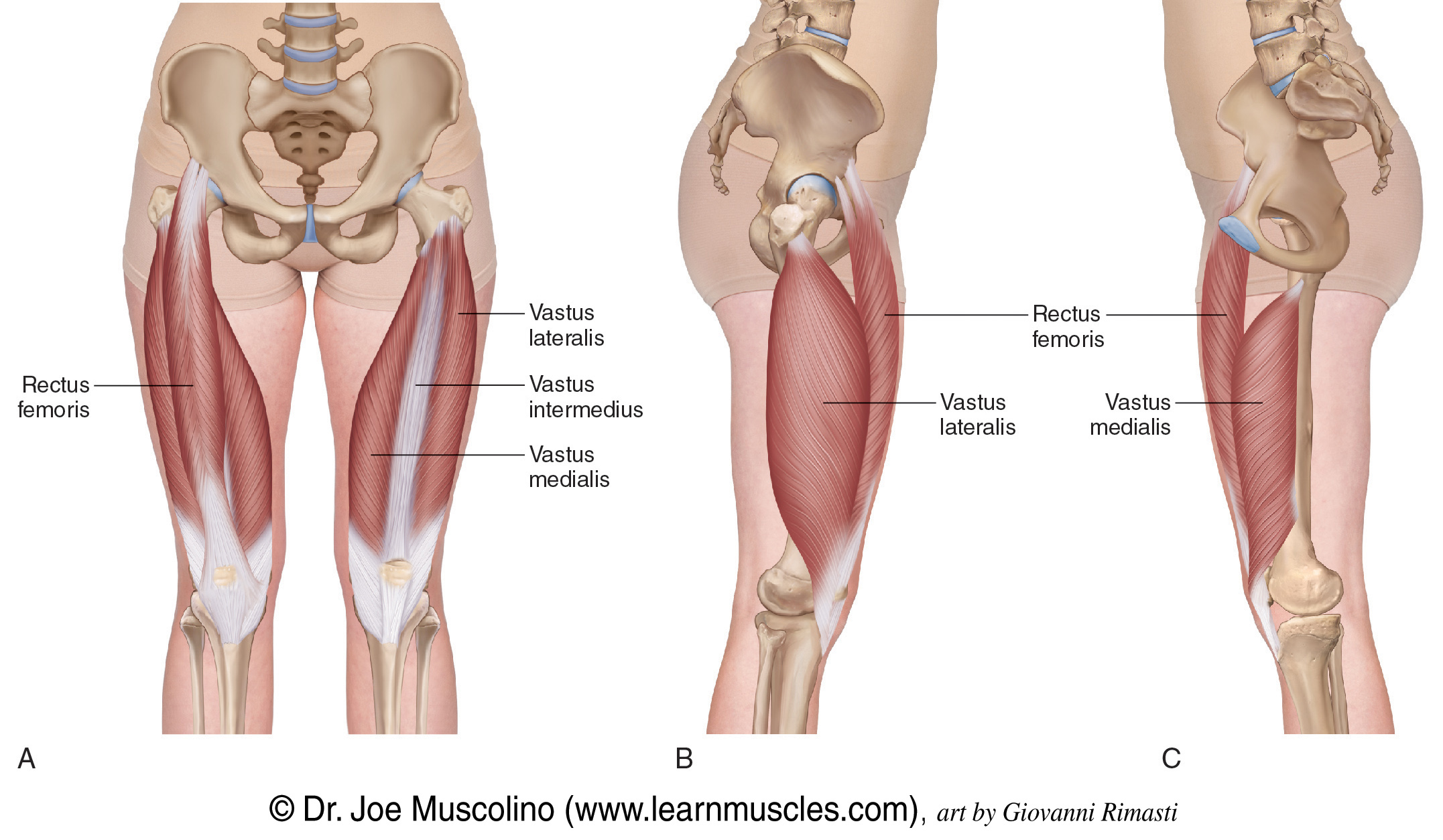
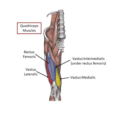
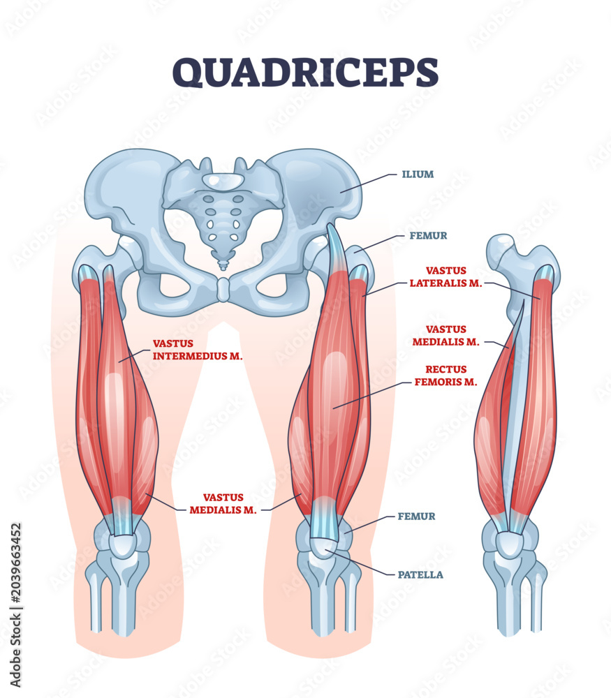
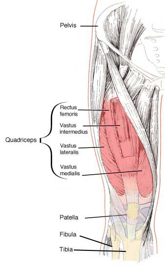
quadriceps femoris
the strongest muscle of the human body
consists of 4 individual muscles:
rectus femoris
vastus medialis
vastus intermedius
vastus lateralis
out of the 4, only rectus femoris crosses both the hip and knee joints, the others only cross the knee joint
they vary in origin but all 4 share a common quadriceps femoris tendon which inserts into the patella
extends the leg at the knee joint and flexes the thigh at the hip joint
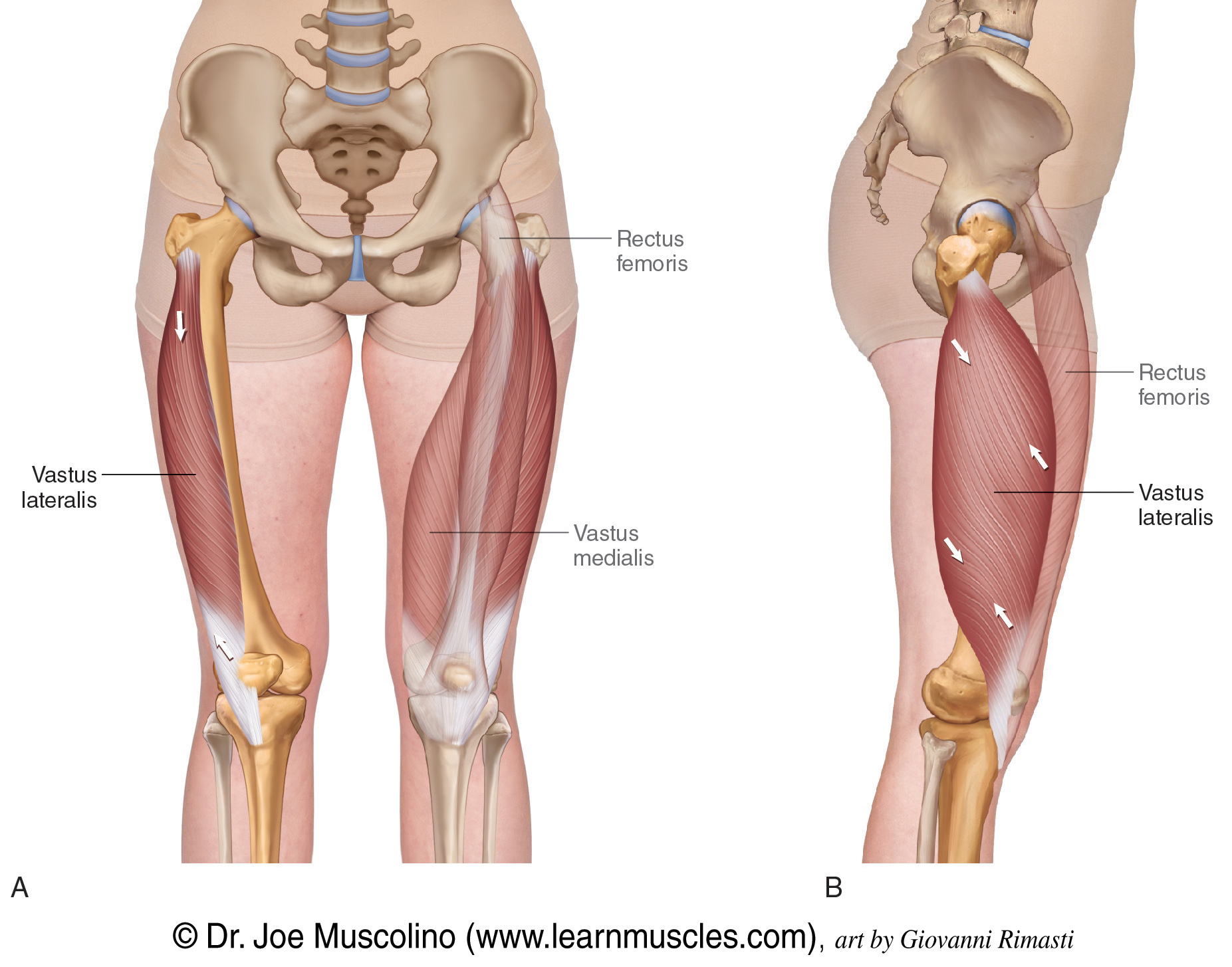
rectus femoris
origin → anterior inferior iliac spine, supraacetebular groove
insertion → tibial tuberosity via patellar ligament, patella
action → thigh flexion at the hip joint, leg extension at the knee joint
innervation → femoral nerve (L2-L4)
blood supply → femoral, lateral femoral circumflex, superficial circumflex iliac arteries
vastus lateralis
origin → intertrochanteric line, greater trochanter, gluteal tuberosity, linea aspera of femur
insertion → tibial tuberosity via patellar ligament, patella, lateral condyle of tibia
action → leg extension at the knee joint
innervation → femoral nerve (L2-L4)
blood supply → lateral circumflex and deep femoral arteries; artery to quadriceps
vastus intermedius
origin → anterior surface of femoral shaft
insertion → tibial tuberosity via patellar ligament, patella
action → leg extension at the knee joint
innervation → femoral nerve (L2-L4)
blood supply → artery to quadriceps, deep femoral arteries
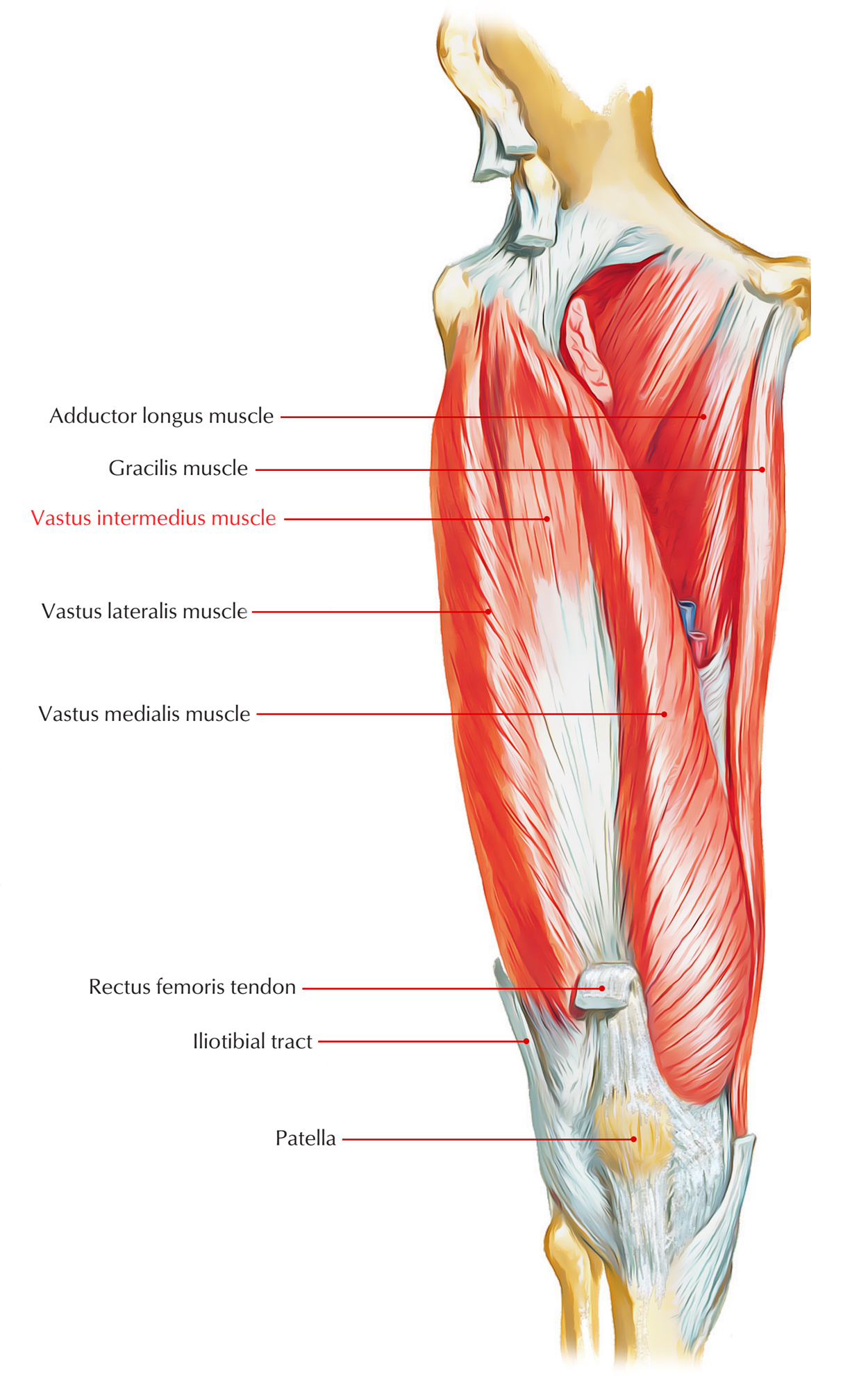
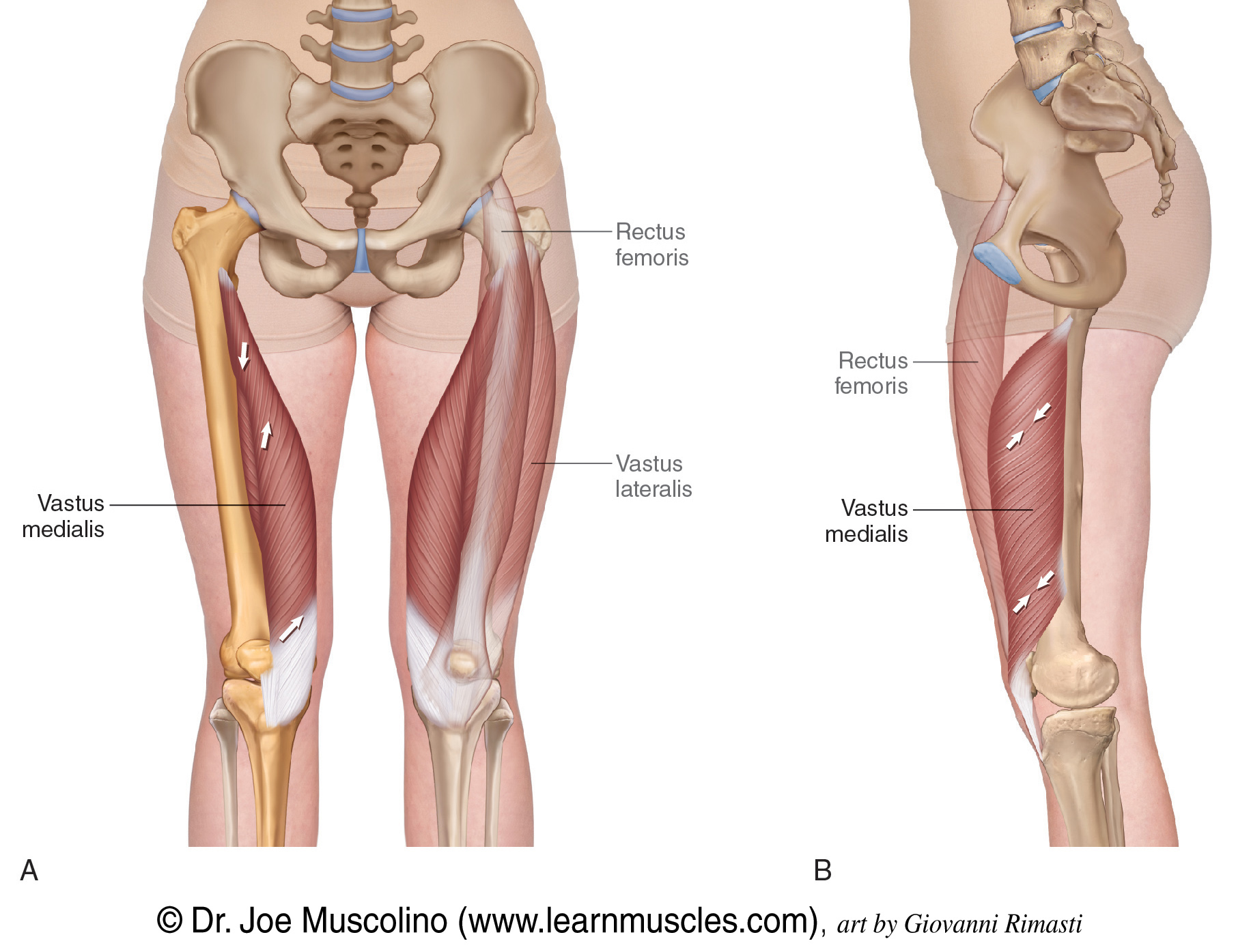
vastus medialis
origin → intertrochanteric line, pectineal line of femur, linea aspera, medial supracondylar line of femur
insertion → tibial tuberosity via patellar ligament, patella, medial condyle of tibia
action → leg extension at the knee joint
innervation → femoral nerve (L2-L4)
blood supply → femoral, deep femoral, descending genicular arteries
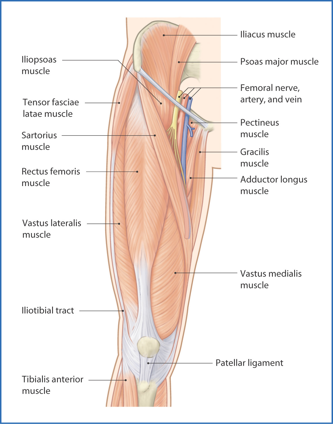
pectineus
a flat muscle found in the superomedial part of the anterior thigh
typically classified as a muscle of the adductor compartment
adducts, flexes, externally rotates, internally rotates the thigh at the hip joint and stabilizes the pelvis and balances the trunk on the lower extremity during walking
origin → superior pubic ramus (pectineal line of pubis)
insertion → pectineal line of femur, linea aspera of femur
action → thigh flexion, adduction, external rotation, internal rotation at the hip joint; pelvis stabilization
innervation → femoral nerve & obturator nerve (L2, L3)
blood supply → medial femoral circumflex artery, obturator artery
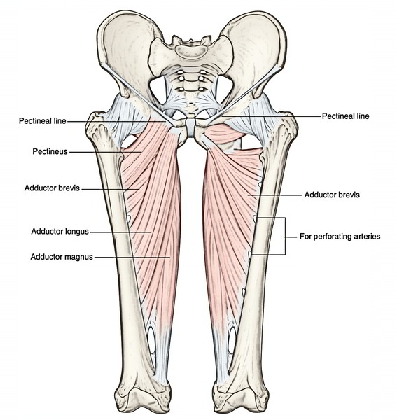
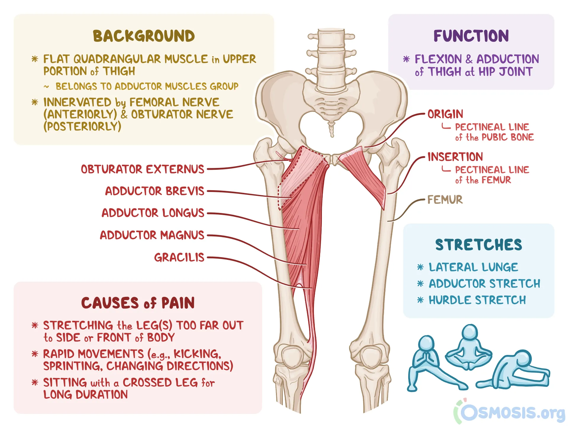
adductor longus
a long, fan shaped muscle in the medial aspect of the thigh
adducts the thigh at the hip joint
origin → body of pubis, inferior to pubic crest and lateral to pubic symphysis
insertion → middle third of linea aspera of femur (medial lip)
action → thigh flexion, adduction, external rotation at the hip joint; pelvis stabilization
innervation → obturator nerve (L2-L4)
blood supply → deep femoral artery, obturator artery
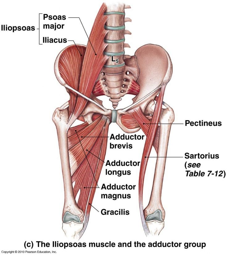
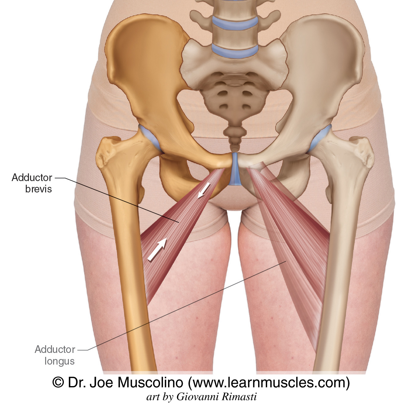
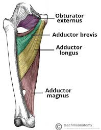
adductor brevis
a flat, triangular muscle running from the pubis to the medial aspect of they femur
produces thigh adduction and participates in flexion, internal rotation of the thigh and pelvis stabilization when standing or walking
a weak adductor, as it is the shortest muscle in the adductor group
origin → anterior body of pubis, pubic ramus
insertion → linea aspera of femur (medial lip)
action → thigh flexion, adduction, external rotation at the hip joint; pelvis stabilization
innervation → obuturator nerve (L2-L4)
blood supply → arteria profunda femoris
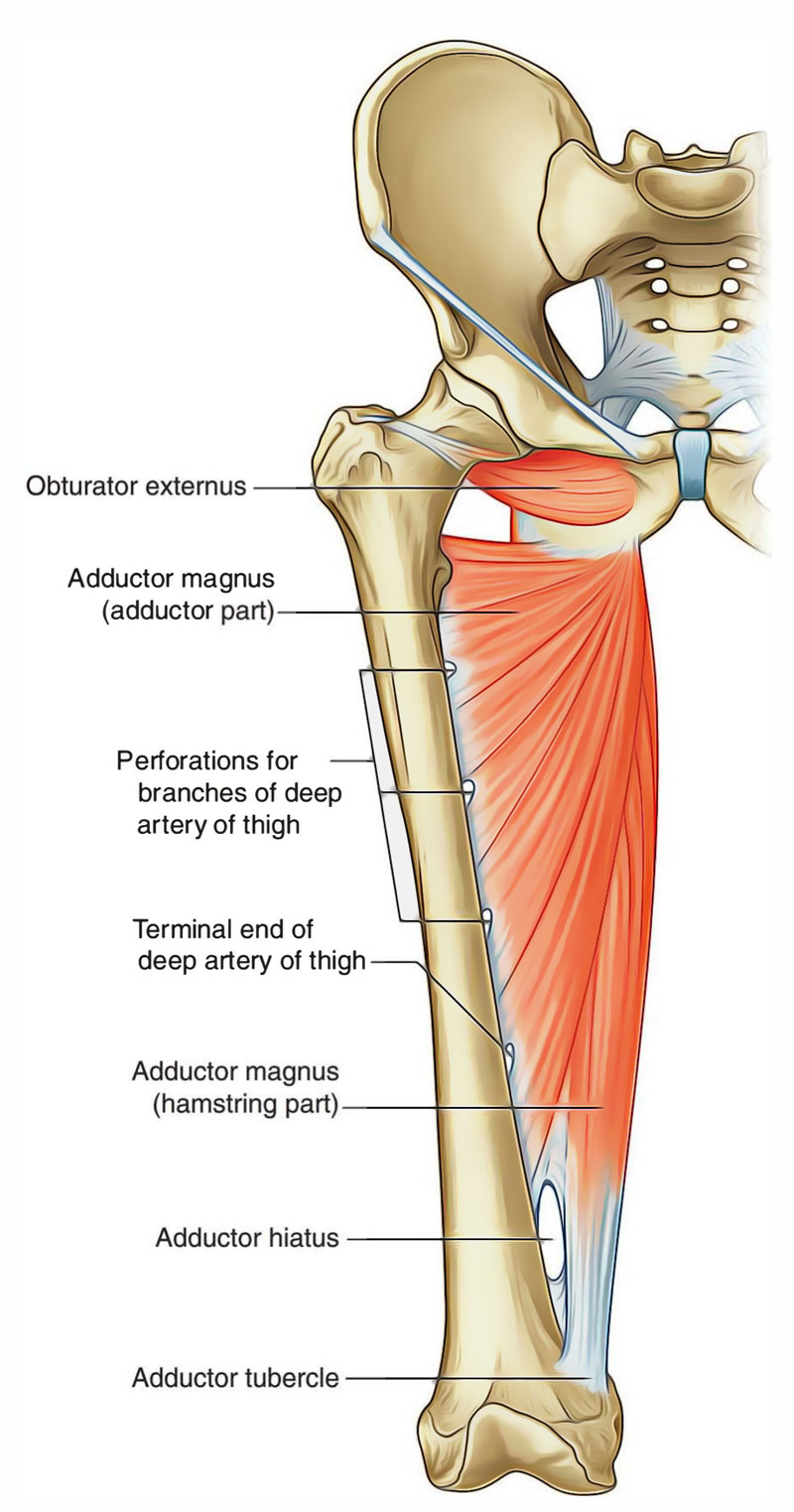
adductor magnus
a large, triangular adductor with its apex situated on the hip bone and its base on the linea aspera of the femur
situated in both the posterior and medial compartments of the thigh but is classified as an adductor
the largest and strongest in the adductor compartment
forms the adductor hiatus, which transmits the femoral artery and vein to the popliteal fossa
contains 4-5 oseo-aponeurotic openings as passage for perforating branches of the deep femoral artery
divided into 2 parts: adductor part and ischiocondylar part
adductor part acts as an adductor, the ischiocondylar part is part of the posterior compartment
origin → adductor part: inferior pubic ramus, ischial ramus
→ ischiocondylar part: ischial tuberosity
insertion → adductor part: gluteal tuberosity, linea aspera (medial lip), medial supracondylar line
→ ischiocondylar part: adductor tubercle of femur
action → adductor part: thigh flexion, adduction, external rotation at the hip joint
→ ischiocondylar part: thigh extension, internal rotation at the hip joint
innervation → adductor part: obturator nerve (L2-L4), ischiocondylar part: tibial division of sciatic nerve (L4)
→ mnemonic: African Mouse Sneaks Out (refers to Adductor Magnus Sciatic Obturator)
blood supply → deep femoral artery; femoral, popliteal, genicular arteries
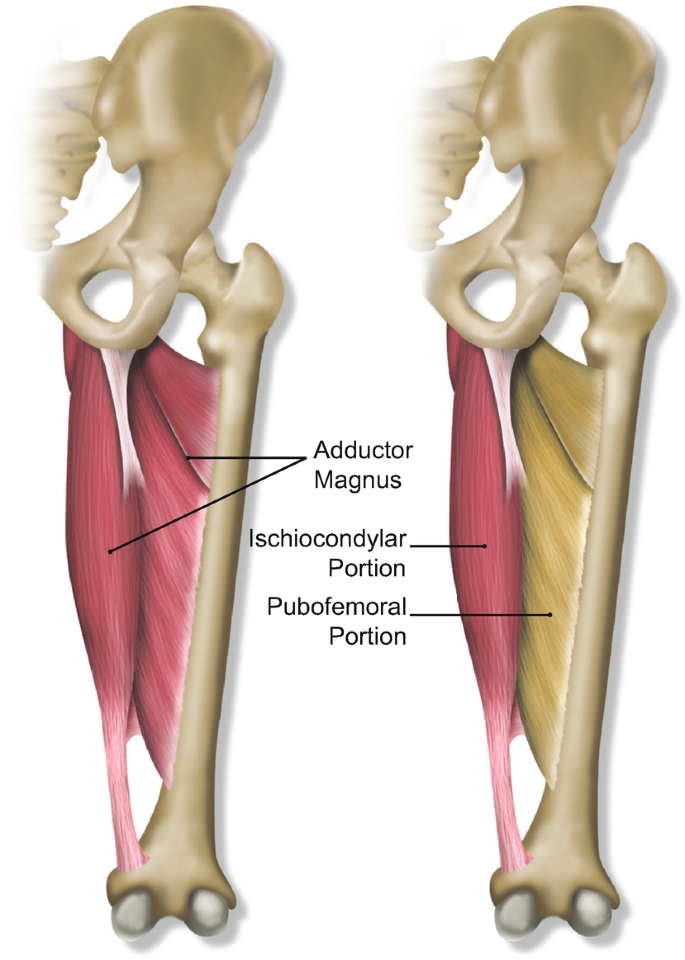
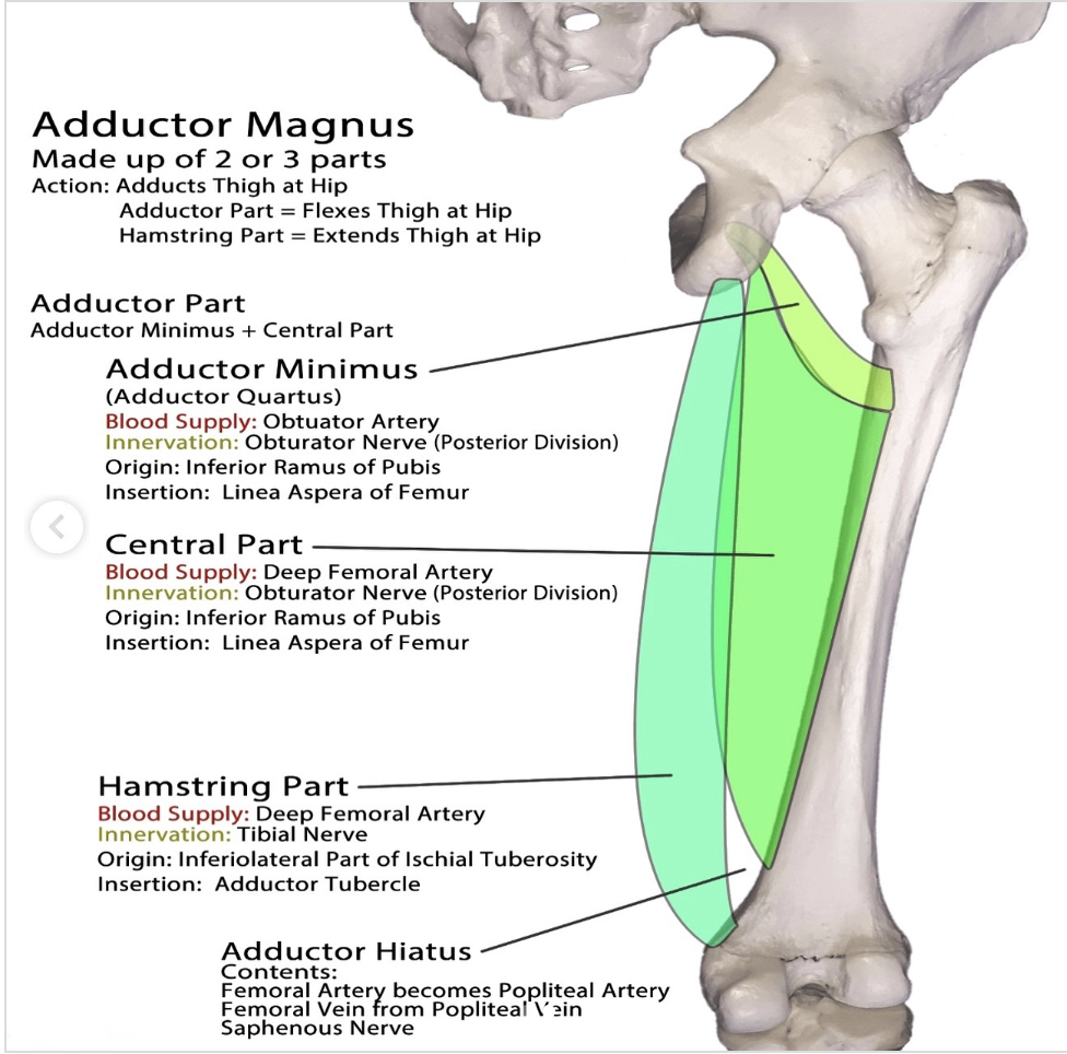
adductor minimus
the uppermost portion of the adductor magnus
origin → inferior ramus of pubic bone
insertion → medial lip of linea aspera
action → adductos the thigh at the hip joint, assists in external rotation of thigh at hip joint
innervation → posterior division of obturator nerve (L2-L4)
blood supply → medial circumflex femoral artery, first perforating branch of deep femoral artery (profunda femoris)
gracilis
a long, slender muscle, the most superficial muscle in the adductor compartment
the weakest adductor, but the only adductor that crosses and acts on both the hip and knee
extends from the coxal bone to the tibia
origin → anterior body of pubis, inferior pubic ramus, ischial ramus
insertion → medial surface of proximal tibia via pes anserinus
action → thigh flexion and adduction at the hip joint, leg flexion and internal rotation at the knee joint
innervation → obturator nerve (L2-L3)
blood supply → deep femoral artery (via artery to the adductors)
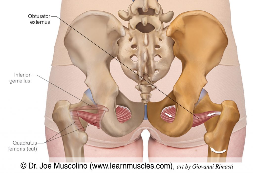
obtutator externus
a flat, triangular, paired muscle of the gluteal region found on the anterior aspect of the obturator foramen, attached to the obturator membrane and the adjacent margin of the obturator foramen
externally rotates the femur when the hip is extended
abducts the thigh when the hip is flexed
** anatomiclly locate in the adductor compartment, but functionally considered a deep gluteal lateral rotator
origin → anterior surface of obturator membrane, bony boundaries of obturator foramen
insertion → trochanteric fossa of femur
action → thigh external rotation and thigh abduction (from a flexed hip) at the hip joint; stabilizes the head of the femur in the acetabulum
innervation → obturator nerve (L3, L4)
blood supply → obturator and medial circumflex femoral arteries
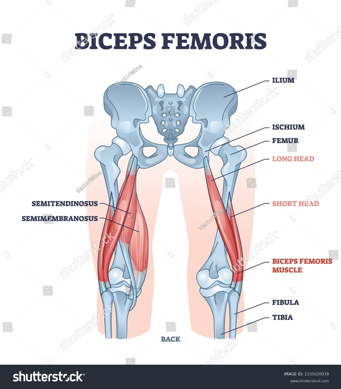
biceps femoris
a long muscle of the posterior compartment, makes up the hamstrings along with semimembranosis and semitendinosus
runs from the ischial tuberosity to the proximal part of the fibula, crossing two joint (hip and knee)
flexes and externally rotates at the knee joint, extends and externally rotates at the hip
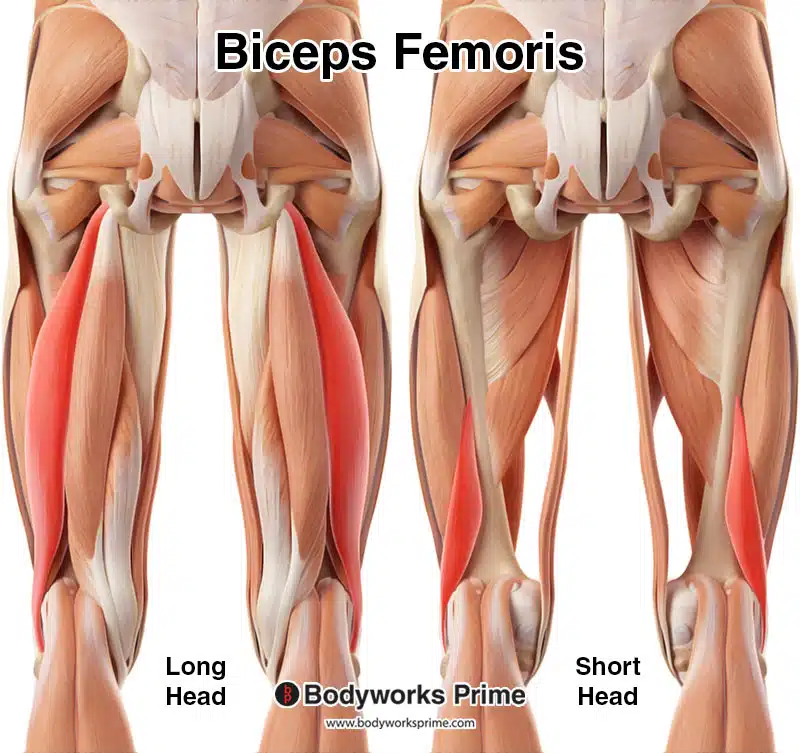
consists of a long and short head
origin → long head: inferomedial impression of the ischial tuberosity, sacrotuberous ligament
→ short head: linea aspera of femur (lateral lip), lateral supracondular line of femur
insertion → lateral aspect of head of femur
action → thigh extension and external rotation at the hip joint; leg flexion and external rotation at the knee joint; stabilizes pelvis
innervation → long head: tibial division of sciatic nerve (L5-S2)
→ short head: common fibular division of sciatic nerve (L5-S2)
blood supply → inferior gluteal artery, perforating arteries, popliteal artery
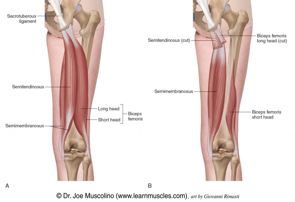
semimembranosis
part of the hamstring group
crosses both hip and knee joints
part of the hip extensor group with the other 2 hamstring muscles plus glute max
occupies the medial aspect of the posterior thigh
origin → superolateral impression of ischial tuberosity
insertion → medial condyle of tibia
action → thigh extension and internal rotation at the hip joint; flexion and internal rotation of the leg at the knee joint; stabilizes pelvis
innervation → tibial division of sciatic nerve (L5-S2)
blood supply → perforating branches of femoral and popliteal arteries
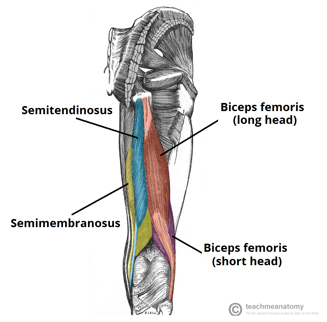
semitendinosus
a fusiform muscle of the posterior compartment of the thigh, part of the hamstring group
a prime mover that extends and internally rotates the thigh, flexes and internally rotates the leg
crosses both hip and knee joints
origin → posteromedial impression of ischial tuberosity
insertion → proximal end of tibia below medial condyle (via pes anserinus)
action → thigh extension and internal rotation at the hip joint; leg flexion and internal rotation at the knee joint; pelvis stabilization
innervation → tibial division of sciatic nerve (L5, S2)
blood supply → first perforating branch of femoral artery (profunda femoris), medial femoral circumflex artery, inferior gluteal artery, inferior medial geniculate artery