4.2.3-Microscopic Mucosa
1/13
There's no tags or description
Looks like no tags are added yet.
Name | Mastery | Learn | Test | Matching | Spaced | Call with Kai |
|---|
No analytics yet
Send a link to your students to track their progress
14 Terms
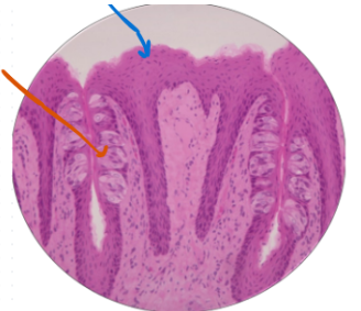
What is this Histology slide? What is the blue arrow? What is the orange arrow?
Taste Bud
Orange arrow-Taste bud:Sensory receptors for detecting basic tastes.
Blue arrow-Foliate Papillae: Folded tissues houses more tastes buds.
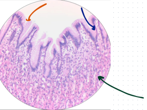
What is this histology slide? What is the orange arrow? What is the blue arrow? What is the green arrow?
Fundic stomach
blue arrow:Gastric pits-Shallow surface openings leading to deeped glands.
green arrow:Fundic glands- Secrete acid and enzymes for chemical digestion.
Orange arrow:Columnar Epithelium- Secretes protective mucus for the stomach lining.
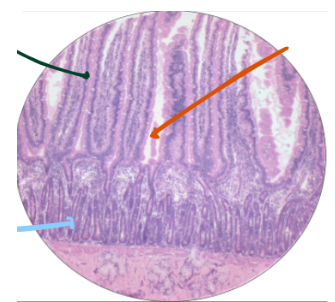
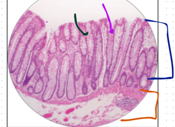
What histology is this?
Duodenum
Orange Arrow:Enterocytes-Primary absorptive cells with brush borders.
Green Arrow:Villi-Finger-like projections increasing surface area
Blue Arrow:Mucosa-Innermost layer specialised for nutrient absorption.
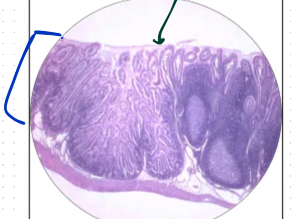
What histology is this?What is the orange arrow? What is the blue arrow? What is the green arrow?
Jejunum
Green bracket-Mucosa:Primary site for nutrient absorption and secretion.
orange arrow-Villi:Increase surface area to maximize nutrient uptake.
Blue arrow-Crypts:produce new cells and secrete intestinal juice.
What is this histology?
Ileum
blue arrow:Mucosa-Absorbs nutrients and acts in immune defense.
green arrow:Villi-Increases surface area for nutrient absorption
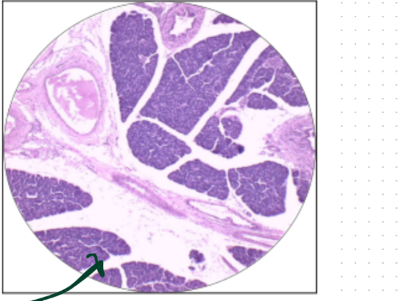
What is this histology slide?
Pancreas
Green arrow-Islet of Langerhans:Secretes insulin and glucagon into the blood.
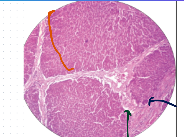
What is this histology slide?
Liver
green arrow:Portal Vein-Carries nutrient-rich blood to the liver.
blue arrow:Bile Duct-Transports bile towards the gallbladder.
Orange Bracket:Lobules-Functional unit for filtering blood.
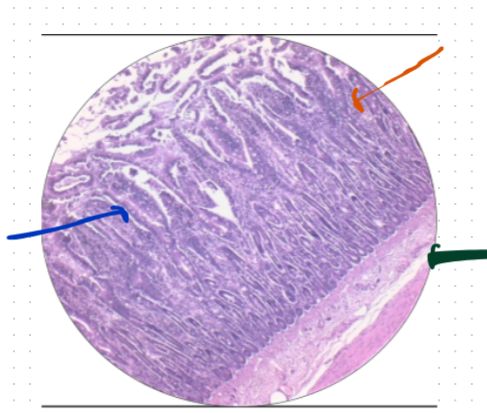
What is this histology slide?
Colon
Blue:Mucosa-Absorbs water and lubricates stool.
Green:Columnar Epithelium-Absorbs water and electrolytes.
Purple:Connective Tissue-Provides structural support the lining.
Orange:Submucosa-Houses blood vessels and nervous plexus.
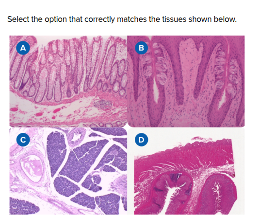
What is A, B, C, and D?
A-Colon
B-Taste Bud
C-Pancreas
D-Fundic Stomach
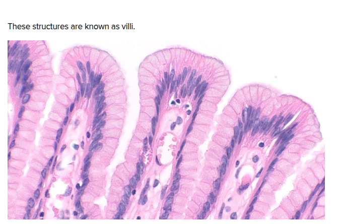
True of False
True-It’s villi
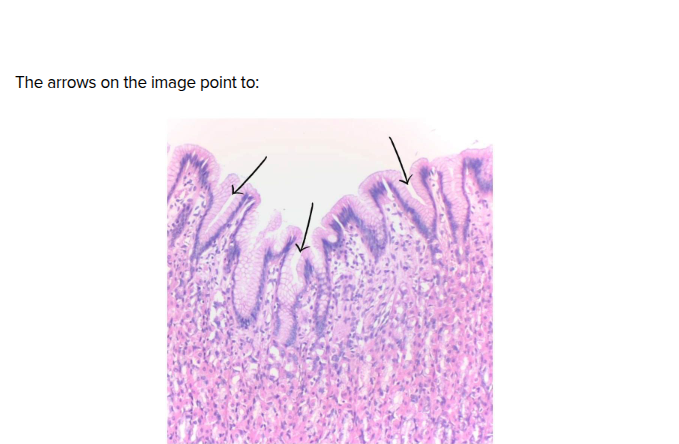
The arrows on the image show?
Gastric pits
Found only in the pancreas, these special cells secrete insulin and glucagon.
Islets of Langerhans
Q1-What are the functions of the smooth muscle in the stomach and the villi in the small intestine? How do the structures help with function?
The stomach and small intestine utilize specialized structures optimized for digestion and absorption. The stomach relies on three layers of smooth muscle to dynamically churn and mechanically smash food into chyme from multiple angles. Further down, the small intestine uses millions of finger-like villi to maximize surface area for nutrient absorption. Each villus features a single-cell thick wall for rapid diffusion, an internal capillary network to absorb water-soluble nutrients directly into the bloodstream, and a lymphatic vessel to transport larger fat molecules.
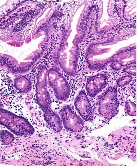
Q2-Abetalipoproteinemia is a rare, inherited disorder that is caused by changes (mutations) in the microsomal triglyceride transfer protein (MTTP) gene. The disorder affects fat absorption by the intestine and mobilization by the liver. Analyze Figure 7, Abetalipoproteinemia Biopsy from the Duodenum. What changes do you see in the tissue? Explain how those changes would cause impaired fat absorption.
HINT: Look closely at the villi and enterocytes
In the duodenal biopsy of a patient with abetalipoproteinemia, the primary tissue change is marked lipid vacuolation within the enterocytes lining the intestinal villi, which gives their cytoplasm a clear, foamy, or distended appearance. This structural change causes impaired fat absorption because mutations in the MTTP gene prevent the proper assembly and secretion of chylomicrons. While the enterocytes can still successfully take up dietary fats from the intestinal lumen and convert them into triglycerides, they completely lack the functional machinery to package and export these fats into the lymphatic lacteals. Consequently, the absorbed lipids become trapped inside the epithelial cells, engorging the enterocytes and preventing the body from utilizing dietary fats and fat-soluble vitamins.