L36: foot and claw
1/118
There's no tags or description
Looks like no tags are added yet.
Name | Mastery | Learn | Test | Matching | Spaced | Call with Kai |
|---|
No analytics yet
Send a link to your students to track their progress
119 Terms
what is the equine hoof?
horny enclosure of the distal phalanx (epidermal hoof)
what is the equine foot?
equine hoof and everything inside
what re the three layers of the equine hoof?
epidermis
dermis
subcutaneous tissue
what is the epidermis of the hoof also known as?
horn
what is the dermis of the hoof also known as?
corium
which layer of the hoof is insensitive and non-innervated?
epidermis
which layer of the hoof is soft, sensitive, and innervated?
dermis
which layer of the hoof has digital and coronary cushions?
subcutaneous tissue
what are the topographical regions of the hoof wall?
toe
quarters
heels
bars
what is the most dorsal region of the hoof wall?
toe
what is the most palmar part of the hoof wall?
heels
which of the quarters in the hoof wall has a thicker wall?
lateral (outer) has thickest wall
what do the bars offer to the hoof wall?
stability for the heels
what seperates the bars from the frog?
paracuneal grroves
describe the thickness of the hoof wall?
wall thickest at toes and thins progressively towards the heel
how do we see the bars of the hoof wall?
must lift hoof off the ground as it is on the sole surface
what is the sole?
space between the wall and the frog
describe the nature of the sole in the equine foot
concave
what is the angles of the sole?
parts between the bars and quarters
what is “corns”
trauma at the angles of the sole in the equine foot
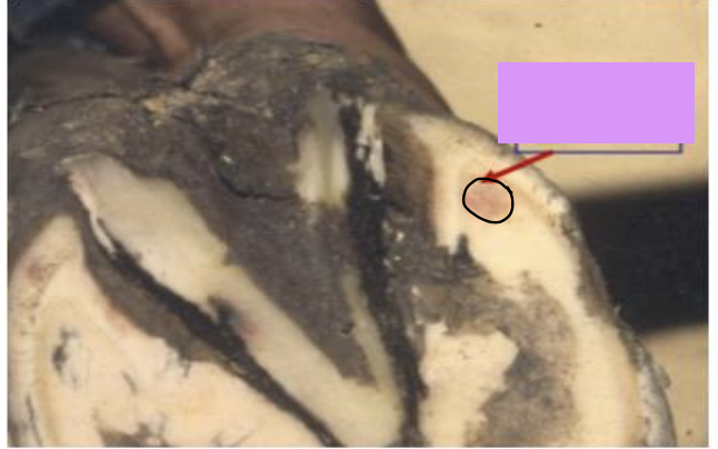
what does the image show?
corns

what region of the hoof is the black box?
hoof
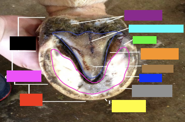
what region of the hoof is the pink box?
quarters

what region of the hoof is the red box?
toe

what structure is the yellow box?
hoof wall

what structure is the gray box?
white line

what structure is the blue box?
sole

what structure is the orange box?
collateral sulcus

what structure is the brown box?
frog

what structure is the green box?
bars

what structure is the light blue box?
central sulcus

what structure is the purple box?
heel bulbs
what is the function of the frog?
weight bearing of body
schock absorption
what is the role of the frog when the foot hits the ground?
yields pressure forces and dissipates most of the impact
what is the central groove/sulci of the frog?
the space between the bases on the side of the frog
what does the frog stay correspond to?
an internal spine formed by the reflection of the central groove
what are the components of the frog?
apex
base
crura
central groove/sulci
what is the intratoric fossa?
groove that divides the heel bulbs of the equine foot into lateral and medial parts
what is the digital cushion in the equine foot?
extensive fibrous fatty tissue above the crura of the frog and below the DDFT that helps the frog with shock absorption
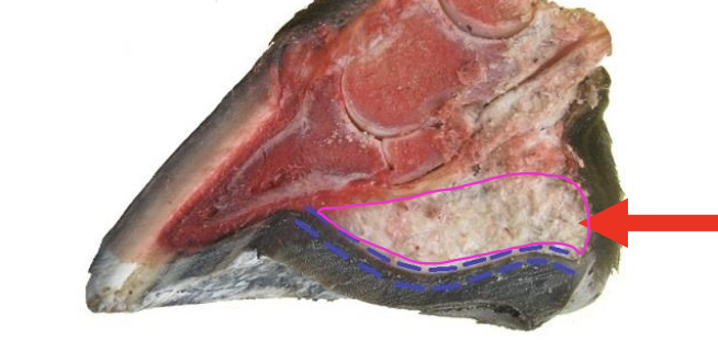
what is the red arrow pointing at?
digital cushion of the equine foot
what bacteria causes thrush in the equine foot?
fusobacterium necrophorum
what is thrush in the equine foot?
degenerative condition of the central sulci and/or collateral sulci of the frog caused by bacteria or fungi
what are the characteristics of thrush in the equine foot?
black, necrotic, foul-smelling material
what surrounding tissues can be infected by thrush if it spreads from the frog?
digital cushion and skin at the heel bulbs
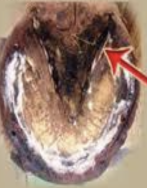
what is occurring in the image?
thrush
hoof capsule
skin derivative that entirely encloses the third phalanx
what is the hoof capsule a product of?
the dermis
what does the dermis of the hoof capsule continue with?
the common dermis of the skin at the coronet
what does the hoof capsule NOT have?
blood vessels or nerves
MCQ: what structure is the most important for shock absorption in the equine foot?
the frog
what is the coronet in the equine foot?
the junction between the skin and the hoof
what are the three layers of the epidermis in the equine hoof wall?
stratum externum
startum medium
startum internum
which area of the epidermis in the hoof wall is predisposed to fissures leading to cracks in the foot potentially?
boundary between the startum internum and startum medium
what connects the distal phalanx to the hoof wall?
dermis
what is the outer layer of the epidermis?
startum externum
what is the inner layer of the epidermis?
startum internum
what are the parts of the stratum externum?
periople
stratum tectorium
what is the function of the periople horn?
acts as a fluid reservoir to keep the underlying coronary horn moist
where does the periople of the stratum externum extend to?
onto the heel bulbs and blends with frog dermis
which structure is the proximal part of the stratum externum?
periople (closer to coronary band)
what is the stratum tectorium?
the rest of the covering of the hoof wall that is impervious to water
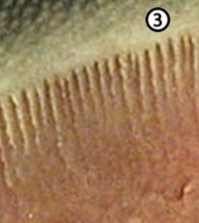
what does parallel proximo-distal lines of the stratium tectorium mean?
shows the growth of the horny tubules
what does horizontal smooth ridges of the stratium tectorium mean?
evidence of nutritional issues or laminitis
where does the stratum medium begin?
distal to the coronary groove
what does hte stratum medium possess?
openings for the papillae of the underlying dermis
describe the structure of the stratum medium
horny tissue with tubular structure formed by tubules and intertubular horn
what layer makes up the majority of the hoof wall?
stratum medium
what produces the stratum medium?
coronary dermis
what else can the stratum internum be referred to as?
epidermal laminae
insensitive laminae
what does the stratum internum fuses to?
stratum medium and connects hoof wall to dermis
what does the inner surface of the stratum internum have?
keratinized primary laminae and non-keratinized secondary laminae
what else does the stratum internum form?
laminae of the bar
what does the secondary laminae of the stratum internum interlock with?
dermal lamina
which layer of the epidermis no longer has tubules?
stratum internum
what is the function of the dermis?
attaches hoof wall to internal foot structures
produces various parts of the hoof wall via their papillae
what part of the dermis does not produce the tubules of the hoof wall?
laminar dermis
what is another name for the dermal laminae?
sensitive laminae
what does the papillae of the sole and frog do?
arranges the rest of the hoof capsule in tubules
what are the 5 areas of the dermis from most external to internal?
perioplic dermis
coronary dermis
laminar dermis
frog dermis
sole dermis
what do all parts of the dermis produce and connect to?
epidermis
what layer of the epidermis does the perioplic dermis produce?
stratum externum
what layer of the epidermis does the coronary dermis produce?
stratum medium
what does the laminar dermis produce?
produces insensitive laminae that will connect to sensitive laminae
what is important to understand about the structure of the laminar dermis layer?
no tubules present
what layer of the epidermis does the laminar dermis produce?
stratum internum
what portion of the dermis produces most of the hoof wall?
coronary dermis
how are the papillae arranged within the coronary dermis?
papillae fit into holes in strata medium and internum of the coronary groove
what happens if there is trauma that affects the coronary dermis of the equine foot?
causes horn defects that will descend within the wall and reach the ground in about 8 months
how will the corresponding part of the hoof capsule in relation to the dermal papillae be arranged?
epidermal part will be arranged as tubules
what else can you call the primary laminae of the larminar dermis layer?
dermal laminae
what does the laminae of the laminar dermis layer interdigitate with?
the laminae of the walls and bars (epidermal laminae)
what does weight of the horse bare down on?
wall of hoof
when horse is bearing down weight, what connection in the hoof needs to be strong?
connection between the wall, dermis (connective tissue) , and P3

the following image is a section of the horse hoof. what is the white layer showing?
epidermal lamina
the following image is a section of the horse hoof. what is the pink layer showing?
dermal layer
what is the sole dermis in contact with?
direct contact with the sole surface of the distal phalanx
what makes up the frog dermis?
densely covered with plump papillae (shorter than sole)
what is the white line in the equine foot?
the thin soft pale horn between the hoof wall and sole
what forms the white line that binds hoof wall to the sole?
distal aspect of sensitive lamina reaching ground surface via terminal papaillae
what is the clinical importance of the white line?
a point of weakness due to it being a mixture between the har laminar and soft tubular horn