11 Crystalline Lens
1/85
There's no tags or description
Looks like no tags are added yet.
Name | Mastery | Learn | Test | Matching | Spaced | Call with Kai |
|---|
No analytics yet
Send a link to your students to track their progress
86 Terms
What is the shape of the crystalline lens?
transparent biconvex structure
What does the crystalline lens look like when it is cut in half?
an onion cut in half - layers
Where is the lens located?
Behind the iris & in front of the vitreous humor
How much accommodative power does the lens contribute when it’s UNaccommodated?
18 to 20 D
How much accommodative power does the lens contribute when it’s accommodated?
58 to 60 D
What is the diameter of the lens? How does this change with age?
grows throughout life increases 0.023 mm per year
6.5 mm diameter at birth
10 mm diameter as adult
What’re the 4 functions of the lens?
transmit visible light
absorb UV light (3-hydroxykynurenin)
refract
focus
What is the transmittivity (what light get thru) of a young vs. old lens?
young lenses act as a UV blocker (transmittance under 400 nm is zero aka the lens absorbs all the light & none of it reaches the retina)
older lens act as a UV & blue blocker (we can only see crystalline lens in people who are 80 years old because before it ages it is so opaque & there will be a right shift in the transmittance graph
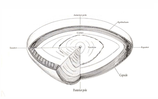
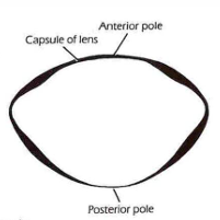
What is the center point of the anterior & posterior sides of the lens?
anterior pole & posterior pole
What is the “axis” of the lens? What is the axial thickness?
The “axis” is the line that connects the anterior & posterior pole. The axial thickness is around 4mm.
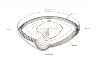
How does the anterior & posterior surface of then lens very in curvature?
anterior surface radius of curvature is 10 mm (it’s less convex than the posterior surface aka flatter)
posterior surface radius of curvature is 6 mm
Where is the equator located in the lens?
At the outer edges
What is the concentration of water & crystalline proteins in the lens composition?
60% water & 40% protein
The lens composition has a high concentration of what compound?
Glutathione (GSH, an organic compound)
What does glutathione maintain in the lens?
maintains protein sulfhydryl group in reduced form
What does glutathione protect against in the lens?
protects from oxidative damage by detoxification of peroxide
What does glutathione remove from the lens?
removes xenobiotics by conjugating with hydrophobic compounds having an electrophilic center
Which protein in the lens are soluble?
alpha, beta & gamma crytallins
What do lens crystallins help maintain in the lens?
maintains spacing of the lens fiber to promote destructive interface, minimize light scattering & maximize transparency
What do alpha crystallin do in the lens?
alpha crystallins act as a molecular chaperone to help beta & gamma crystallins recover from injury
What happen to the crystallin proteins in the lens as we age?
become insoluble
form aggregates
contribute to loss of transparency & cataract formation
What causes a cataract development in the lens?
lens proteins becoming insoluble with age
What’re the layers of the lens?
lens capsule (outermost elastic layer)
epithelium (only on anterior surface)
lens fibers: cortex & nucleus (innermost)
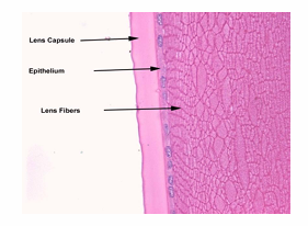
What is the lens capsule?
an acellular basement membrane that completely envelopes the lens
Where is the lens capsule the thickest & thinnest?
thinnest at the poles & equator
thickest in the mid region
What is the lens capsule formed by?
anteriorly by lens epithelium
posteriorly by lens fibers
What does the lens capsule consist of?
consist of 40 lamella made up of reticular fibers (net-like or entangled) embedded in sulfate glycoaminoglycan
What does the lens capsule composed of?
collagen IV
What does the lens capsule do?
forms a barrier to bacteria & inflammatory cells
allow diffusion of smaller molecules
What is a capsulorhexis?
its an anterior capsulotomy & its a surgical technique to create a window in the anterior capsule of the lens during cataract surgery
What is the histology of the lens epithelium?
single layer of simple cuboidal epithelium on anterior surface UNDER the capsule & is NOT found posteriorly
becomes columnar towards the equator & convert into lens fiber
How does the capsule effect the lens epithelium?
The lens epithelium can’t shed because the lens capsule does not allow it to do so
What cell junction is found in the lens epithelium?
sodium potassium pumps (low in Na+ & high in K+)
strands of zonule fuse with the outer surface of the capsule; anchoring junction on the inner surface bind it to the epithelium
What’re the lens epithelium zones?
central zone (ion transfer)
germative or proliferative zone (replication)
transition zone & equator (growth)
What’re the characteristics of the central zone of the lens epithelium?
cells are flattened & hexagonal
What’re the characteristics of the germative/proliferative zone of the lens epithelium?
cells are columnar & smaller in area but higher in density
new cells are generated here, nuclei divide & migrate posteriorly to become new lens fibers in the cortex
layers are laid down in close contact& the lens fibers are interlocked through “ball & socket” like joints
What’re the characteristics of the transitional zone & equator of the lens epithelium?
cells elongate & rotate so long axis is parallel to cortical surface
Which lens fibers/cells are nucleated vs. anucleate?
new superficial fibers are nucleated
deeper fibers are anucleate (true fibers)
How is the nuclear bow formed?
in order for a epithelium cells to become a lens fiber it must change chape (elongated) & lose it’s nucleus. there is a region around the nucleus where those early lens fibers still have there nucleus & thats called the lens bow!
forms as nuclei move anteriorly & fiber pass deeper into the lens to form the cortex
What happens to the lens cells preceding new generations?
they’re pushed deeper into center of lens
Which direction do lens fibers grow?
from the equator towards the pole
How are lens fibers interlocked with each other?
ball & socket joint (forms loops like a puzzel)
How are lens sutures formed?
lens fiber grow from the equator towards the poles & when the fiber meet, they form a suture
What is the difference between an anterior & posterior lens suture?
anterior lens suture form an upright Y
posterior lens suture form an inverted Y
How does the lens accommodate? What theory does this involve?
Helmholtz theory
ciliary muscle contracts moving ciliary body inward & forward
zonular tension is released which releases the tension from the lens (it is no longer being pulled flat & will become more curved - especially the anterior surface)
the capsule molds to the lens
What’re the 2 categories of lens metabolism?
50% oxidative metabolism (lens epithelium)
50% anaerobic via glycolytic pathway (lens fiber)
How much energy does the lens use? When?
uses only SMALL amount of energy
growth
synthesis
pumping nutrient in & waste out
maintaining water balance
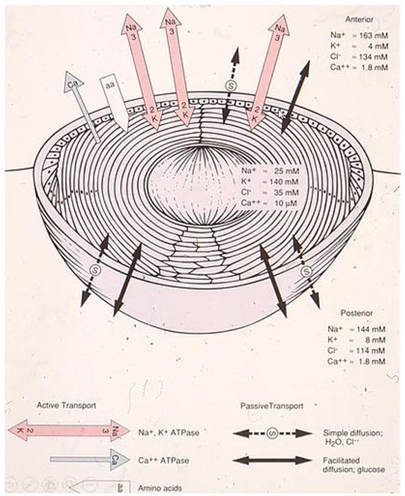
What is the ion movement thru the lens?
Active transport:
Na+,K+ ATPase
Ca2+ ATPase
AA (entering ant. lens)
Passive transport:
simple diffusion (water & chlorine)
facilitated diffusion (glucose)
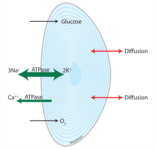
What’re the varying concentrations in the lens?
High potassium & calcium
Low sodium & chlorine

What is the varying concentrations outside the lens?
Low potassium & calcium
High sodium & chlorine
What is the largest component of the eye? %?
vitreous humor; 80% of internal volume
What’re color is the vitreous humor?
colorless (transparent gel)!
What is the vascularization of the vitreous humor?
Avascular
What does the vitreous humor consist of? (4 bullets)
99% water
salt (some)
soluble proteins
hyaluronic acid contained within meshwork of insoluble protein collagen
What structure helps the eye to hold its shape?
vitreous humor
How does the VH components aid the eye?
help eye to transmit light, there is minimal light scattering due to low concentration of particles & wide interfibrillar spacing
How does the VH properties aid the eye?
acts as a shock absorber due to viscoelastic properties, protecting the retina in rapid movements & physical activity
How does the VH specifically provide to the retina & lens?
storage area for metabolites for retina & lens, providing avenue for movement of these substances in the eye
What is the organization of the VH?
hyaloid membrane - outermost layer of vitreous
cortex - around the periphery, closer to the retina
center - towards the middle of the globe
face - anterior surface (i.e. anterior hyaloid membrane)
How long in the vitreous chamber?
15 to 16 mm
How is the vitreous chamber measured? Why is this important?
can be measured with a-scan (like immersion) ultrasound, b-scan or optical biometer
important when following myopia progression
What is lenstar biometer useful for?
low-coherence optical biometer
often used for myopia research (viewing vitreous chamber)
uses partial coherence interferometry
How much vitreous is in an adult eye?
4 to 5 mL
What is the refractive index of the VH?
1.3349
What precent of light gets transmitted by the VH? What is the wavelength of this light?
transmits 90% of light between 300 & 1400 nm
How does the VH consistency change with age? Pathology?
100% gel at birth, 40% gel at age 65
posterior vitreous detachment is common with age
What component of the VH are you looking at when you look directly at your patient?
vitreous face (anterior surface of vitreous aka anterior hyaloid)
What is patellar fossa?
the indentation of the vitreous face where the lens sits
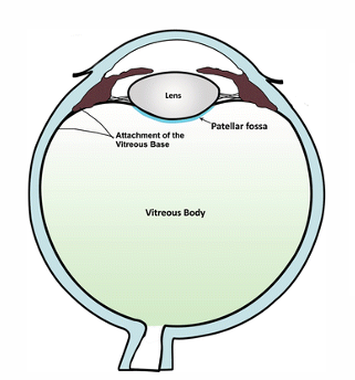
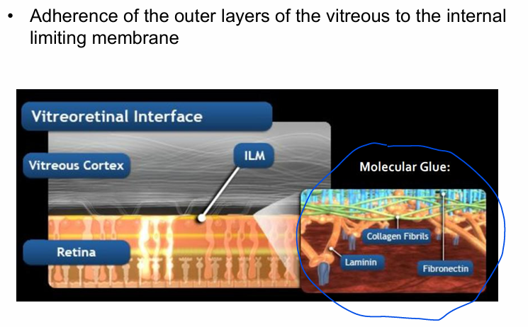
draw what makes up the vitreoretinal interface?
What is the composition of the vitreous?
water (99%)
collagen (mainly type II)
hyaluronic acid (a type of GAG)
glycoproteins
A few types of cells:
hyalocytes
fibrocytes
glial cells
What is the function of hyalocytes in the vitreous cells?
produces hyaluronic acid
synthesis of extracellular matric (ECM)
modulator of immune reaction
modulator of inflammation
What is the function of fibrocytes in the vitreous cells?
primarily located near optic disc & ciliary processes
involved in collagen synthesis, especially in pathology
What is the function of glial cells in the vitreous cells?
unknown purpose, possibly misplaced cell
no neural element in the vitreous
Is the vitreous metabolically active?
yes - for an avascular tissue w few cells
In terms of vitreous metabolism, where is the different oxygen tensions?
lowest oxygen tension in the center
greatest in the posterior cortex (which is where most cells are & closest to the blood supply)
How are vitreous metabolism & retinal metabolic waste related?
Vitreous metabolism serves as a temporary repository for retinal metabolic waste
Can vitreous regenerate?
no, but it can fill the space with fluid
What drops relieve dry eye & discomfort?
0.2 to 0.4% HA eye drops and as slow release contact lenses
What is a vitrectomy?
the removal of VH (if there’s an oxygenation problem to the eyes, VH buffers the amount of oxygen reaching the skin)
Vitreous aging (birth, 2, 25, 65)?
birth: 0% liquid & 100% gel
age 2: 5% liquid & 95% gel
age 25: 20% liquid & 80% gel
age 65: 60% liquid & 40% gel
What is the pathology associated with age & VH?
liquefaction causes traction & 65% of individuals have posterior vitreous detachment
What the vitreous attachments?
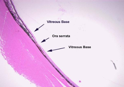
vitreous base (spanning the ora serrata)
hyaloid capsular aka retrolental ligament of Weigert (posterior lens in the midperiphery)
area of martigiani (around optic nerve)
at the fovea
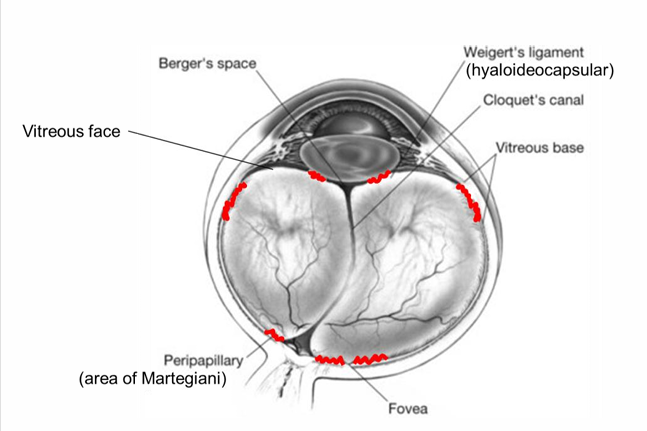
What is the strongest attachment of vitreous to the eye wall?
Vitreous base - very adherent
Where does the vitreous base span? What is the consequence of this?
spans from 2mm anterior to ora serrata & 2-4mm behind ora serrata
peripheral retina is susceptible to detachment from continued traction from the vitreous
Where is the area of martegiani?
end of hyaloid canal in front of optic disc (very adherent)
What is a symptom of posterior vitreous detachment?
occurs when vit pulls away & detaches from retina
collagenous condensation leave Weiss’s ring & often floaters