Chapter 24- Thoracic Wall
 Call Kai
 Learn
Learn Practice Test
 Spaced Repetition
 Match
Match Flashcards
 Knowt Play
1/128

There's no tags or description
Looks like no tags are added yet.
Name | Mastery | Learn | Test | Matching | Spaced | Call with Kai |
|---|
No analytics yet
Send a link to your students to track their progress
129 Terms
what is the function of the thoracic wall?
to form a protective cavity for the:
1. heart
2. lungs
3. for the organs of the superior aspect of the abdomen
what are the bones of the thoracic wall?
1. sternum
2. ribs
sternum is also known as
breast bone
what is the sternum?
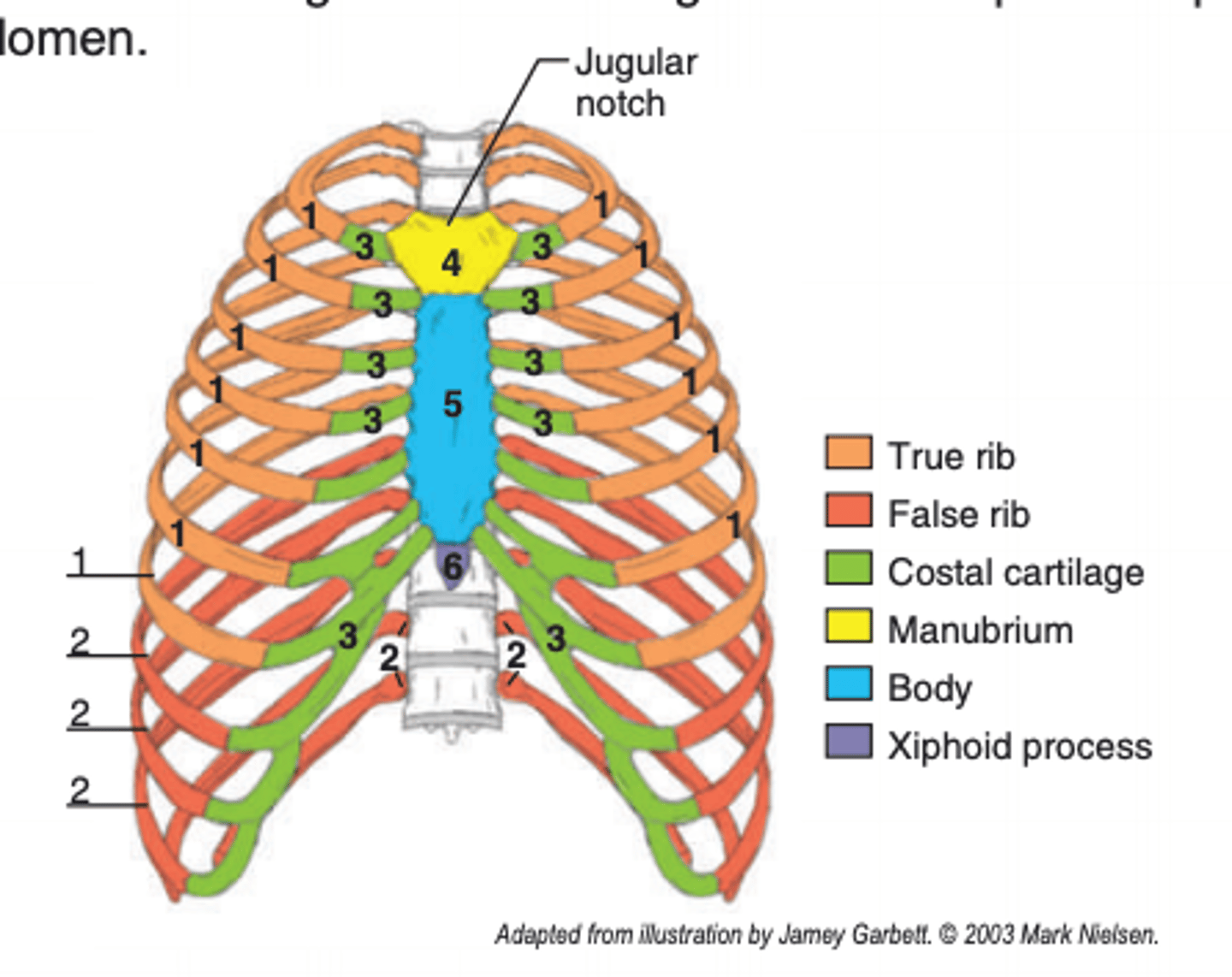
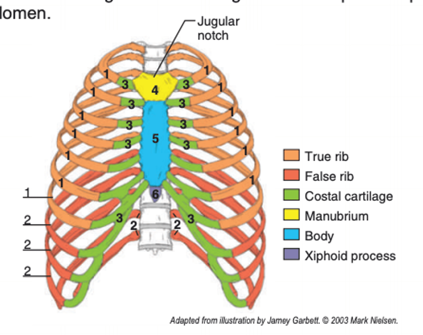
a flat bone (4, 5, 6)
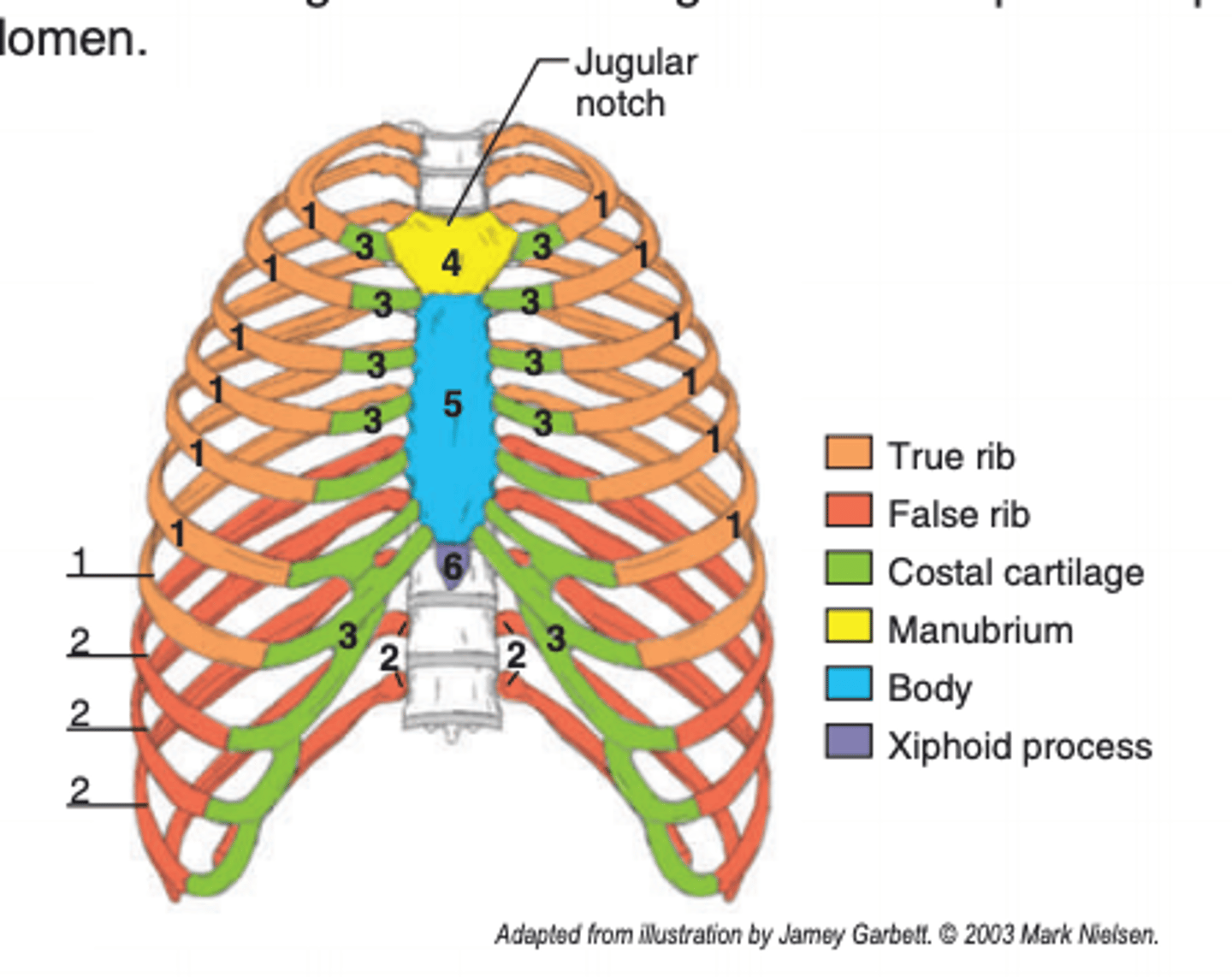
where is the sternum located?
anterior aspect of the thorax in the midline
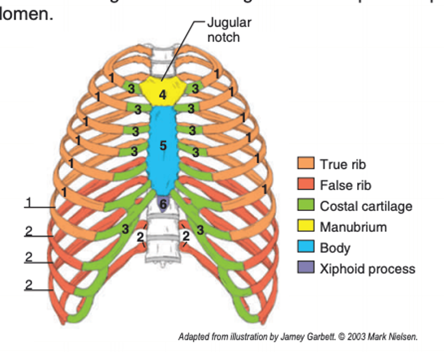
from Greek, sternon means what?
breast of man
how many parts is the sternum divided into? what are they?
3 parts:
1. manubrium (4)
2. body (5)
3. xiphoid process (6)
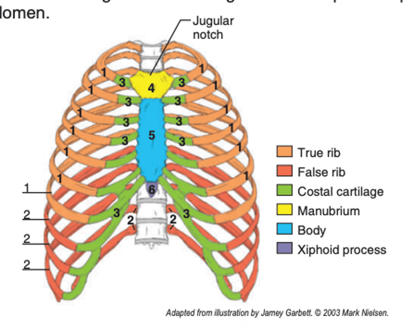
what is the manubrium?
the superior portion of the sternum (4)
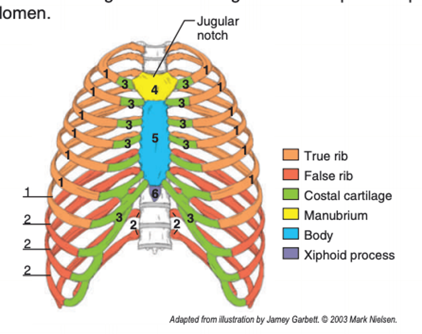
from Latin, manus means what?
hand
what is the sternum fancied to resemble?
a sword, with the manubrium being the part of the sword held in the hand
what is the jugular notch?
found on the superior border of the manubrium
what does the manubrium articulate with?
the clavicle and first 2 pairs of costal cartilage (hyaline cartilage between ribs and sternum)
what is the body?
middle portion of the sternum (5); long, narrow, and thinner than the manubrium
what does the body articulate with?
the costal cartilages of ribs 2-7
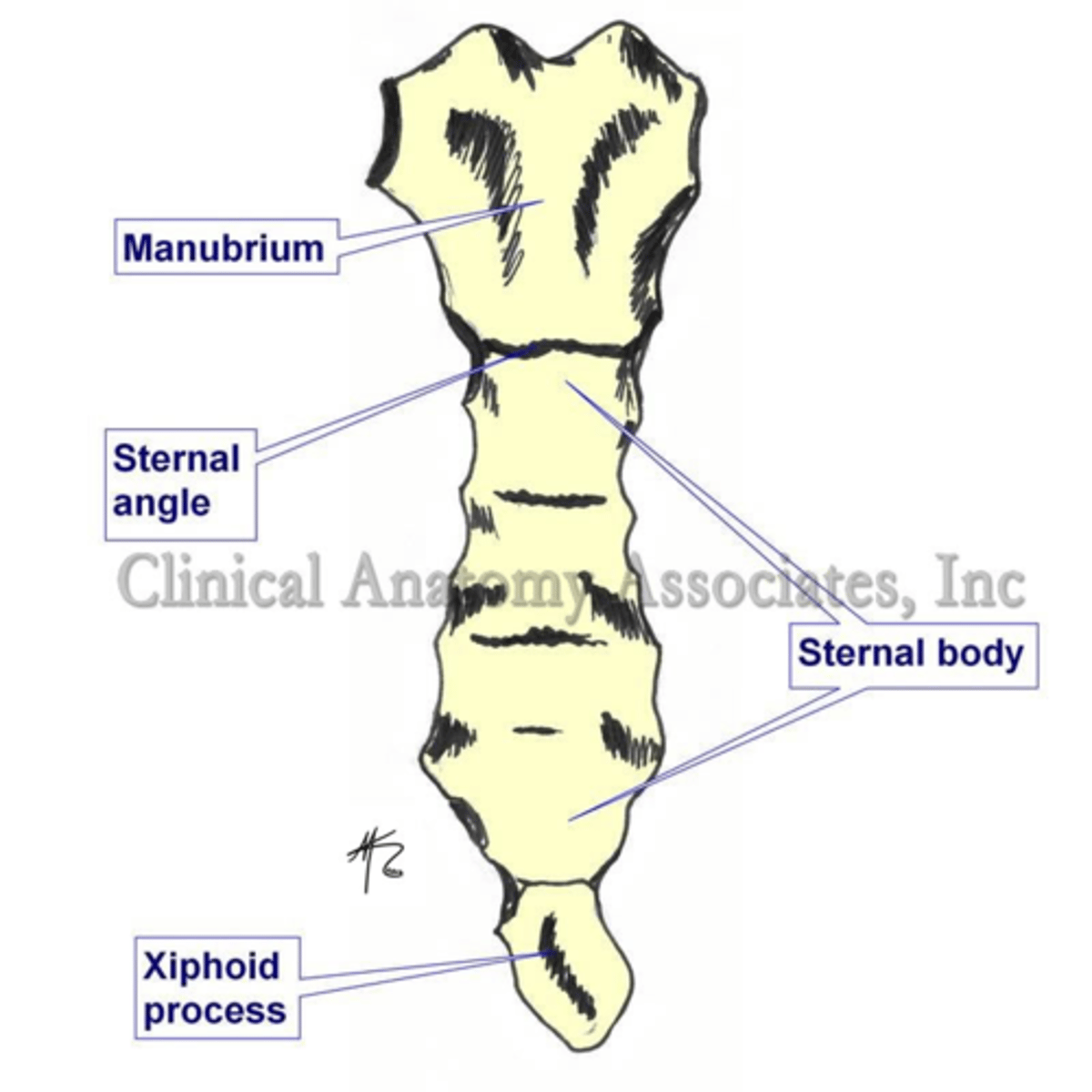
what is the sternal angle?
the junction of the manubrium and body
why is the sternal angle important?
it is a palpable landmark for counting ribs
what is the xiphoid process?
SMALLEST, most variable, and inferior portion of the sternum (6)
from Greek, xiphon and oeides means what?
sword shaped
what is the epigastric fossa?
(pit of the stomach) depression on the ANTERIOR aspect of the xiphoid process; it is palpable and often visible
what is the xiphisternal joint?
found between the xiphoid process and the body at the superior aspect of the epigastric fossa
from Latin, costa means what?
rib
what are ribs (aka ____)?
-costae
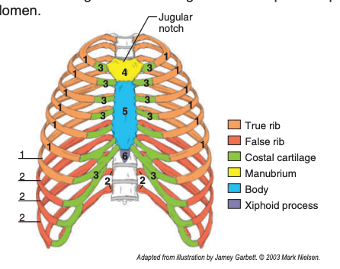
-12 elongated pairs of flat bones that FORM MOST OF THE THORACIC WALL (1, 2)
how many ribs do humans have?
12 ribs on each side of the body (variation include cervical ribs at CV7 or lumbar ribs at LV1)
the elongated flattened bones course where?
anteriorly and inferiorly from the thoracic vertebrae
what does the head portion of the rib articulate with?
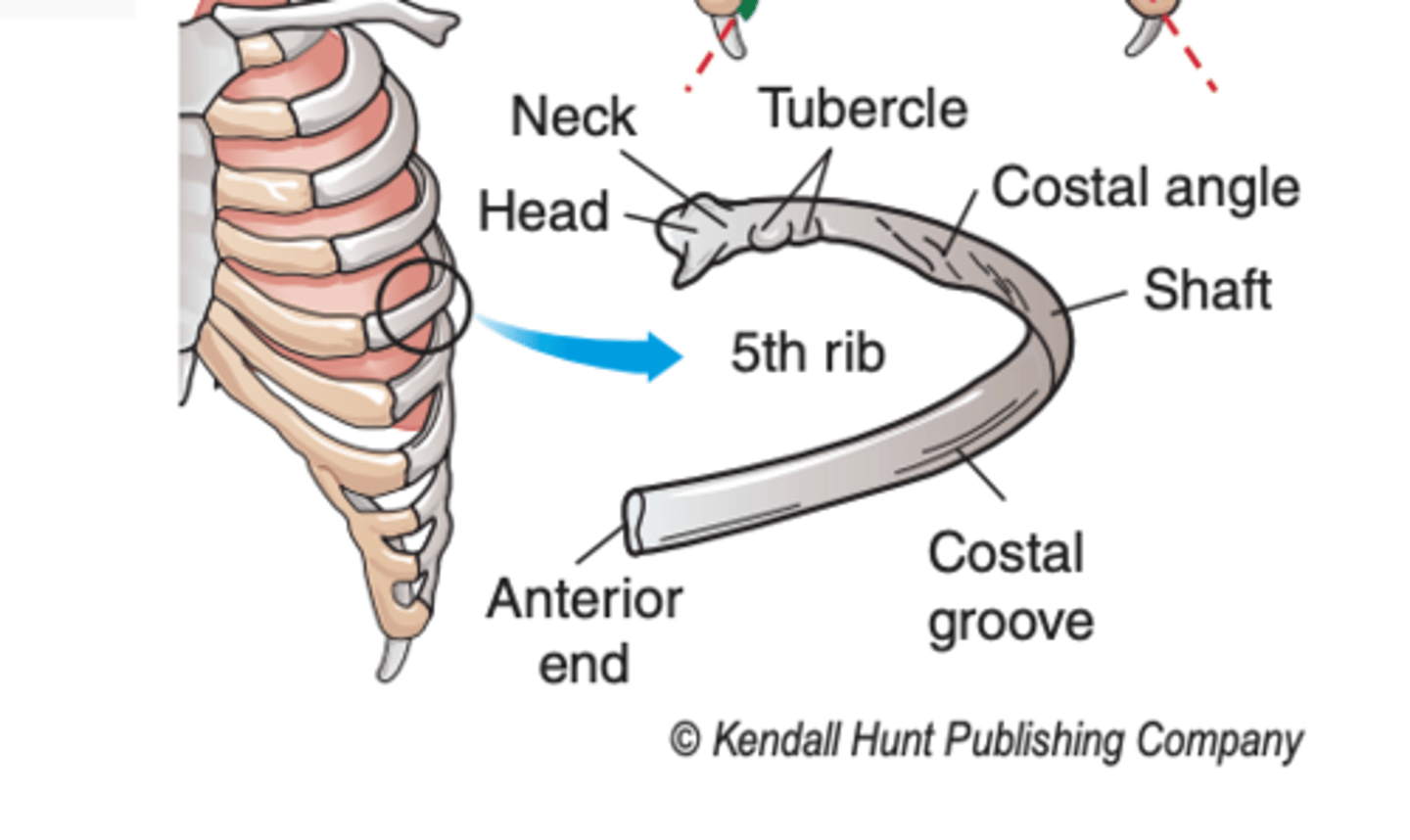
the bodies of the thoracic vertebrae
what is the neck portion of the rib?
constricted portion lateral to the head
where is the tubercle of the rib located?
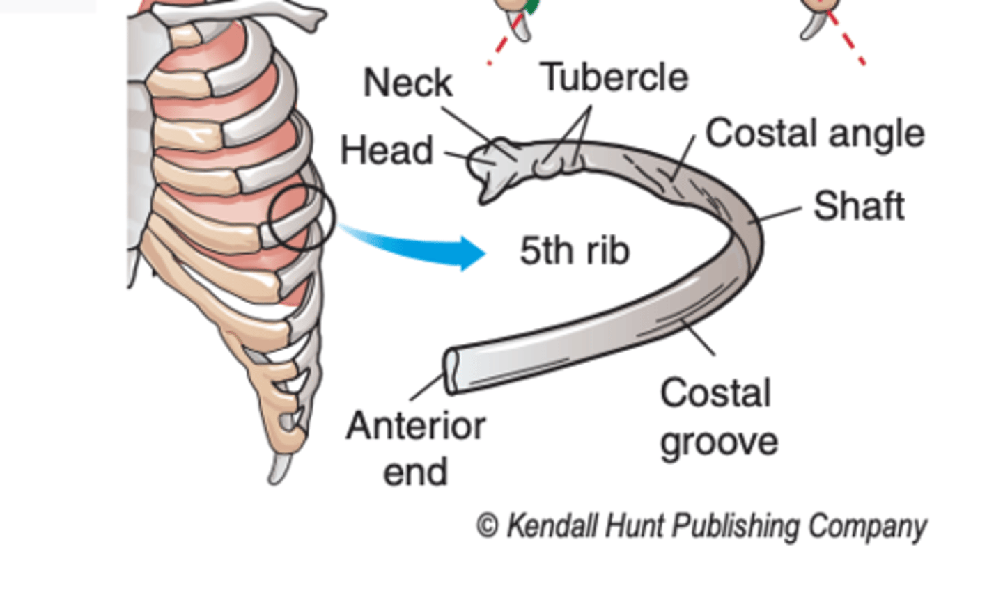
posterior projection lateral to the neck
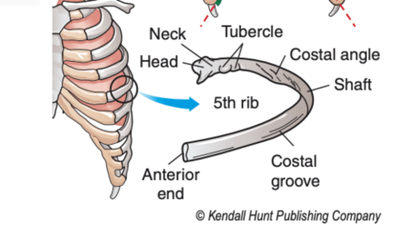
what does the tubercle articulate with?
the transverse process of its respective thoracic vertebrae
what is the body of the rib?
the shaft of the rib
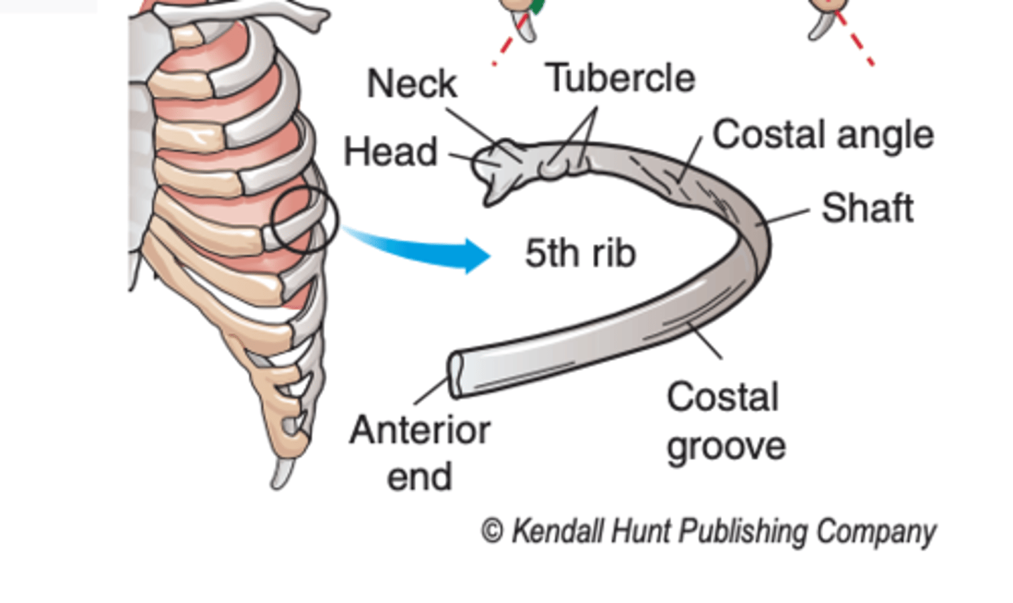
what is the angle of the rib?
the point where the curve (or angle) of the rib is the sharpest
what does the anterior end of the rib articulate with?
costal cartilage
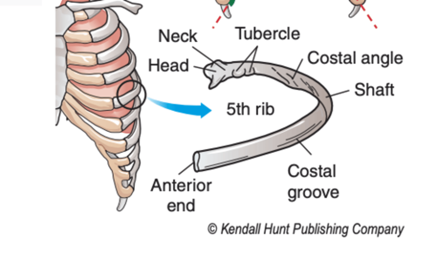
what is the costal groove?
the ridge that lies on the inferior surface of the shaft of the rib
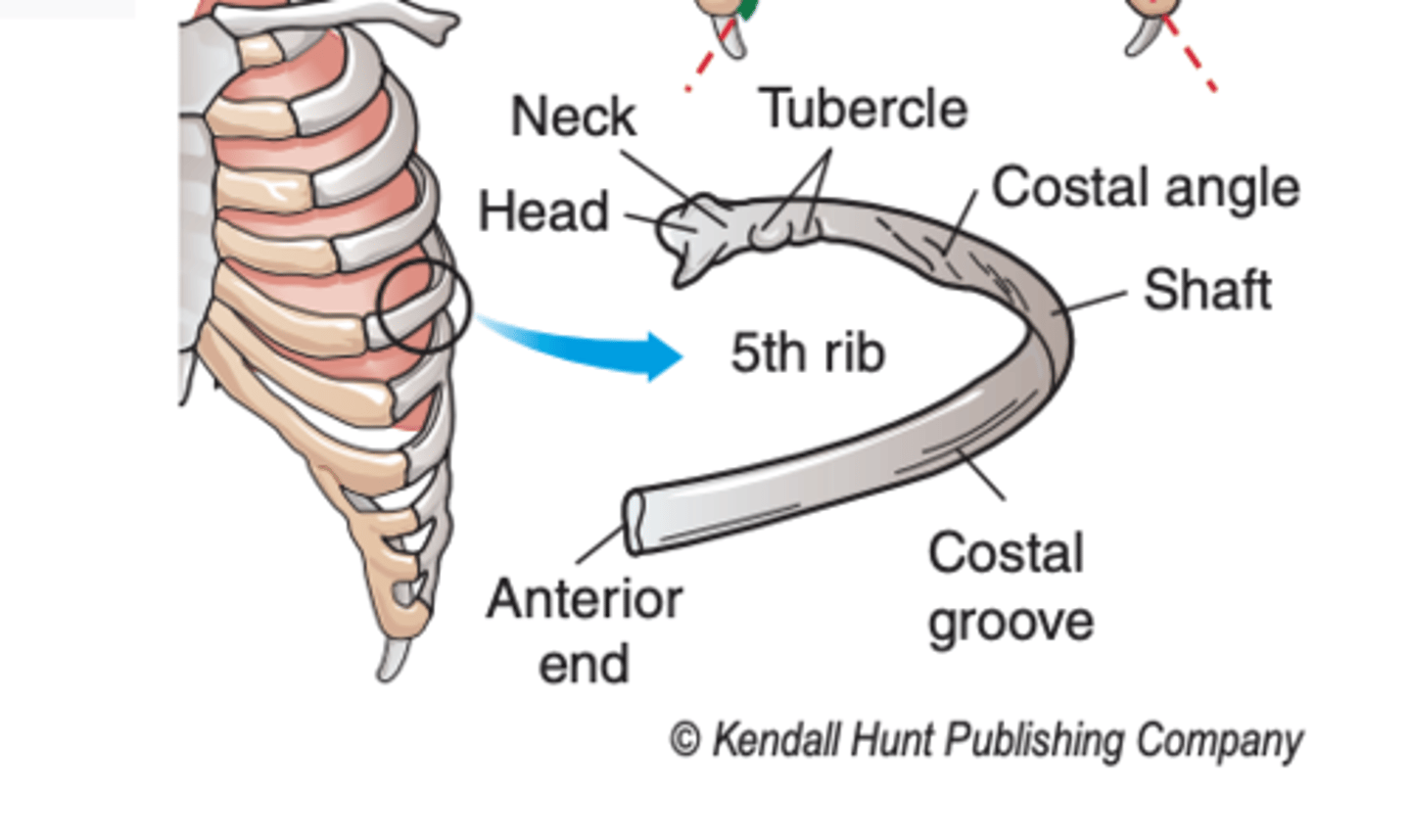
what lies in the costal groove?
from superior to inferior:
the intercostal vein, artery, and nerve
where does the length of the rib increase? where does it decrease?
1st to 7th rib; to the 12th rib
what are the first seven ribs called?
true ribs (1)
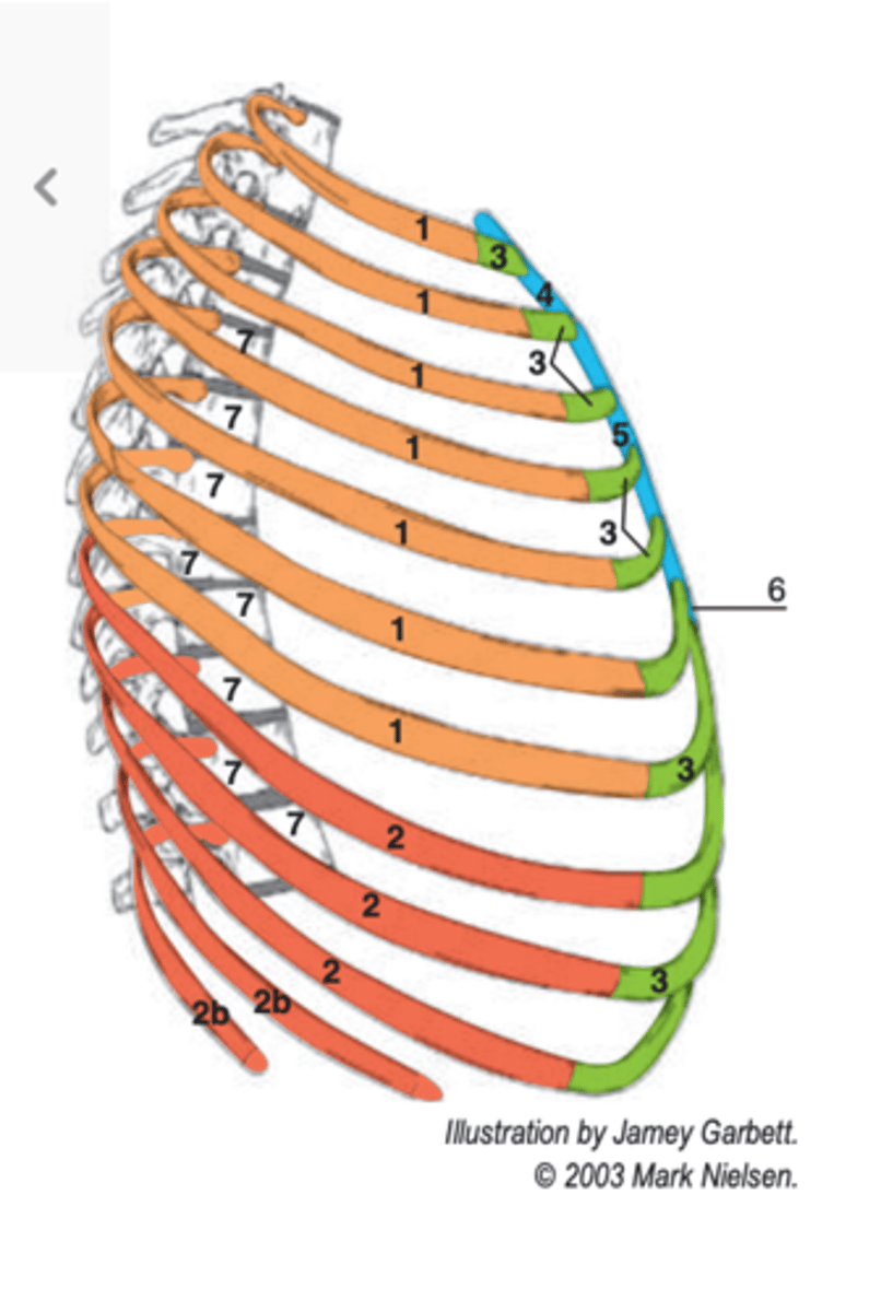
where are the true ribs directly connected?
to the sternum via their costal cartilages (3)
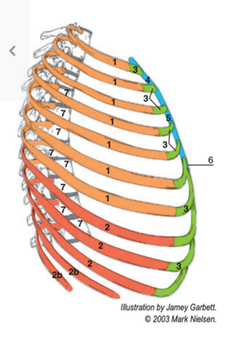
what are the last five ribs called?
false ribs (2)
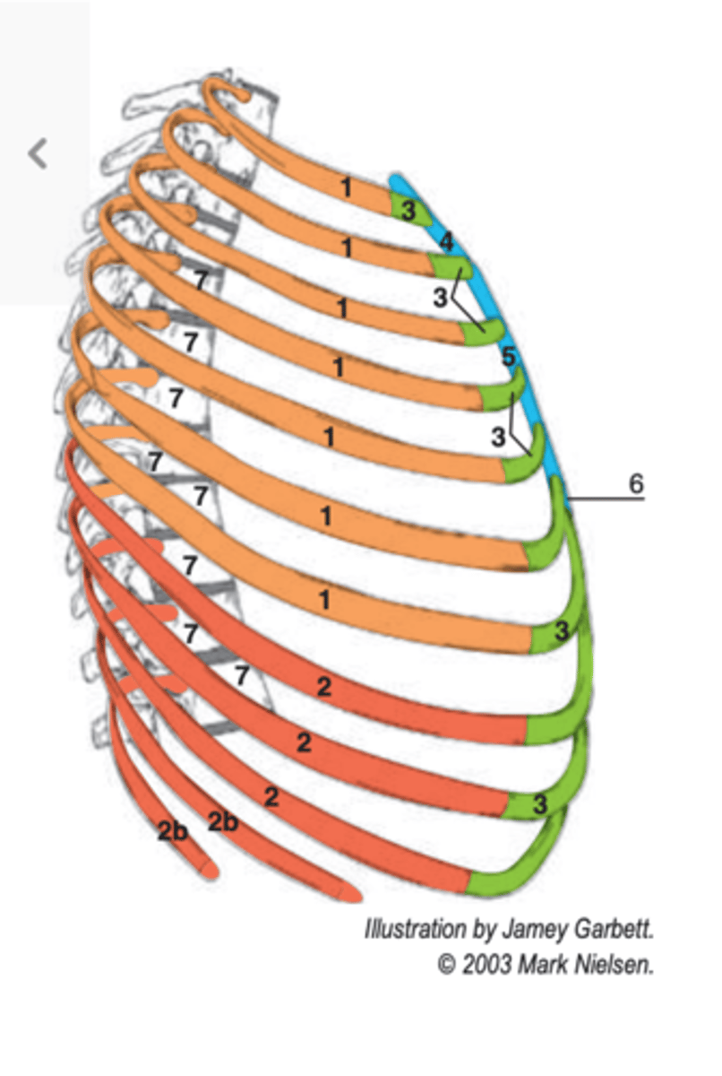
each costal cartilage of ribs 8, 9, and 10 joins what?
its respective suprajacent costal cartilage (8 to 7, 9 to 8, 10 to 9)
where do the costal cartilages of ribs 11 and 12 end? what are these ribs (11 and 12) also classified as?
blindly in muscle with no anterior articulation; floating ribs
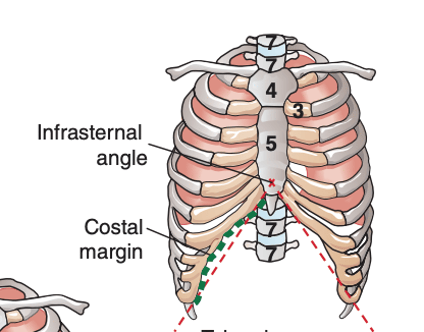
what is the costal margin formed by?
costal cartilages of ribs 7, 8, 9, and 10, which course medially and superiorly
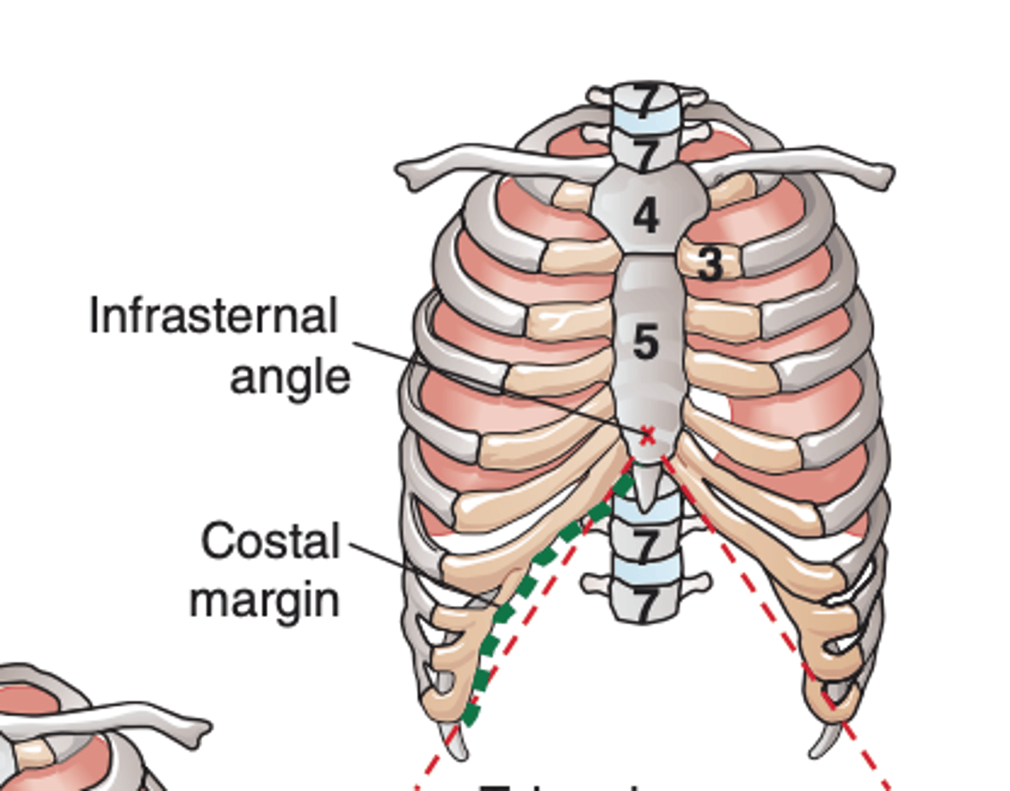
-where do the costal margins intersect?
-what is the intersection of the costal margins termed?
-at the xiphisternal joint
-infrasternal angle
what is the superior thoracic aperture bounded by?
upper border of the sternum and by the first ribs
what is the superior thoracic aperture?
an opening through which the great vessels and visceral tubes ascend or descend between the thorax and the neck
what is the inferior thoracic aperture normally closed by?
the diaphragm
how many openings are found in the diaphragm?
3 major openings
what do these 3 major openings allow for?
the passage of the inferior vena cava, esophagus, and aorta
what are the three layers the muscles of the thorax are arranged in?
1. external layer
2. middle layer
3. internal layer
which muscles are part of the external layer?
external intercostal muscles
which muscles are part of the middle layer?
internal intercostal muscles
which muscles are part of the internal layer?
innermost intercostal musles
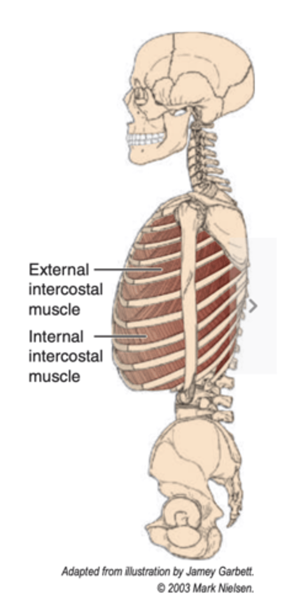
what is the origin of the external intercostal muscles?
lower margin of the rib above
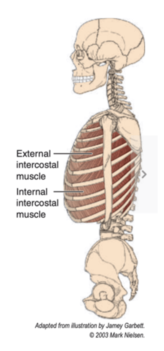
what is the insertion of the external intercostal muscles?
upper margin of the rib below
what is the action of the external intercostal muscles?
elevation of the ribs during inspiration
what is the innervation of the external intercostal muscles?
-intercostal nerves T1 to T6
-thoracoabdominal nerves T7 to T11
how many pairs of external intercostal muscles are there?
11
from Latin inter= and costa=
between; rib
from their point of origin, the fibers of the external intercostal muscles pass where?
medially and inferiorly to their point of insertion
what happens to the external intercostal muscles in the intercostal spaces?
the muscles extend from the tubercle of the rib posteriorly to the region of the costochondral junction anteriorly
what happens at or near the costochondral junction?
the muscles thin out and become membranous where they are then referred to as the external intercostal membrane
what is the origin of the internal intercostal muscles?
upper margin of the rib below and its costal cartilage
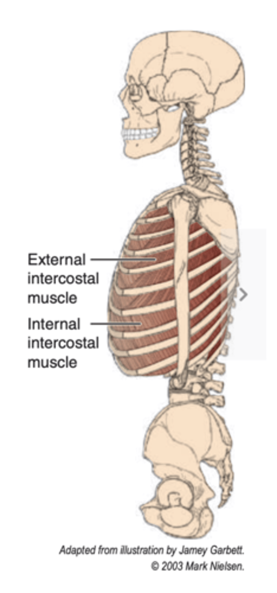
what is the insertion of the internal intercostal muscles?
lower margin of the rib above and its costal cartilage
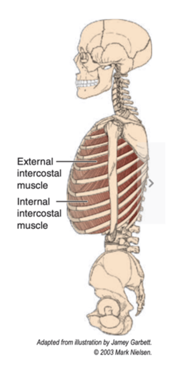
what is the action of the internal intercostal muscles?
depression of the ribs during expiration
what is the innervation of the internal intercostal muscles?
-intercostal nerves T1 to T6
-thoracoabdominal nerves T7 to T11
how many pairs of internal intercostal muscles are there?
11
from internal intercostal muscles point of origin, the fibers of these muscles pass where?
medially and superiorly to their point of insertion
in the intercostal spaces, the muscles extend where?
from the sternum anteriorly to the angles of the ribs posteriorly where they thin out and form the internal intercostal membrane
how many pairs of innermost intercostal muscles are there?
11
innermost intercostal muscles have the same?
origin, insertion, action and innervation as the internal intercostal muscles
review:
origin = upper margin of the rib below and its costal cartilage
insertion = lower margin of the rib above and its costal cartilage
action = depression of the ribs during expiration
innervation = intercostal nerves T1-T6; thoracoabdominal nerves T7-T11
innermost intercostal muscles are separated from what? by what?
-the internal intercostals
-by the intercostal vessels and nerve
what is the area between the internal and innermost intercostal muscles named?
neurovascular plane
what other muscles are also associated with the thoracic wall?
transversus thoracis and subcostales
how many thoracic spinal nerves are there?
12
where do all the thoracic spinal nerves traverse?
where do they divide?
-an intervertebral foramen
-divide into a dorsal ramus and ventral ramus
where do the dorsal rami course?
posteriorly and divide into medial and lateral branches
what are the ventral rami?
larger branches of the spinal nerves that course forward into the intercostal spaces
what are the regional names of the ventral rami?
1. T1-T6: Intercostal nerves (proper)
2. T7-T11: Thoracoabdominal nerves
3. T12: Subcostal nerves
how does a typical intercostal nerve course?
1. it passes below the neck of the rib numerically corresponding to it; EX: T4 courses inferior to the fourth thoracic rib
what is the second step? (where does it now course?)
it courses between the innermost intercostal and the internal intercostal muscles in the neurovascular plane
what is the third step? (where does it pass forward in? where does it course?)
it passes forward in the costal groove on the lower surface of the rib, where it courses inferior to the intercostal vein and artery
what is the fourth step? (what does it give off?)
it gives off a lateral cutaneous branch
what does the lateral cutaneous branch pierce?
the internal intercostal, external intercostal, and serratus anterior muscles
what does the nerve then divide into/supply?
anterior and posterior branches that supply the skin of the chest wall
what is the last step? (what happens at the anterior end of the intercostal space?)
at the anterior end of the intercostal space, it turns forward through the internal intercostal muscle, the external intercostal membrane, and the pectoralis major muscle
what is the intercostal nerve now distributed as?
the anterior cutaneous branch to the skin of the front of the chest
what are breasts?
SWEAT GLANDS modified to produce MILK
what is the major function of the breast (sweat gland)?
lactation --> secrete milk for the nourishment of the child
Latin for breast = ______, thus the name ________
mammae; mammary gland
what is the main constituent of the mammary gland?
the 15-20 lobes of the glandular tissue (alveoli)
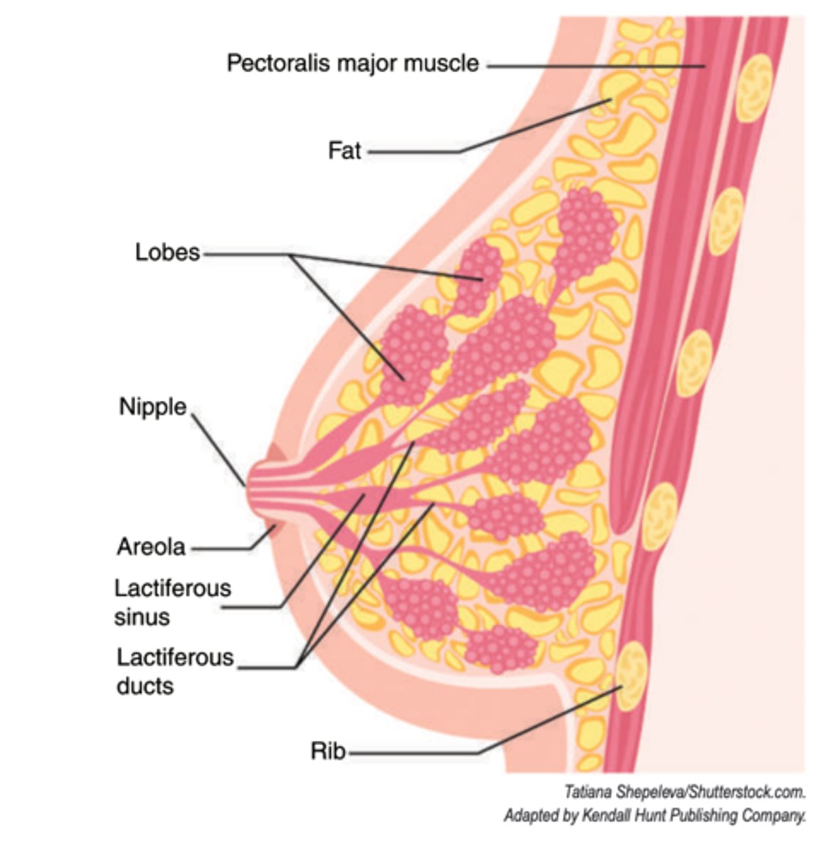
what do the lobes contain?
alveoli or milk-secreting mammary epithelial cells
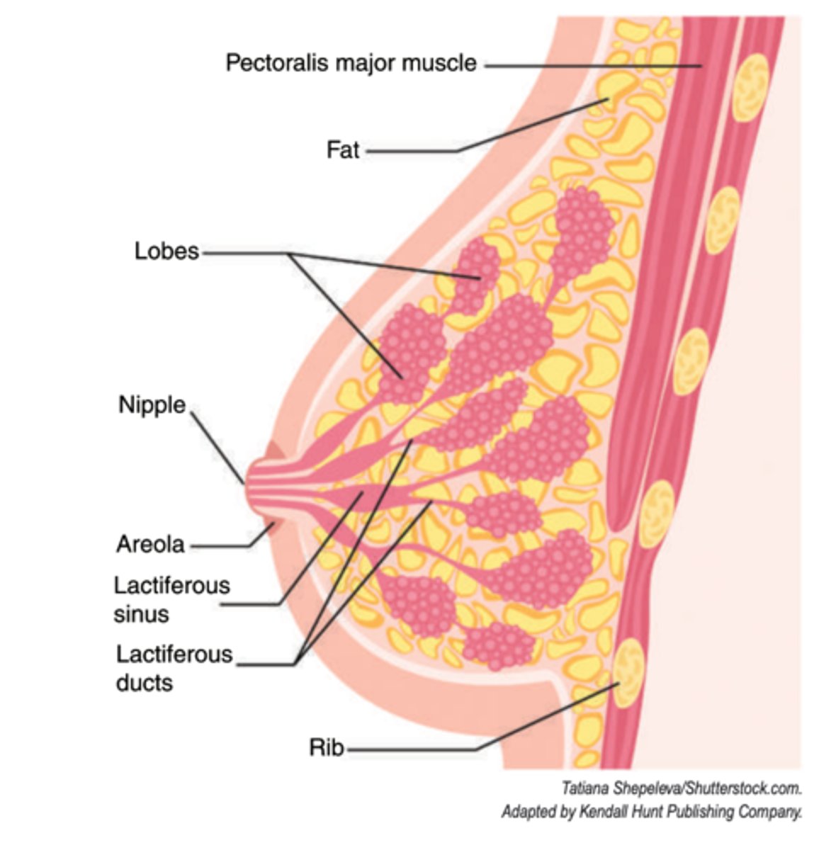
how are the lobes arranged?
grape-like clusters
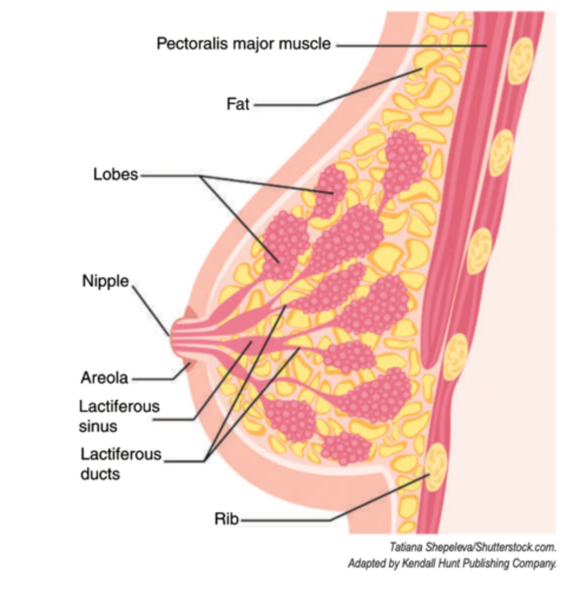
of these lobes, how many assume the entire functional load?
6-8
what happens to the remaining majority?
small and poorly developed
what is surrounding and overlying the glandular tissue?
a large amount of FAT, which makes up the majority of the organ
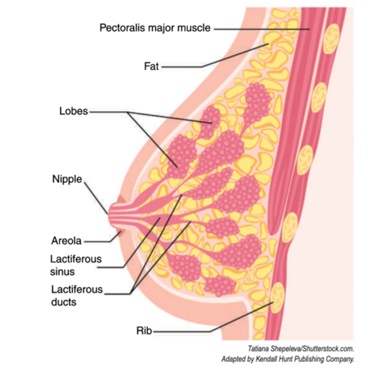
what is each lobe drained by?
a lactiferous duct that dilates near its termination
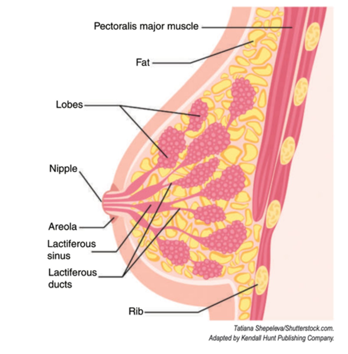
what is the dilation called?
lactiferous sinus
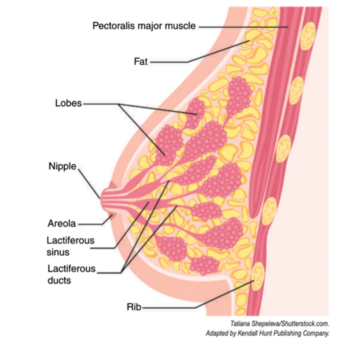
where does the duct empty?
at the nipple
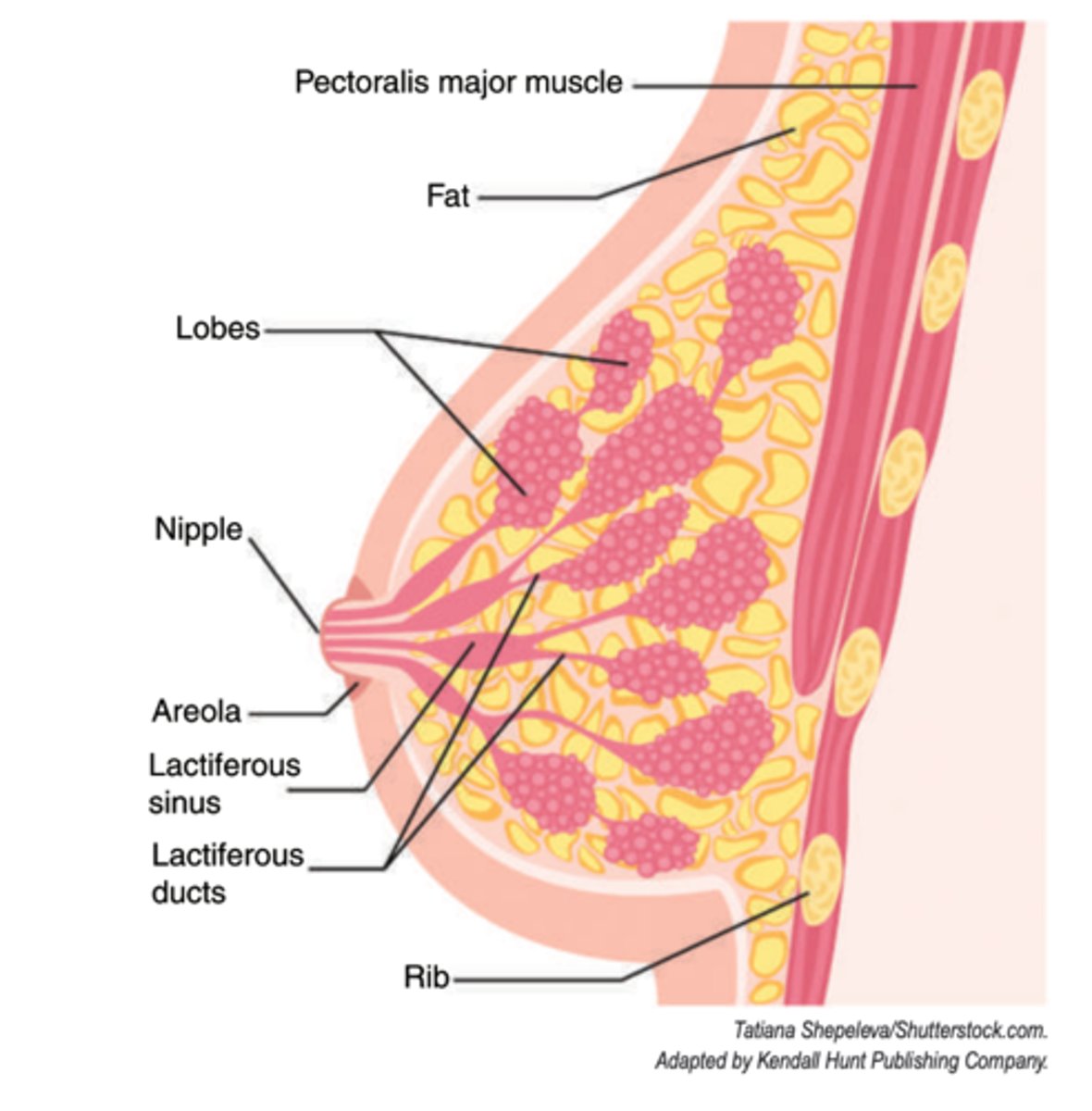
where does the axillary tail extend?
into the axilla
where is breast tissue firmly attached to?
skin over much of the remainder of the organ
what is the breast tissue attached to skin by? where is this especially represented in?
heavy ligamentous bands of connective tissue; in the well-developed "retinacula cutis"
what do the well-developed "retinacula cutis" do?
traverse and subdivide the fat and anchor the glandular tissue to the skin