Transport in Animals
1/33
There's no tags or description
Looks like no tags are added yet.
Name | Mastery | Learn | Test | Matching | Spaced | Call with Kai |
|---|
No analytics yet
Send a link to your students to track their progress
34 Terms
why do multicellular organisms require transport systems?
high metabolic demands
large SA:V ratio
hormones made in one place but needed in another
food digested in one place but needed in another
waste products of metabolism need to be removed from everywhere into excretory organs
what do circulatory systems contain?
transport medium
vessels for transport medium
pumping mechanism to move the transport medium
mass transport system
when substances are tranpsorted in a mass of fluid with a mechanism for moving the fluid around the body
open circulatory systems (insects)
transport medium is haemolymph
haemolymph is not enclosed in vessels
haemolymph is always under low pressure
haemolymph flowing to a tissue cannot be varied
haemolymph returns to heart via an open ended vessel
closed circulatory systems
transport medium is blood
blood is enclosed in vessels
blood supplied to organs can be varied
blood returns to heart
single closed circulatory system (fish)
blood travels once through the heart for each complete circulation of the body
blood passes through two sets of capillaries: in the first, oxygen and carbon dioxide is exchanged, in the second, substances are exchanged between the blood and cells
this causes the blood pressure to drop, limiting the efficiency of exchange processes
however, in fish, their countercurrent exchange mechanism, their weight being supported by water, and no need for temperature regulation reduces the metabolic demands allowing them to be active even with a single closed circulatory system
double closed circulatory system
two types of circulation: pulmonary and systemic
pulmonary circulation: blood being pumped from the heart to the lungs to pick up oxygen and unload carbon dioxide and then returning back to the heart
systemic circulation: blood being pumped from the heart to the body to provide oxygen to cells and then returning back to the heart
what do elastic fibres do?
can stretch and recoil to provide vessel walls with flexibility
what does smooth muscle do?
contracts or relaxes, changing the size of lumen
what does collagen do?
provides structural support to the vessel
arteries
elastic fibres
smooth muscle
collagen
smooth endothelium for easy blood flow
arterioles
more smooth muscle than arteries
less elastin than arteries as less pulse surge
same amount of collagen
capillaries
microscopic
lumen only 10µm wide
made of endothelial cells
adaptations of capillaries
large surface area for the diffusion of substances into and out of the blood
slow movement of blood in capillaries gives more time for exchange by diffusion
single endothelial cell thick therefore thin diffusion distance
veins
valves to prevent backflow of blood
lots of collagen
less elastic fibre
wide lumen
smooth endothelium for easy blood flow
what are the adaptations for blood in veins to move against gravity under low pressure?
valves to prevent backflow
muscle contractions in arms and legs force blood back to the heart
chest movements act as a pump to send blood back to the heart
positive cooperativity
erythrocytes enter capillaries in lungs with relatively low oxygen level, making a steep concentration gradient between the inside of the erythrocytes and the air in the alveoli
one oxygen molecule bind to a haem group of the Hb molecule, and the Hb molecule changes shape, making it easier for the next oxygen molecules to bind
the oxygen is bound to the Hb, with free oxygen concentration staying low, therefore a steep diffusion gradient is maintained until all the Hb is saturated with oxygen
adaptations of erythrocytes
biconcave shape for large surface area
no nucleus for more space for oxygen
bohr effect
in active tissues with higher partial pressures of carbon dioxide, haemoglobin gives up oxygen more readily
what is the affect of the bohr effect in the lungs?
less carbon dioxide in the lungs, so Hb will not give up oxygen easily, instead it will bind easily
describe the relationship between fetal and adult haemoglobin (hint: fainting during pregnancy)
fetal Hb has a higher affinity for oxygen than adult Hb, so when the mother’s oxygenated blood flows past the deoxygenated fetal blood, the fetal Hb removes the oxygen from the maternal blood
where is carbon dioxide transported to?
5% in plasma
10-20% converted into carbaminohaemoglobin
75-80% converted into hydrogen carbonate ions
journey of deoxygenated blood into heart and lungs to oxygenated blood to body
inferior vena cava (from lower body) + superior vena cava (from upper body) → right atrium → tricuspid valve → right ventricle → semi lunar valves → pulmonary artery → lungs → pulmonary veins → left atrium → bicuspid valve → left ventricle → semi lunar valves → aorta → body
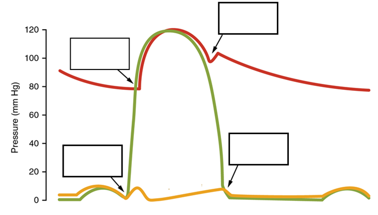
what valves open/close at A, B, C, and D? (A - bottom left, B - top left, C - top right, D - bottom right)
A - AV valves close, B - SL valves open, C - SL valves close, D - AV valves open

what is diastole?
relaxation
what is systole?
contraction
myogenic
initiates its own rhythmic contractions without the need of electrical signals from the brain
journey of a heartbeat
wave of excitation from the SAN, causing atrial systole, initiating heartbeat - (non-conducting tissue prevents excitation passing directly to ventricles)
AVN picks up this electrical activity and stimulates the Bundle of His (conducting tissue made of Purkyne fibres) after a short delay
this tissue conducts a wave of excitation to the apex of the heart, spreading out to the ventricles, therefore initiating ventricle systole
why does the AVN impose a slight delay?
to ensure the atrial systole has finished before ventricular systole begins
tachycardia
rapid heartbeat
bradycardia
slow heartbeat
ectopic heartbeat
extra heartbeat
atrial fibrillation
arrhythmia - fast fibrillations
describe an ECG trace with reference to P, Q, R, S and T
at P, SAN sends a wave of excitation causing atrial systole (atrial depolarisation)
in the short PR interval, AVN stimulates the Bundle of His and Purkyne fibres
in QRS, the Purkyne fibres send a wave of excitation to across the ventricles and down to the apex (ventricular depolarisation)
at T, ventricular systole occurs (ventricular repolarisation)