PQs - short answers & MCQs | Quizlet
1/59
There's no tags or description
Looks like no tags are added yet.
Name | Mastery | Learn | Test | Matching | Spaced | Call with Kai |
|---|
No analytics yet
Send a link to your students to track their progress
60 Terms
What is faraday cage and what is is used for?
A. Lead plates in the wall of x-rays
B. a grid of conductors in the walls isolating the room from radio waves
C. lead plates in the walls isolating the room for radio waves
D. conductor grids in the walls to absorb x-rays
B. a grid of conductors in the walls isolating the room from radio waves
Why can't ketamine be used in patients with suspected cranial trauma?
- Can ↑ intracranial pressure (ICP)
- Causes ↑ cerebral blood flow + brain oxygen demand
- May worsen brain edema/neurological damage in cranial trauma
In computed tomography, the amount of radiation dose received by the patients is dependent on
a. Test time
b. set acquisition parameters (anode voltage, KV nr)
c. the degree of advancement of the computed tomography model
d. all answers are correct
d. all answers are correct
In what species is the ovum pickup laparoscopically
- Sheep 🐑
- Due to small size of ovaries/reproductive tract
Indication of cystotomy?
- Opening of urinary bladder
Indications:
- Stones (uroliths)
- Tumours
- Rupture
- Obstruction
- Foreign body removal
What structures are hypo/hyper-echogenic in USG?
- Hypo: kidney → liver → spleen (dark to light)
- Hyper: bone
- Anechoic: fluid
What is a radiopharmaceutical?
- Biologically active compound containing a radionuclide
- Used for diagnostic imaging or therapy
- Consists of → radionuclide, vector, connector
In the magnetic field of magnetic resonance:
- Hydrogen protons align in an orderly manner with the magnetic field (B₀)
- Creates net magnetization used to generate MRI signals
In CT
Number of layers + rows are independent of each other
In computed tomography, it is possible to work the following modes
- Axial
- Helical/spiral
- Contrast or non-contrast
Phlebography is:
- Imaging of veins using contrast medium
- Radiographic procedure assessing venous structure + blood flow
- Used to detect thrombosis, obstruction or vascular abnormalities
Sort the tissues visible on a classic CT image in grayscale: blackest → whitest:
Lungs → fat → soft tissue/blood → bones
Radiation per one CT exam, depending on the available apparatus + the scanner's area, partial or whole body examination, should be in the range:
From 0.9 to 15 mSv (milli sieverts)
Functional MRI
Perfusion
When + why can metal elements inside the patient's body be an obstacle in performing a diagnostic examination using a CT scanner?
- Image artefacts → misinterpretation, distortion, loss of detail
- Signal interference
True or false:
it’s forbidden to take metal objects into room where CT scanner is located
False
True or false:
Arthroscopic procedures in horses are usually performed on a standing horse (under local anaesthesia)
False
Notes:
- General anesthesia
True or false:
Linear probe is best for tendons
True
True or false:
Typical procedures preformed with flexible endoscope
True
True or false:
The name of radiopharmaceutical is the same as the radioisotope
False
Notes:
- Radiopharmaceutical = radioisotope + carrier/vector molecule
True or false:
The PET examination uses the fact that certain lesions are accompanied by a change in the metabolism of cortina chemical compounds, e.g. sugars, because energy in the body is obtained mainly by burning them
True
True or false:
The most common indication for use of magnetic resonance imaging is the diagnosis of a wide range of diseases of the NS, inflammation + soft tissue injuries
True
True or false:
MRI is a painless + non-invasive imaging technique, the basis of which is the removal of the magnetic spin vector of a single proton from its equilibrium position as a result of the action of a fast, alternating magnetic field
False
Notes:
- The magnetic field is constant
- Radio waves are alternating
True or false:
During the OPU procedure, the USG apparatus is inserted transvaginally, + then follicular fluid with the oocyte is collected through ovarian puncture
True
Notes
- OPU → ovum pick-up
True or false:
Contrast agents used in magnetic resonance imaging are paramagnetic agents whose basic opponent is gadolinium
True
True or false:
The operation of the USG scanner is based on the analysis of the echo created as a result of the reflection of the USG beam from the structures along their course
True
True or false:
The most common cause of USG artefact is gas in the intestines or lungs, which is a barrier to the USG wave, + close contact of the gel-coated transducers with the skin of the body being examined
False
Notes:
- Gas causes artefacts
- Gel contact ↓ artefacts by removing air between probe + skin
What cyclotron isotope is used in PET?
- Fluorine-18 (18F)
- Produced in a cyclotron from Oxygen-18
- Most commonly used in PET imaging (e.g. 18F-FDG)
What generator isotope is I used in PET?
- Ga-68
- Produced from Ge-68
- Used in PET imaging without requiring a cyclotron
Rank the following diagnostic methods according to the harmfulness of performing the examination (not incl anesthetic reasons, from the safest to the most harmful)
- USG
- MRI
- CT
- PET/CT
Rank the following diagnostic methods in order of increasing examination time (not incl patient prep time)
- X-ray
- CT
- USG
- MRI
- PET/MRI
Routine patient preparation for PET/CT examination with the use of 18FDG does not include:
limit excerise
Physiological accumulation of 18FDG occurs in the following organs
ALL
What is the power range (in tesla) of permanent magnet MRI machines
0.5-3 T
What are the properties of gadolinium contrast
Shortens T1 relaxation times
What tests allow to assess the condition of BVs
- MRI
- Angiography
- USG Doppler
- CT angiography (contrast CT)
Sedatives
A2 agonist: dexmedetomidine, detomidine
opioids: fentanyl, butorphanol
Benzodiazepines: diazepam, midazolam
Post-contrast enhancement in the area of inflammation in 🐱 due to viral infection of the brain should be in the T1 sequence
- Hyperintense enhancement on T1-weighted images
- Due to gadolinium crossing disrupted BBB in inflamed tissue
Hydrated structure such as edema in MRI image will be:
Strongly hyperintense in T2 dependent sequence
In CT image:
Intervertebral discs are invisible
What part of the endoscope can be sterilised
- All parts can be sterilised
- Only by different methods
What is 18FDG, what is it used for?
- Radioactive glucose analogue used in PET
- Taken up by metabolically active cells
- Tumors show ↑ uptake ("hot spots")
- Used for cancer detection + monitoring
What is the most common radionuclides in SPECT imaging
99mTc
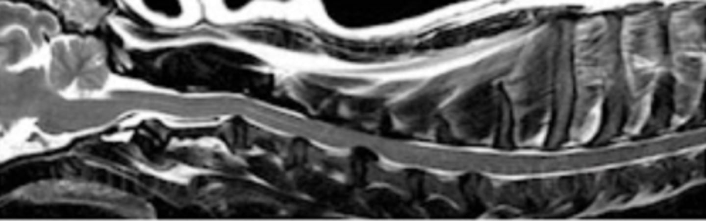
What plane is shown on the image:
sagital cross section

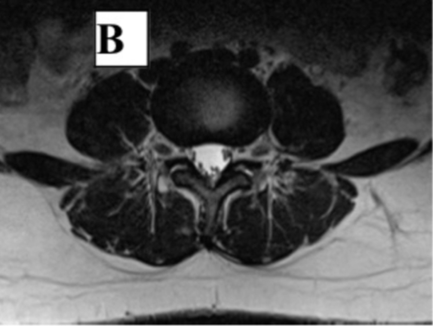
What plane is shown on the image:
axial cross section
The MRI image shows:
Extrusion at the level of the cervical C4/5 in T2-dependent sequence
Name the planes on an MRI scan of a brain:
- Sagittal
- Coronal (dorsal normally)
- Axial (transverse)
Parts of the cerebrum
- 2 cerebral hemispheres
- Outer grey matter → cortex
- Inner white matter
- 4 lobes: frontal, parietal, temporal, occipital
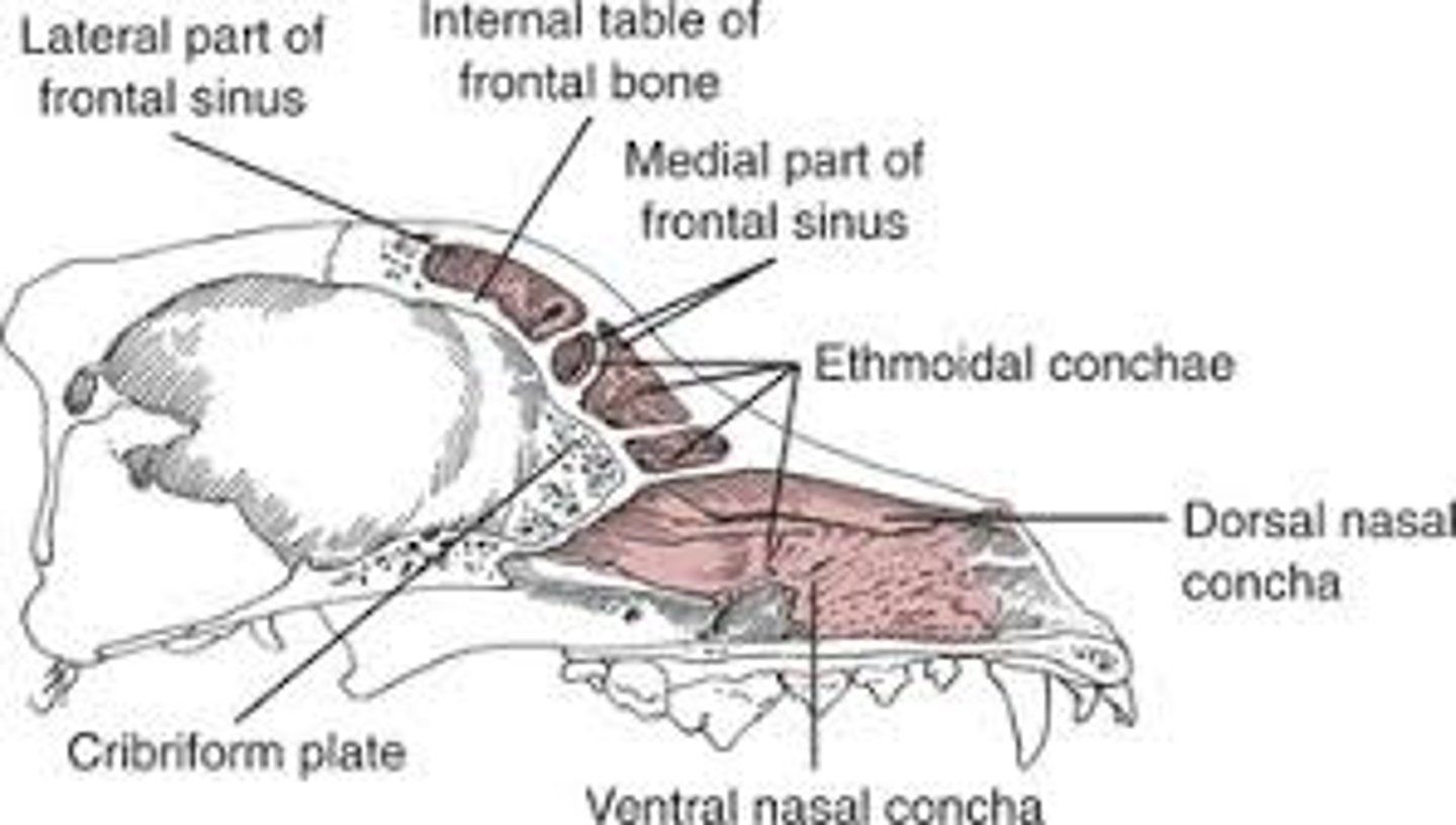
Paranasal sinuses in dog
- Frontal sinus
- Maxillary sinus
- Ethmoidal sinus
- Sphenoidal sinus
Low + high frequency probes, how do they penetrate the body?
Low frequency probes:
- ↑ penetration
- ↓ resolution
- Used for deep structures
High frequency probes:
- ↓ penetration
- ↑ resolution
- Used for superficial structures/tendons
Which probe is used for tendon exam, pregnancy confirmations in large animals, and other reasons?
Linear probe
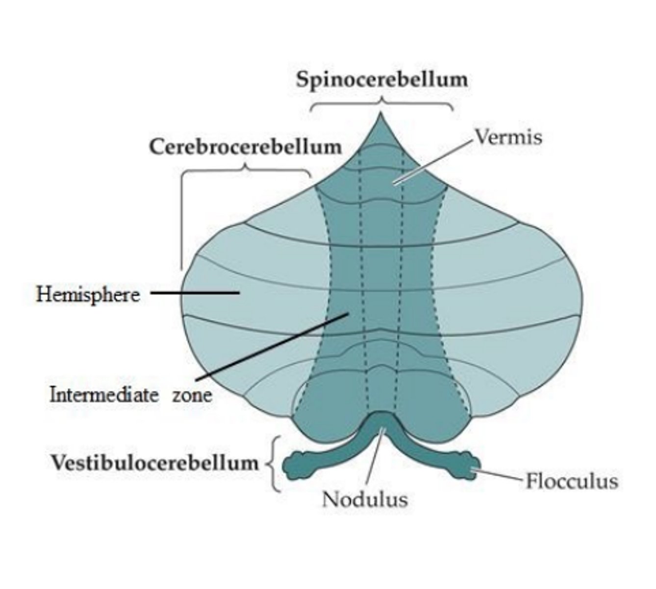
Parts of the cerebellum
1. Cerebrocerebellum
2. Spinocerebellum
3. Vestibulocerebellum
What is radio tomograph
Technology which locates objects using radiofrequency signals in a wireless network
What are the different types of endoscopy
Rigid + flexible
What is a gantry
- Circular frame of a CT scanner
- Houses X-ray tube + detectors
- Rotates around the patient during scanning
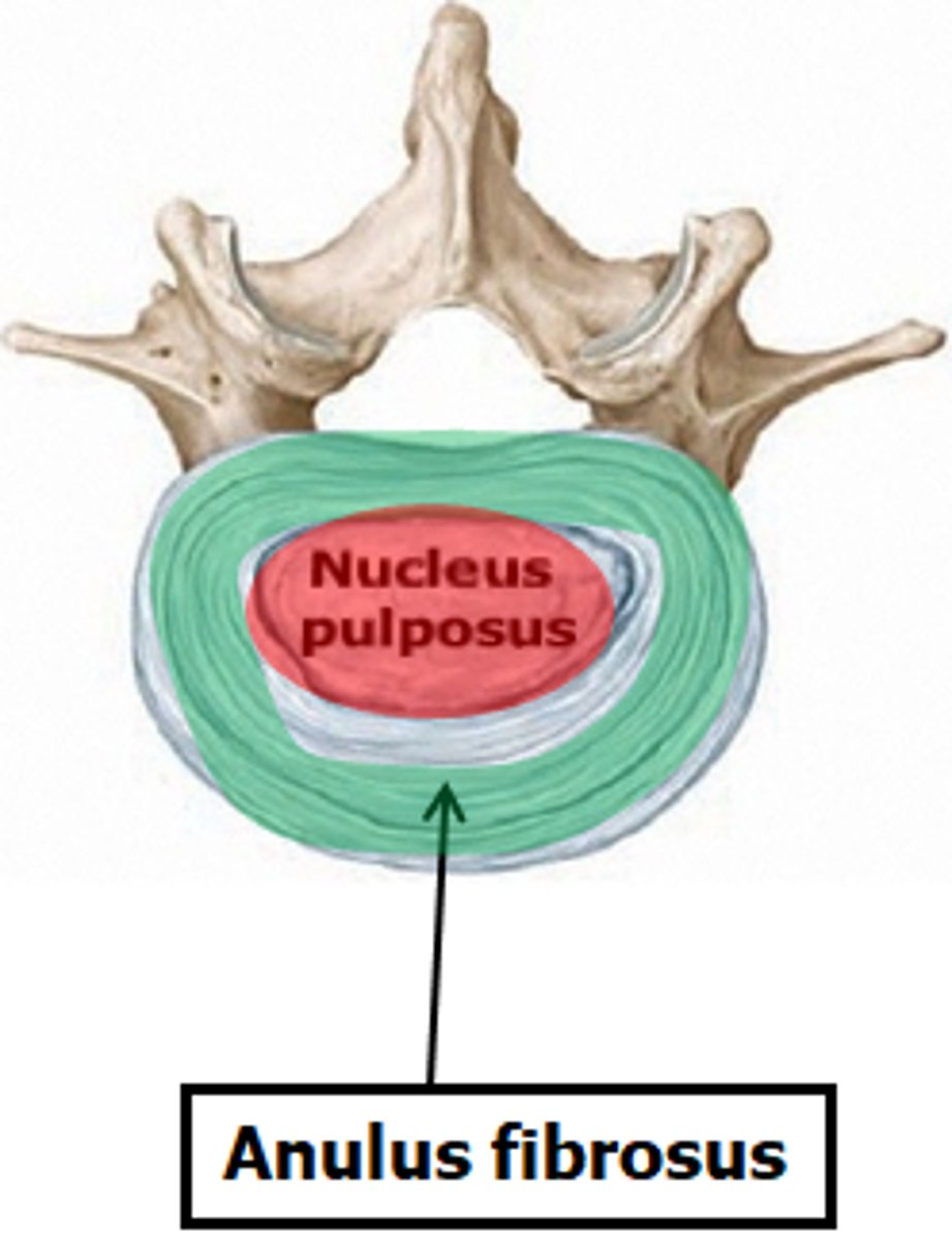
Anatomy of intervertebral disc
- Outer anulus fibrosus → fibrocartilage layers
- Inner nucleus pulposus → gel-like center
- Functions as shock absorber between vertebrae
Fluid in FLAIR
- CSF/free fluid → dark
- Pathological fluid (edema/inflammation) → bright
What is a Faraday cage + what is it for?
- Conductive enclosure around MRI room
- Blocks internal/external electromagnetic interference
- Prevents radiofrequency signal disturbance during MRI scanning
Which structures/organs cannot be visualised using USG
- Brain
- Lungs
- Bones
- Solid organs deep in abdomen
Choose which of the features of the description of mass (about 2 cm in dm) found in the brain of a Labrador matches the description of glioma (malignant, metastatic):
a. Shape; irregular. margins; irregular + noncircumsribed. signal intensity; hypointensive + heterogeneous in the T1 sequnece, after contrast administration the signal enhanced, heterogenous with the central hypointense area
b. Shape; irregular. margins; regular. signal intensity; hypointense + homogenous in the T1 sequence, after contrast administration the signal intensity is homogeneously enhanced
c. Shape; regular. margin; circumscribed. signal intensity: hypointensive + homogeneous in the T1 sequence, after contrast administration, the signal strength is homogenously enhanced
a. Shape; irregular. margins; irregular + noncircumsribed. signal intensity; hypointensive + heterogeneous in the T1 sequnece, after contrast administration the signal enhanced, heterogenous with the central hypointense area