Fetal Head
1/34
There's no tags or description
Looks like no tags are added yet.
Name | Mastery | Learn | Test | Matching | Spaced | Call with Kai |
|---|
No analytics yet
Send a link to your students to track their progress
35 Terms
list the intracranial parts
cerebrum
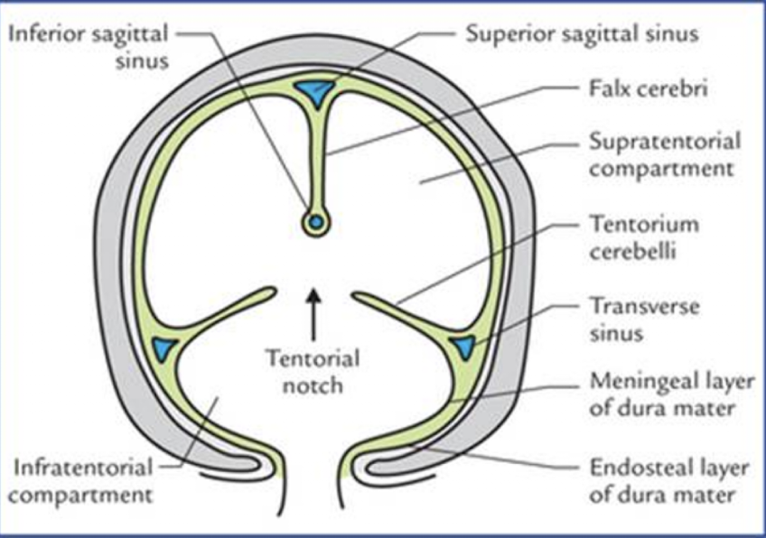
falx cerebri
lateral ventricle
choroid plexus
thalamus
cerebral peduncles
3rd ventricle
cave septum pellucidum
cerebellum
cisterna magna
list the extracranial parts
nuchal fold
profile
nasal bones
lips/nares
orbits
Cerebrum
large hemispheres of brain tissue
what is the cerebrum separated by?
the falx cerebri
cerebrum on US
is hypoechoic
when can you see the sulci?
late in pregnancy
what view?
long
what view?
coronal
what view
axial
falx cerebri
fold of dura matter that divides the cerebral hemispheres
hyperechoic line
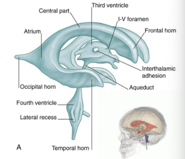
lateral ventricle (LV)
two largest cavities of the ventricular system
contain CSF made by the choroid plexus
what are the sections of the lateral ventricle?
frontal horn
body
atrium
occipital horn
temporal horn
the frontal horn is…to the thalami
anterior
the frontal horn is…to the CSP
lateral
the frontal horn is prominent with?
ventricular dilation
the lateral ventricle is… and…to the thalami
superior and lateral
the lateral ventricle is usually…during the 2nd/3rd trimester?
collapsed
the lateral ventricle is not well visualized with US unless…
dilated
occipital horn is…and…to the thalami
posterior and inferior
temporal horn is not well visualized in US unless?
dilated
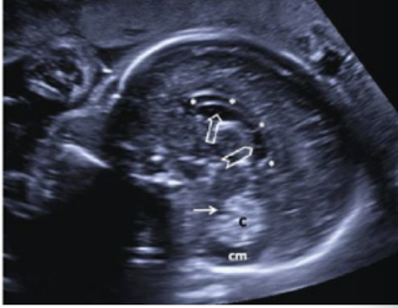
atrium
junction of the LV body, occipital, and temporal horns
located inferior to the mid skull
posterior/lateral to the thalamus and 3rd ventricle
usually filled with hyperechoic choroid plexus
measurement:
across posterior portion
posterior LV
less than 10mm is normal
Choroid plexus
located within the ventricles
produces CSF
appears hyperechoic
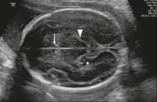
Thalamus
located centrally in the brain
inferior to the falx
superior to midbrain
consists of two lobes
encase the 3rd ventricle
diamond or heart shaped
hypoechoic relative to the cerebrum
cerebral peduncles
2 symmetric anterior protrusions of the midbrain
located centrally in the brain and lie slightly inferior to the 3rd ventricle and thalamus
lie posterior to the circle of willis
appears hypoechoic and smaller than the thalamus
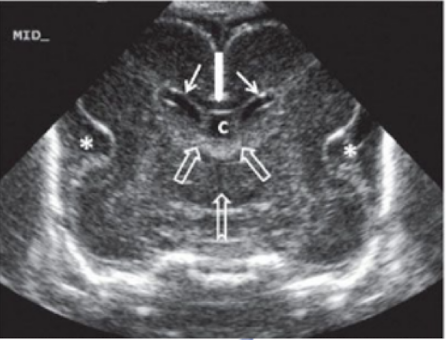
3rd ventricle
located centrally in the brain
inferior to the lateral ventricles
typically collapsed and only enlarged with pathology
cave septum pellucidum (CSP)
fluid filled, midline structure
located between the anterior flax and 3rd ventricle
two echogenic lines lateral to the falx
cerebellum
divided into two hemisphere that are connected by the vermis
hypoechoic and barbell butterfly shaped
measurement should correspond to GA after 16w
evaluate shape (banana shape associated with Arnold chair II malformation)
Cisterna Magna
fluid filled cavity in the posterior skull
posterior to the cerebellum and anterior to the occipital bone
normal 3-10mm
measure after 16w
nuchal fold
skin that covers the inferior mid occipital bone and posterior neck
normal measurement is 18w-20w: <6mm
measure outside occipital bone outer skin edge
evaluate profile for?
abnormal contour
note shape of lips, nose, forehead, mandible
Nasal bone
assess for presence or shortening of the nasal bone
normal length > 2.5mm
Evaluating Lips
check for cleft lip or palate abnormalities
coronally
evaluate orbits
assess size of orbits and distance between orbits
orbital diameter (OD)
measure a single fetal orbit
measure inner diameter
Interocular distance (IOD)
measure the distance between the two orbits