A&P II- Digestion I
1/9
There's no tags or description
Looks like no tags are added yet.
Name | Mastery | Learn | Test | Matching | Spaced | Call with Kai |
|---|
No analytics yet
Send a link to your students to track their progress
10 Terms
The Digestive System
Structure
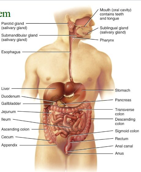
Gastrointestinal/digestive system,
Alimentary canal: extends from the mouth to the anus through the ventral body cavity (approximately 9 m, or 30 ft.).
Accessory organs: teeth, tongue, salivary glands, liver, gallbladder, and pancreas
Function
ABSORPTION.
Digestion
◦ Chemical
◦ Mechanical
ingestion,
secretion,
mixing and propulsion,
defecation
Role in metabolic processes.
Catabolism: Larger molecules are broken into smaller molecules (mouth, stomach, duodenum).
◦ In the GI tract, this is called Digestion and can occur by either mechanical or chemical means.
Anabolism: Smaller molecules are used as building blocks for larger molecules (liver)
Digestion
Mechanical digestion: all Movements that facilitate catabolic processes
Mastication
Mixing
◦ Increase contact of food with digestive chemicals
Other movements:
Swallowing
Peristalsis
◦ Movement of muscles within the GI tract that facilitates movement of food
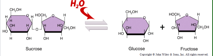
Chemical digestion: breaking large molecule into smaller ones, mainly through hydrolysis.
Fats -> fatty acids and glycerol.
Carbohydrates: polysaccharides -> monosaccharides.
Proteins-> polypeptides -> amino acids.
Requires specific enzymes
GI Tract Anatomy Overview
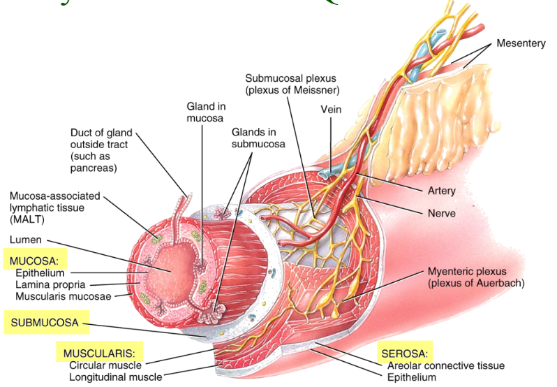
Four layers of tissues
Mucosa (deepest), Submucosa, Muscularis, Serosa/adventitia (superficial) lumen is the inside of the tube.
Mucosa:
mucous membrane
Epithelial tissue
Stratified squamous (mouth, pharynx, esophagus, anus)
Simple columnar (stomach and intestines)
Includes glandular cells that secrete mucus and fluid into the lumen
Areolar connective tissue (lamina propria.)
Smooth muscle (muscularis mucosae)
Lamina propria contains “MALT”, mucosa-associated lymphatic tissue that protect against disease.
Muscularis mucosae: creates folds in the lining of the stomach and small intestines
Submucosa
irregular connective tissue that binds the mucosa to the muscularis.
blood and lymphatic vessels (to receive absorbed substances)
submucosal plexus: a network of neurons
Muscularis:
mouth, pharynx, superior and middle parts of the esophagus, and anal sphincter contain skeletal muscle
Rest of the tract: smooth muscle
Inner circular sheet
Outer longitudinal sheets, myenteric nerve plexus between them
Serosa/adventitia
is the outermost layer.
adventitia - fibrous connective tissue attached to surrounding tissues (e.g. esophagus).
serosa - fibrous connective tissue in the peritoneal cavity, with a mesothelium surface layer
Serosa covers the intra-abdominal organs as the visceral peritoneum
Anatomy Overview
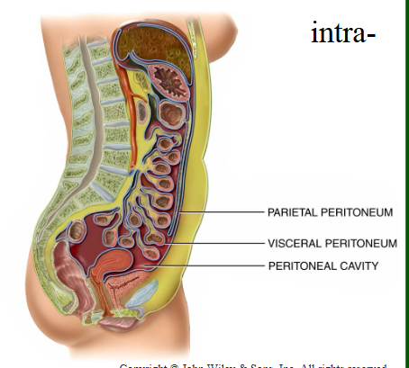
Peritoneum
the body’s largest serous membrane, wraps around most abdominopelvic organs.
Visceral peritoneum: the serosa of the alimentary canal and covers other intra- abdominal organs.
Parietal peritoneum: the abdominal wall, connects to visceral peritoneum.
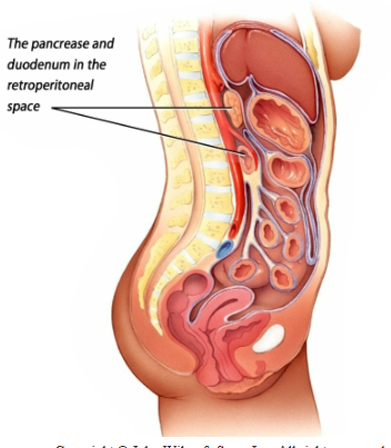
Retroperitoneal organs
are covered by visceral peritoneum only on their anterior surfaces. The portion of the organ that lies behind the peritoneum is said to be “retroperitoneal”.
Organs in the retroperitoneal space include:
The kidneys and ureters
Most of the pancreas
The adrenal glands
The aorta and inferior vena cava
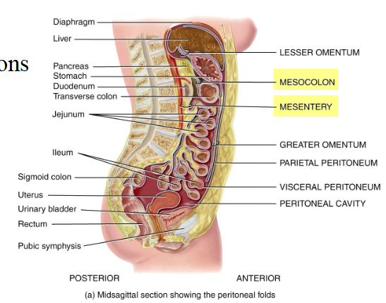
Peritoneal Folds
five major peritoneal folds that bind the organs to one another and to the cavity walls
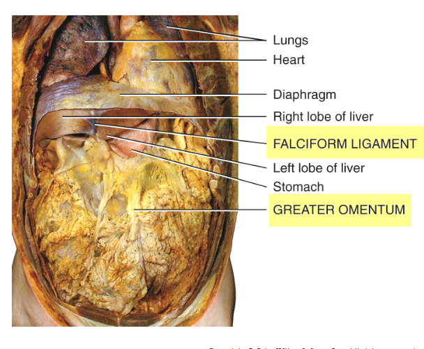
Greater omentum
largest peritoneal fold.
Connects the stomach to the transverse colon
Drapes over the transverse colon and the anterior coils of the small intestine.
Contains: lymph nodes and a large amount of adipose tissue that can greatly expand
Falciform ligament
attaches the liver to the anterior abdominal wall and diaphragm.
Lesser omentum
suspends the stomach and duodenum from the inferior edge of the liver.
pathway for blood vessels to enter the liver
contains the common bile duct
Mesentery (small) and Mesocolon (large)
attach intestine to posterior abdominal wall
Loose attachment so muscular contractions can mix and move the contents along the GI tract
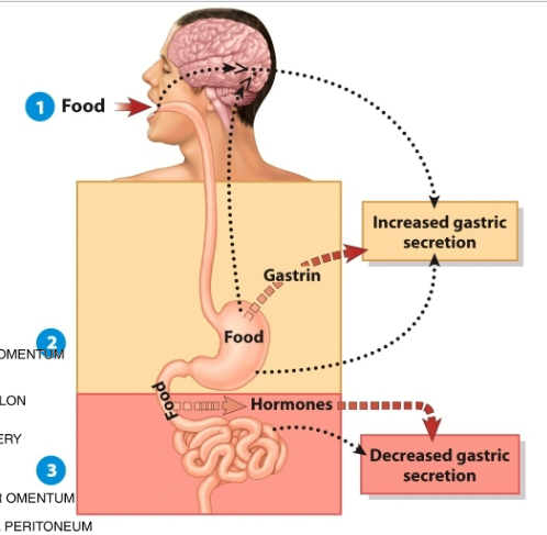
Physiology Overview
Digestive activities are controlled in three overlapping phases:
1. The cephalic phase
2. The gastric phase
3. The intestinal phase
Cephalic phase:
smell, sight, thought, or initial taste of food neural centers in the CNS to prepare for digestion.
stimulate secretion of saliva and gastric juice
Gastric phase:
food enters the stomach.
Nervous and endocrine systems, gastrin is a key hormone
Promotes secretion of gastric juice and gastric motility.
Intestinal phase:
acidic food enters the small intestine.
Neural response decreases gastric motility
Hormones (secretin, CCK) increases intestinal secretions and decreases gastric secretions and motility
The Mouth
oral or buccal cavity:
formed by the cheeks, hard and soft palates, and the tongue.
Mechanical digestion: mastication (chewing)
Saliva
mixes with food to soften it so it can be easily swallowed.
Starts the process of Chemical digestion
Carbohydrates: salivary amylase
Fats: lingual lipase active in acidic stomach (limited activity)
Salivary regulation is under the control of the ANS
Parasympathetic stimulation promotes secretion
This is enhanced by the cephalic phase.
Sympathetic stimulation decreases saliva secretions.
The tongue
Moves food in the mouth for chewing and swallowing (deglutition)
Provides a sense of taste
composed of skeletal muscle
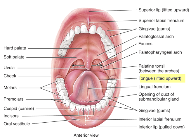
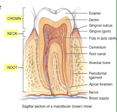
Teeth
Teeth or dentes:
located in sockets of the mandible and maxillae.
Periodontal ligament - a dense fibrous connective tissue that anchors the teeth.
three major external regions: the crown, root, and neck.
The neck of each tooth is covered by the gingivae , or gums, which extend slightly into each socket.
Dentin:
calcified connective tissue that forms most of the tooth.
Enamel: harder-than-bone calcified non-living material, covers the crown
Pulp provides nutrition
20 dentitions as a child
32 as an adult
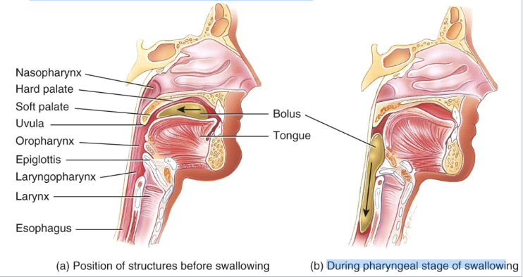
Deglutition
Deglutition:
swallowing food.
Making sure food moves from the mouth through the pharynx to the esophagus and NOT the airway/nasal cavity.
3 stages:
Voluntary: tongue pushing bolus of food into the pharynx,
Pharyngeal: palate rises and epiglottis lowers involuntarily due to presence of food
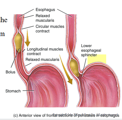
Esophageal: peristalsis moves the bolus down toward the stomach
Peristalsis: a progression of coordinated contractions and relaxations of the circular and longitudinal layers of the muscularis, push the bolus onward.
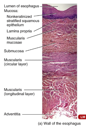
The Esophagus
Esophagus:
Propulsion is only function (moving food into the stomach).
Connects laryngopharynx to stomach posterior to the trachea.
traverses the posterior mediastinum pierces the diaphragm
Mucosa: nonkeratinzed stratified squamous epithelium
Muscularis:
superior 1/3: skeletal muscle
intermediate 1/3: skeletal and smooth muscle
inferior 1/3: smooth muscle
Upper and lower esophageal sphincters (UES and LES) at each end of the tube.
The LES regulates the movement of food from the esophagus into the stomach