Named Radiographic Projections Practice
1/12
There's no tags or description
Looks like no tags are added yet.
Name | Mastery | Learn | Test | Matching | Spaced | Call with Kai |
|---|
No analytics yet
Send a link to your students to track their progress
13 Terms
Grashey
AP oblique shoulder projection used to visualize the glenohumeral joint space without overlap from the humeral head or glenoid cavity
Towne
AP axial skull projection used to visualize the occipital bone, petrous pyramids, dorsum sellae, and posterior clinoid processes within the foramen magnum
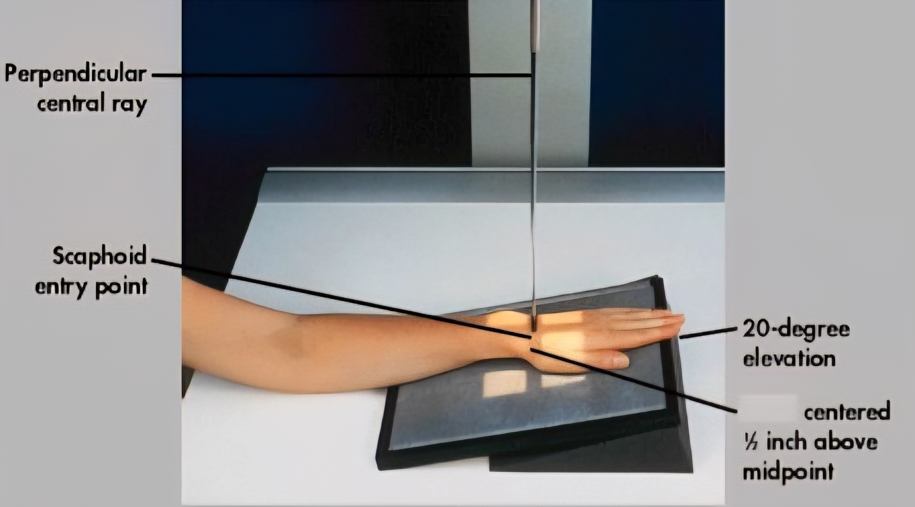
Stecher
PA axial projection, often with 20-degree ulnar deviation, used to visualize the scaphoid bone in the wrist without superimposition, typically to detect fractures
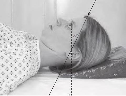
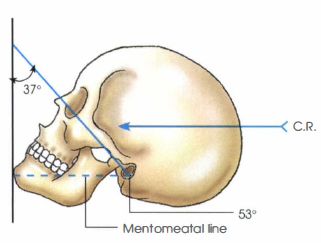
Waters
Skull projection primarily used to visualize the maxillary sinuses, facial bones, zygomatic arches
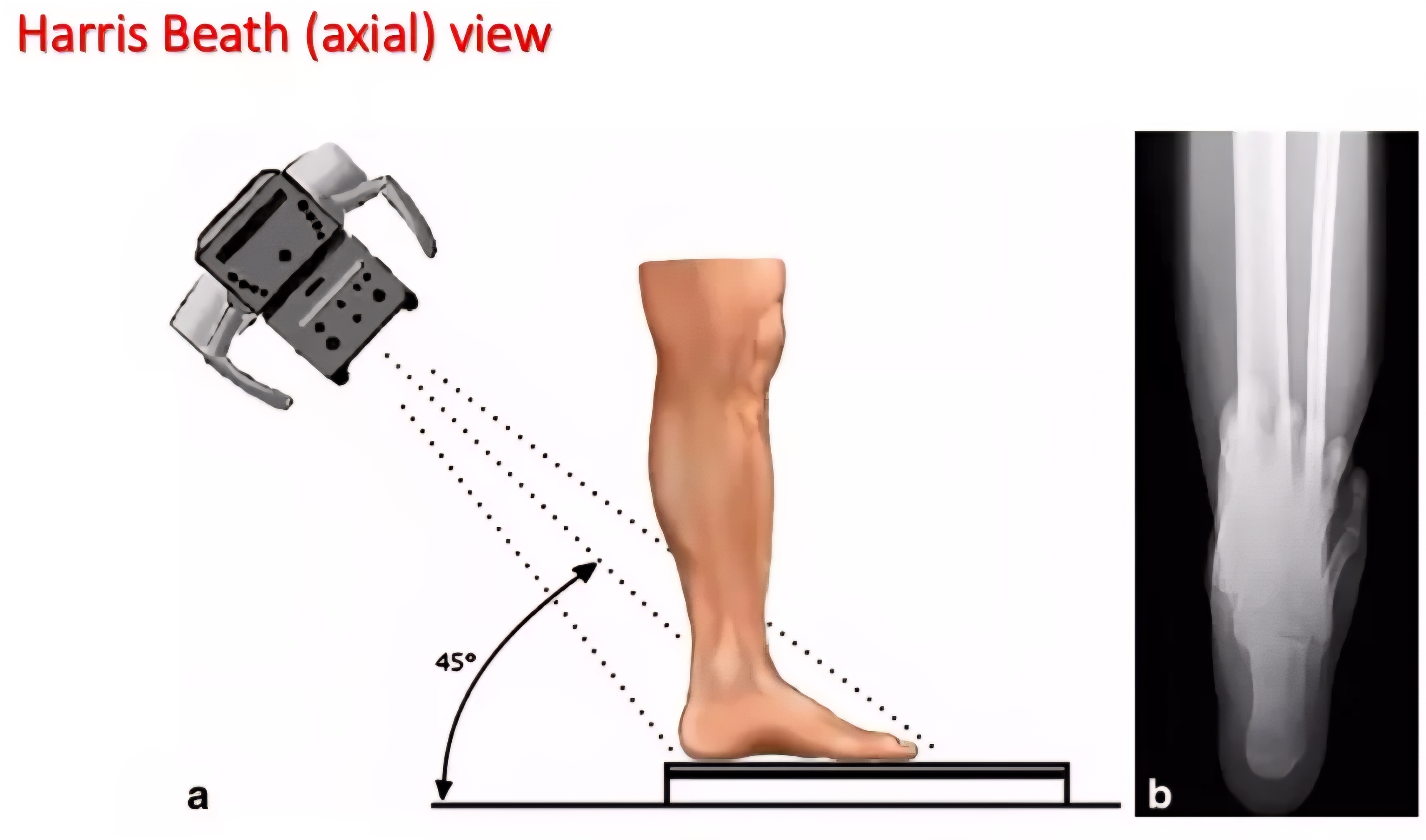
Harris and Beath
Weight-bearing axial projection used to visualize the calcaneus, subtalar joint facets, and sustentaculum tali
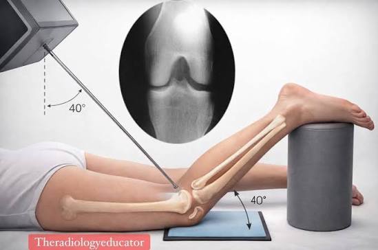
Camp Coventry
PA axial projection used to visualize the intercondylar fossa of the knee. Patient is prone
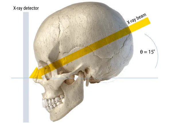
Caldwell
PA axial skull projection used primarily to visualize the paranasal sinuses, orbital rims, and facial bones
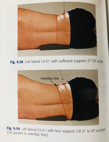
Lateral Spot
Lumbar spine (L5-S1) projection aimed at imaging the lumbosacral junction in a true lateral position to visualize the intervertebral disc spaces
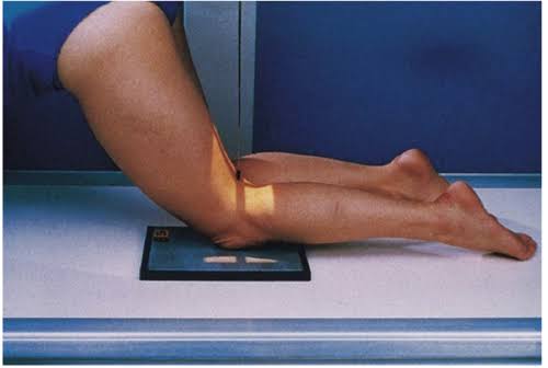
Holmblad
PA axial view of the knee used to visualize the intercondylar fossa, tibial plateaus, and joint space. Patient is knee-kneeling
Lawrence
Inferosuperior axial view of the shoulder used to visualize the glenohumeral joint, humeral head, and coracoid process
Merchant
A superior-inferior axial knee X-ray technique used to evaluate the patellofemoral joint
Coyle Axial Lateral
Gaynor-Hart