Test 3 A&P
1/19
There's no tags or description
Looks like no tags are added yet.
Name | Mastery | Learn | Test | Matching | Spaced | Call with Kai |
|---|
No analytics yet
Send a link to your students to track their progress
20 Terms
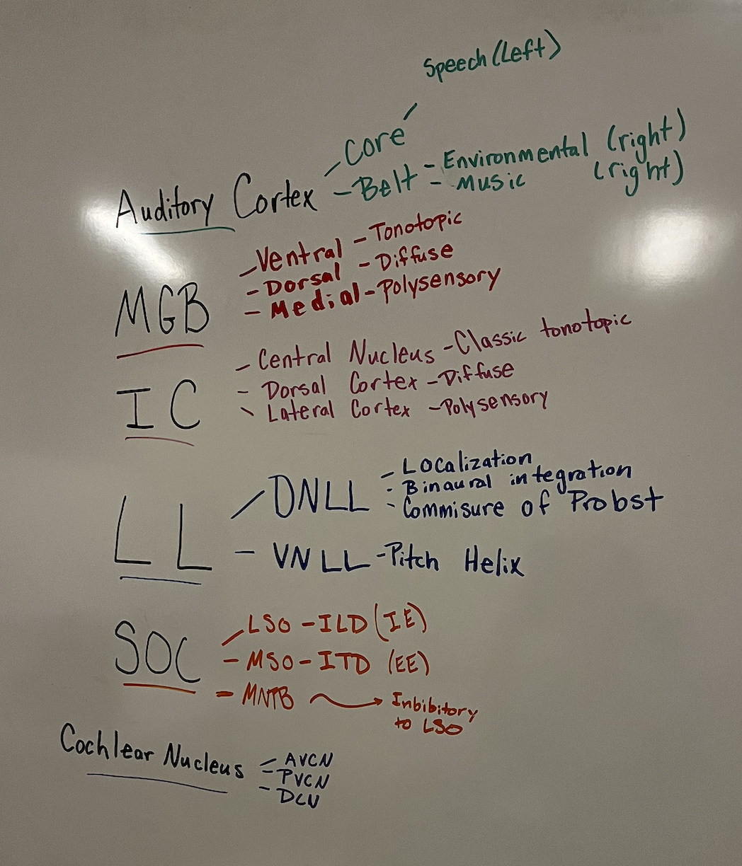
anatomical pathway from cochlea to cortex. note major decussations.
Cochlear nucleus. Decodes duration, intensity, and frequency.
Superior Olivary Complex. Fibers cross over; first stage of Binaural hearing.
Thalamus (MGB). Integration with motor.
Major Decussations in the ACNS: Ventral acoustic stria (primarily projects to SOC on both sides); Intermediate acoustic stria; Dorsal acoustic stria (mainly to contralateral LL and IC).
Give a 1 sentence, general definition for what ‘tuning’ is. What does it mean that the ACNS has multi-dimensional tuning? Give several examples of tuning in the ACNS. State where the cells are and what they respond to.
At different frequencies, how loud does the sound need to be before the cell responds to it.
Multi-dimensional Tuning: Cells can be ‘tuned’ for other important aspects of stimuli.
Examples: 1. Duration (IC/VNLL); 2. Time delay of signals between ears (SOC); 3. Intensity level between ears (SOC)
Name and describe as many maps as you can in the ACNS
Multiple, opposing tonotopic maps: Found in the cochlear nucleus (AVCN, PVCN, DCN) and the ICC; instead of a single place, frequency is represented as a 3D frequency lamina (a sheet of cells tuned to the same frequency).
ITD (Interaural Time Difference) Map: Found in the MSO; binaural cells are lined up anatomically in a way that corresponds to characteristic characteristic delays; phase difference because of long wavelengths in low frequencies
ILD (Interaural Level Difference) Map: Found in the LSO; represents the difference in sound level between the two ears, providing evidence for localization based on intensity.
Timing Map: General representation of timing between ears used for localization
The ACNS enhances features encoded by primary auditory nerve fibers. Give two examples. State where, why and how this happens.
Example 1: Frequency Specificity
Where: Inferior Colliculus (IC).
Why: To make neurons "more frequency specific" and assist in the "identification" of a sound to "define an auditory object".
How: Inhibition narrows the tuning curve by "getting rid of the tails" of the tuning curves found in primary auditory nerve fibers.
Example 2: Precise Timing
Where: AVCN (Anteroventral Cochlear Nucleus).
Why: To "retain and strengthen" temporal encoding for higher levels of the system.
How: Spherical bushy cells produce "precise timing" by firing over a "restricted phase of the signal," giving them "synchronization coefficients that are higher than those of AN fibers"
Inhibition is important in the central nervous system. What is inhibition? How does it influence responses from a neuron in the ACNS? Where is it found in the ACNS?
Several neurons converge on another neuron, making that neuron to have unique selectivities through inhibition.
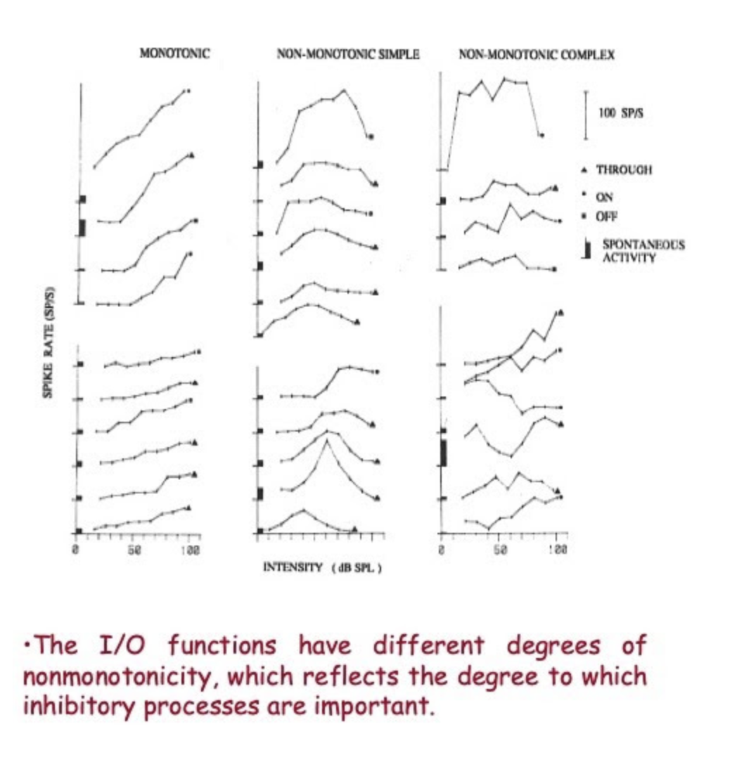
Makes responses of a cell in the CN more complicated or enhanced. I/O functions can be highly non-monotonic and create a cell that has a ‘best intensity’. (Found throughout the ACNS, including CN, SOC, and IC).
Where do binaural interactions first arise within the ACNS?
Superior Olivary Complex (SOC) is the first place in the brainstem that inputs from two ears are combined
What is the duplex theory of sound localization. Does the SOC support this model?
Theory of binaural sound localization.
Low Frequencies (≤700 Hz): Rely on timing differences (ITD); long wavelengths do not cast an ‘acoustic shadow’.
High Frequencies: Use level differences (ILD); wavelengths shorter than distance between ears create an acoustic shadow.
SOC Support: Yes. It is the first stage of binaural hearing and the major center for localization.
Specialized Centers: MSO handles time (ITDs) for low frequencies; LSO handles level (ILDs) for high frequencies.
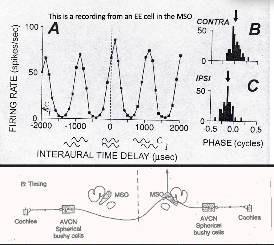
Explain the Jeffress coincidence detector model. Describe cells in the ACNS that support this model.
The brain contains a coincidence detection circuit—cells receive precisely timed excitatory input from the two ears. If the excitatory input does not come at the same time, the neurons do not fire.
Supporting Cells: SPHERICAL BUSHY CELLS FROM AVCN → EE cells (Excitatory-Excitatory) in the Medial Superior Olive (MSO)
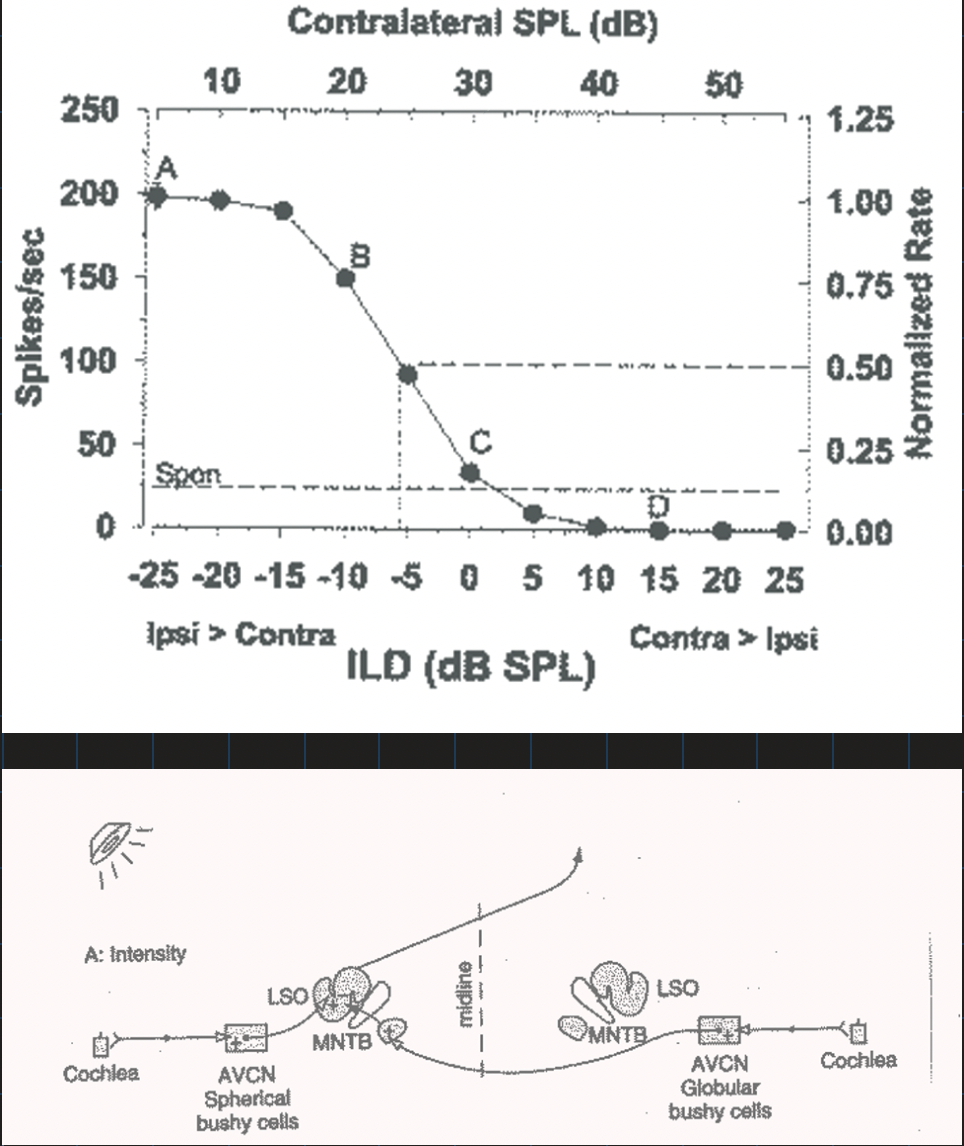
Explain the cellular support for ILDs in the ACNS.
Be able to draw a graph demonstrating the response from a cell that has an ILD.
Be able to draw the neural pathway for ILDs.
Cellular Support: IE neurons (Inhibitory-Excitatory) found in the LSO (Lateral Superior Olive). Inhibitory comes from MNTB
Response Pattern: These cells are excited by ipsilateral stimuli and inhibited by contralateral stimuli.
The MNTB Role: Contralateral inhibition comes through the MNTB, which serves as an inhibitory relay nucleus and provides a ‘sign’ inversion for contralateral signals.
Precise Timing: The Calyx of Held (largest synapse in mammalian brain) provides potent excitatory drive to the MNTB to maintain speed.
Neural Pathway:
Ipsilateral: Cochlea → CN → LSO (Excitatory).
Contralateral: Cochlea → CN → decussation → MNTB → LSO (Inhibitory)
Explain the cellular support of ITDs in the ACNS.
Be able to draw a graph demonstrating the response from a cell that has an ITD.
Be able to draw the neural pathway for ITDs.
Cellular Support: EE neurons (Excitatory-Excitatory) found in the MSO (Medial Superior Olive).
The Model: Jeffress coincidence detector model; cells behave like coincidence detectors and respond maximally when input spikes from both sides arrive in coincidence.
Mechanism: Afferents project to binaural cells in the form of neuronal delay lines.
Cell Count: 65% of cells in the MSO are EE.
Neural Pathway: Cochlea → AVCN (Spherical bushy cells) → MSO (bilateral excitatory input)
Use ITDs for 1.5 kHz and below
What is the proposed function of the VNLL and DNLL? From where do they receive inputs?
DNLL Function: Enhances the lateralization of sound sources which was initially established in the lower stages of the auditory system.
DNLL Inputs: Ipsi MSO, LSOs from both sides, and Contra CN.
VNLL Function: Has a role in processing complex sounds (used to detect duration of signal); part of the monaural sound identification system.
VNLL Inputs: All cell types of the contralateral AVCN (Bushy, stellate, octopus) and ipsi MNTB
Explain the cellular structure of the central nucleus of the inferior colliculus. From where does it receive inputs? What type of signal processing is occurring in this nucleus? What are the other main nuclei in the IC?
Cellular Structure of the ICC
80% disc-shaped neurons: Dendrites organize in a flat plane, along the plane of the laminations; oriented parallel to lamina.
20% Stellate cells: Organized across laminae; integrate across frequency.
Inputs to the ICC
The ICC receives afferent inputs from Contra CN, SOC and DNLL AND ipsi LSO, MSO, DNLL and VNLL. Also ipsi CN.
VNLL provides the largest single source of input to the ICC.
Signal Processing in the ICC
Neurons show a ‘jump’ in the complexity of responses.
Integration of the sound identification and sound localization streams.
Response to temporally complex stimuli.
The IC is a critical stage in the transformation to those that integrate acoustic properties that begin to define an auditory object.
It performs active work by altering and enhancing information received from lower levels.
Other Main Nuclei in the IC
External Cortex (EC): Also called lateral cortex (LCN); polysensory.
Dorsal Cortex (DC): Also referred to as DCN; diffuse.
Describe the three main nuclei of the MGB and the inputs to each. What are the inputs to each? Is the tonotopic map represented in each? Talk about rate-level functions in the MGB.
Three Main Nuclei: Ventral (MGBv), Dorsal (MGBd), and Medial (MGBm).
Ventral (MGBv):
Inputs: Ascending inputs arise from the ICC.
Tonotopy: Tonotopic; preserves an orderly spatial arrangement of best frequencies. Has SEVERAL tonotopic maps within it.
Structure: Disc-like principle cells with flattened dendritic fields arranged in parallel with afferent fibers.
Dorsal (MGBd):
Inputs: Projection from the dorsal cortex of the IC.
Tonotopy: “Diffuse” pathway; so named because of a lack of obvious tonotopic organization.
Medial (MGBm):
Inputs: Projection from the lateral nucleus of the IC; also receives nonauditory fibers, especially from the somatosensory system.
Tonotopy: “Polysensory” pathway.
Rate-Level Functions: Divided into monotonic and non-monotonic. The proportion of non-monotonic units in MGBv is rather high (75%), reflecting the degree to which inhibitory processes are important. These cells are sensitive to intensity
Discuss the main areas of the auditory cortex. What are the afferent innervations paths?
Level 1 - Cochlear Nucleus (CN): First nucleus in brainstem; decodes duration, intensity, frequency.
AVCN & PVCN: Primary-like and chopper responses.
DCN: Different from VCN; complex processing including horizontal localization.
Level 2 - Superior Olivary Complex (SOC): First stage of binaural hearing; major center for localization.
MSO: Time (ITDs); responds best to low frequencies.
LSO: Level (ILDs); responds best to higher frequencies.
Lateral Lemniscus (LL): Major brainstem pathway.
VNLL: Processing complex sounds; part of monaural sound identification system.
DNLL: Enhances lateralization of sound sources.
Inferior Colliculus (IC): Critical stage that begins to define an auditory object.
Nuclei: ICC (tonotopic), External Cortex (polysensory), Dorsal Cortex (diffuse).
Level 3 - Thalamus (MGB): Integration with motor information.
Subdivisions: MGBv (tonotopic), MGBd (diffuse), MGBm (polysensory).
Auditory Cortex Areas: Primary Auditory Cortex (end of primary pathway) and Polysensory Cortex.
Afferent Paths: 1. Tonotopic (from MGBv); 2. Diffuse (from MGBd); 3. Polysensory (from MGBm).
Describe the core and belt theory for human cortical auditory processing. State the inputs to each.
The Core: Corresponds to the Primary Auditory Cortex, which is the end of the primary auditory pathway.
Input: Receives ascending projections from the MGBv (Ventral subdivision of the Medial Geniculate Body) via the tonotopic (lemniscal) pathway.
The Belt: Corresponds to the Polysensory Cortex, where non-primary auditory information is integrated with other systems.
Inputs: Receives projections from the MGBd (Dorsal subdivision) via the diffuse pathway and the MGBm (Medial subdivision) via the polysensory pathway
Describe the ‘what’ and ‘where’ stream theory for human auditory cortical processing.
Theory Proposes: Some channels, or pathways help us to decide ‘what’ a sound is, vs ‘where’ it is coming from.
Tonotopic info MGB → core → belt → diffuse MGB gives info straight to the belt → belt creates 2 streams of info to auditory cortex →
Dorsal stream = where information
Ventral stream = what information
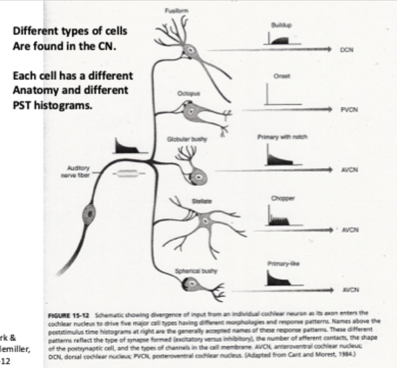
What are the cell types and typical PSTs to cells found in the CN?
Spherical bushy cells: Primary-like, timing info, superior phase locking
Globular bushy cells: Primary with notch, superior phase locking, timing info, slightly weaker than spherical bushy cells
Stellate cells: Chopper: intensity of the signal, poor phase locking, spectral info, wide dynamic range
Octopus cells: Onset; precise onset info, poor frequency tuning
Fusiform cells (DCN): Build up, Narrow frequency tuning, Inhibited by broadband noise
parallel processing starts at the cochlear nucleus: auditory nerve diverges, connecting to different cells that attract different info from same signal, create separate streams of auditory signals
BUSHY CELLS: primary-likem convey TEMPORAL/TIMING info
What is the anatomy of the CN? How is it innervated, where do the outputs go and what cell types are most generally found in each?
How is the DCN different from the AVCN and PVCN in terms of proposed function?
Anatomy: First nucleus in brainstem; located between medulla and pons, under the cerebellum. Consists of AVCN, PVCN, and DCN.
Innervation: Each 8th nerve fiber bifurcates to PVCN and AVCN; the branch to PVCN then bifurcates to DCN. This shows parallel processing of information.
Outputs (The 3 Striae):
Ventral acoustic stria: Primarily projects to SOC on both sides.
Intermediate and Dorsal acoustic stria: Both project mainly to the contralateral LL and IC; involved in sound identification.
Cell Types Found in Each:
AVCN: Bushy and Stellate cells.
PVCN: Stellate and Octopus cells.
DCN: Fusiform cells.
Proposed Functional Differences:
VCN (AVCN/PVCN): Decodes duration, intensity, and frequency. Bushy cells enhance timing; stellate cells convey sound level.
DCN: Complex processing including horizontal localization. Tuning curves match spectral shaping of the pinna; inhibited by broadband sounds.
What is inhibition in the CNS and give examples of what happens to I/O functions or receptive fields when a cell has inhibitory input.
What is Inhibition: Input that results in "less spikes". It makes responses of a cell "more complicated or enhanced".
Impact on I/O Functions: Creates "non-monotonic" functions (slope increases and then decreases) and results in cells with a "best intensity".
Impact on Receptive Fields: Produces "more highly ‘tuned’ responses to frequency" and "specialized response patterns".
Examples of Inhibitory Effects:
Tuning: Creates "narrow tuning curves without a tail" or "closed" response areas.
PSTH Patterns: Reflected in "Off" units, "late" units, and "suppressed" units
Where does the processing of speech mainly occur in the auditory cortex? What about music? What about environmental sounds?
Speech Processing: Mainly occurs in the left auditory cortex.
Music Processing: Mainly occurs in the right auditory cortex.
Environmental Sounds: Mainly occur in the right auditory cortex.
Identification Stream: These sound categories are processed via the ‘what’ stream to help us decide ‘what’ a sound is.
Defining Auditory Objects: The system integrates acoustic properties to define an auditory object and provide an idea of what you are hearing.
Active Work: Neurons at higher levels do active work by altering and enhancing information to assist in understanding language and complex stimuli