anatomy
1/37
There's no tags or description
Looks like no tags are added yet.
Name | Mastery | Learn | Test | Matching | Spaced | Call with Kai |
|---|
No analytics yet
Send a link to your students to track their progress
38 Terms
The heart:
is a hollow muscular organ that is somewhat pyramid-shaped and it is the largest organ.
What is mediastinum?
the region between the two lungs.
What is the weight of the heart?
250-350 grams
How much dose the heart beats a day?
100,000 times per day, pumping roughly 7,500 liters of blood through the circulatory system.
The Pericardium:
The pericardium is a fibro-serous sac that encloses the heart and the roots of the great vessels.
The Pericardium Function:
restrict excessive movements of the heart as a whole and to serve as a lubricated container in which the different parts of the heart can contract efficiently.
Fibrous Pericardium:
The outer strong layer composed of dense connective tissue. This tough, inelastic layer protects the heart from external trauma and prevents overfilling of the heart chambers.
Pericardial Cavity:
A potential space located between the parietal and visceral layers, containing 15-50 mL of lubricating serous fluid that allows the heart to beat with minimal friction.
Serous Pericardium - Parietal Layer:
The outer, parietal layer adheres to the inner surface of the fibrous pericardium, creating a smooth lining that reduces friction during heart contractions.
Visceral Layer (Epicardium):
The innermost layer lies directly on the heart and is considered a part of the heart wall itself, providing protection and reducing friction against surrounding structures
The Four Heart Chambers:
The heart consists of four chambers divided into two atria (receiving chambers) and two ventricles (pumping chambers). Each chamber has a specific role in the cardiac cycle.
Right Atrium:
Small, thin-walled chamber that receives deoxygenated blood. It contracts to push blood through the tricuspid valve into the right ventricle.
Left Atrium:
Small, thin-walled chamber that receives oxygenated blood. It contracts to push oxygen-rich blood through the mitral valve into the left ventricle.
Left Ventricle:
The wall is thicker than the right ventricle and this feature is necessary to pump oxygenated blood at high pressure through the systemic circulation to all body tissues
Right Ventricle:
It pumps deoxygenated blood to the lungs through the pulmonary artery.
Right Side:
Receives deoxygenated
blood
• Lower pressure system
• Thinner myocardial walls
• Pumps to lungs
(pulmonary circulation)
• Two-chambered pathway
Left Side:
Receives oxygenated
blood
• Higher pressure system
• Thicker myocardial walls
• Pumps to body (systemic
circulation)
• Two-chambered pathway
Upper respiratory tract:
The nose, nasal cavity, pharynx and larynx
Lower respiratory tract:
Trachea, bronchi, and lungs
The pharynx:
is a muscular tube about 13 cm long that connects the nasal cavity and mouth to the larynx and esophagus.
The pharynx is divided into three parts:
Nasopharynx, oropharynx and laryngoparynx
The larynx:
hollow muscular organ that forms an air passage to the
lungs and holds the vocal cords.
3- large unpaired cartilages:
Cricoid cartilage
Thyroid cartilage
Epiglottis cartilage
3- pairs of smaller cartilages:
Arytenoid cartilages
Corniculate cartilages
Cuneiform cartilages

Name the green part
Epiglottis

Name the red part
Thyroid cartlige

Name the blue part
Arytenoid cartilages

Name the yellow part
Cricoid cartilage
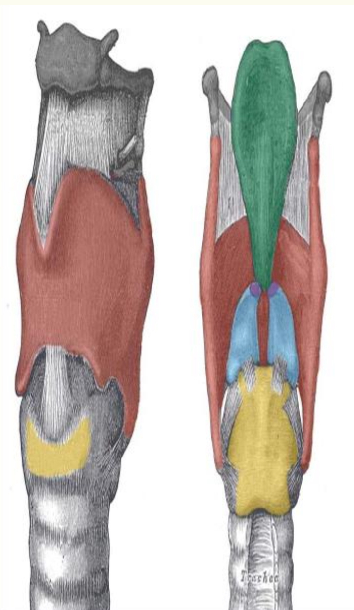
Name the purple part
Corniculate cartilages
The trachea:
A flexible tube running from the larynx, dividing inferiorly into two main bronchi. The wall contains 16–20 C-shaped rings of hyaline cartilage joined by fibro-elastic connective tissue, incomplete posteriorly where the trachealis muscle occurs.
Functions of the trachea:
Air passageway, filters, moistens air
The bronchi:
The trachea divides at the carina into two main (primary) bronchi, each entering a lung at the hilum
Right Main Bronchus:
Wider, shorter, and more vertical than the left. Subdivides into three secondary bronchi. angle.
Left Main Bronchus:
Subdivides into two secondary (lobar) bronchi. Passes under the aortic arch, making it longer and more horizontal
The lungs:
They are soft, spongy, cone-shaped organs located in the thoracic cavity, surrounded by the pleura (a double-layered membrane).
Right Lung:
Three lobes: superior, middle,
inferior
• Divided by oblique and
horizontal fissures
• Shorter (liver below) and
wider.
Left Lung:
Two lobes: superior and inferior
• Divided by the oblique fissure
• Longer and narrower (heart
beside)
• Contains the cardiac notch
where the heart sits
The alveoli:
tiny air sacs at the end of the bronchioles. They are the functional units of the lungs where oxygen enters the blood and carbon dioxide is removed.