Pathology histology - Hepatocellular carcinoma (HCC)
1/10
Earn XP
Description and Tags
Pathology histology
Name | Mastery | Learn | Test | Matching | Spaced | Call with Kai |
|---|
No analytics yet
Send a link to your students to track their progress
11 Terms
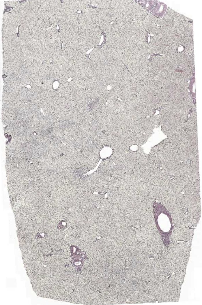
Normal liver
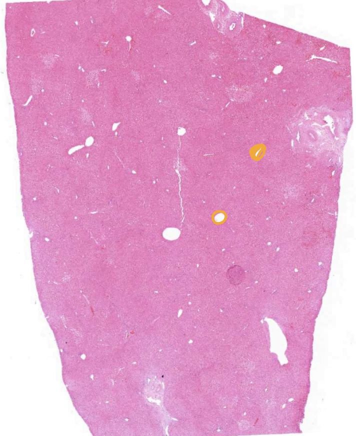
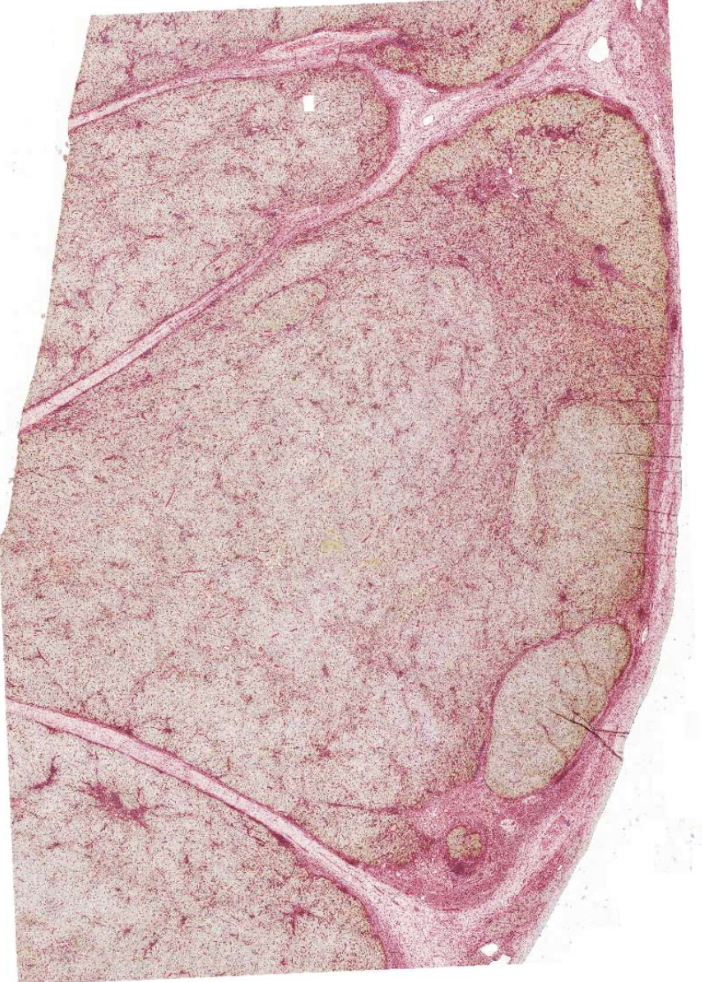
Liver with hepatocellular carcinoma
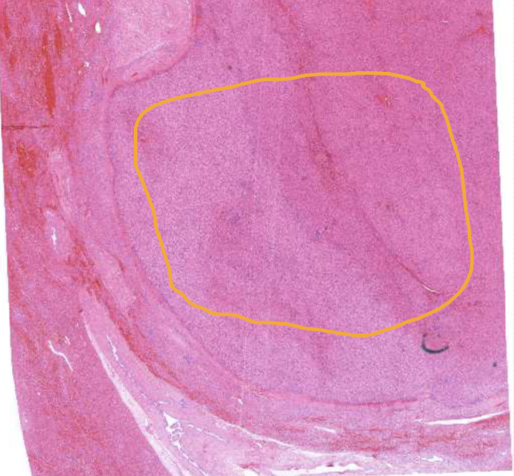
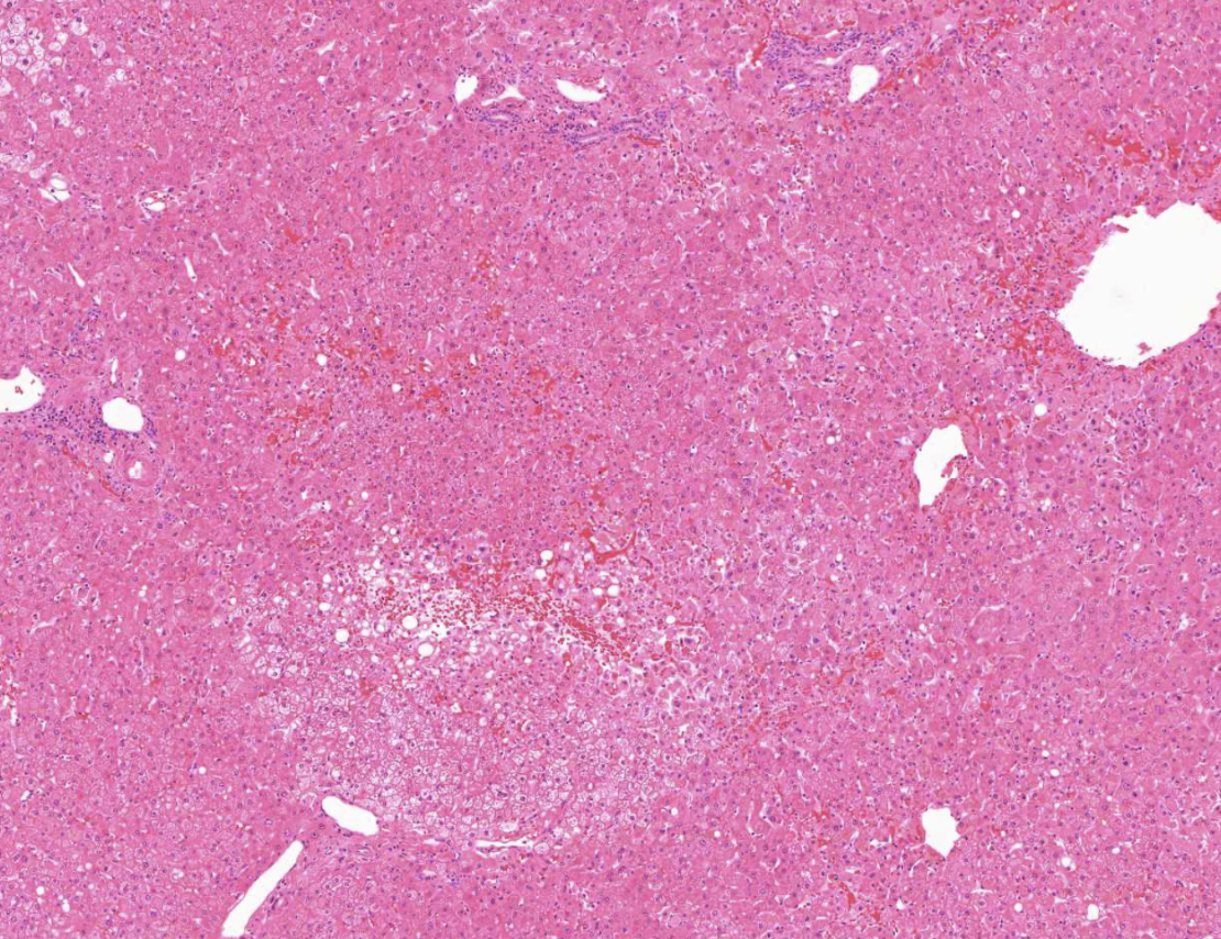
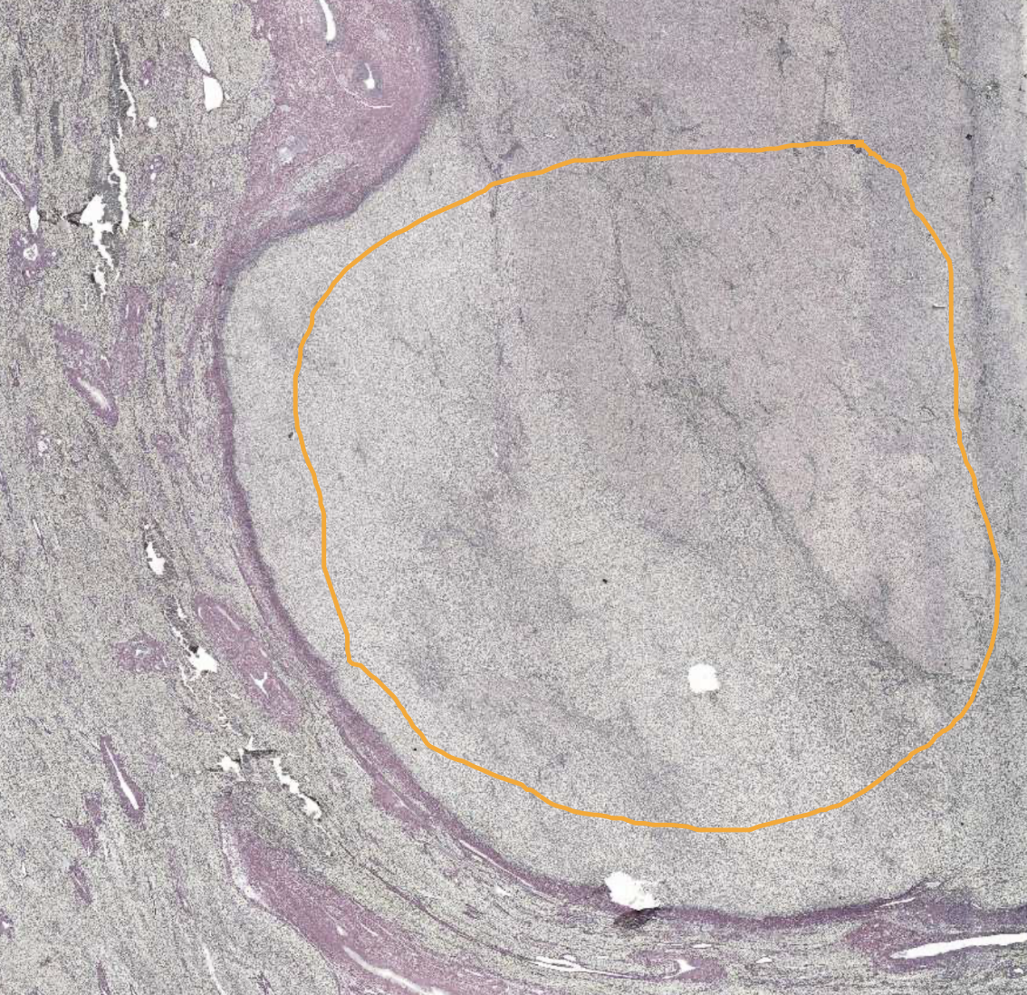
HCC - Tumor area
Surrounding areas are normal (has cells with lipofuscin in cytoplasm)
This area is a tumour surrounded by a capsule
Tumour area shows loss of normal sinusoid pattern
Normal structures can be seen amongst the tumour portion (e.g. bile ducts)
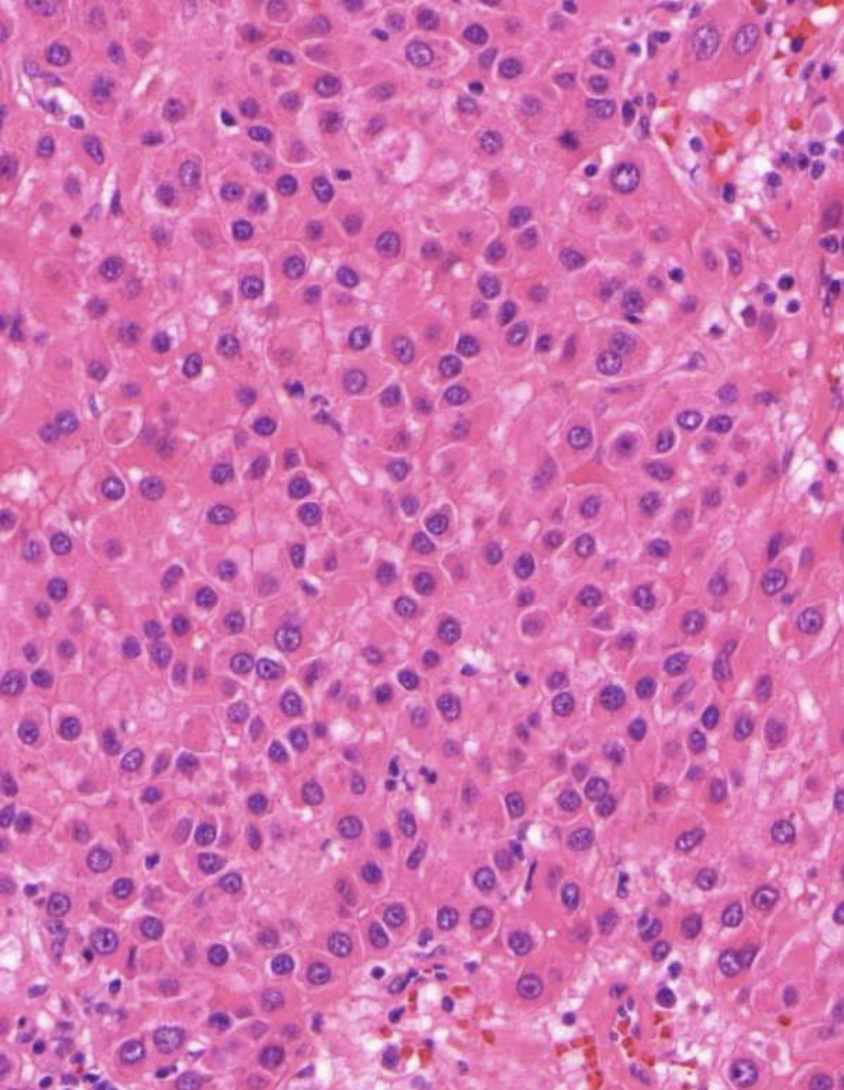
Enlarged nuclei (high nuclei/cytoplasm area)
No portal triads
Less pink, more blue → Enlarged nuclei + Immune cells
Nuclei are very close → Less cytoplasm
Large variations of nuclei size
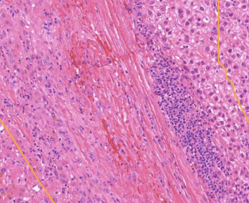
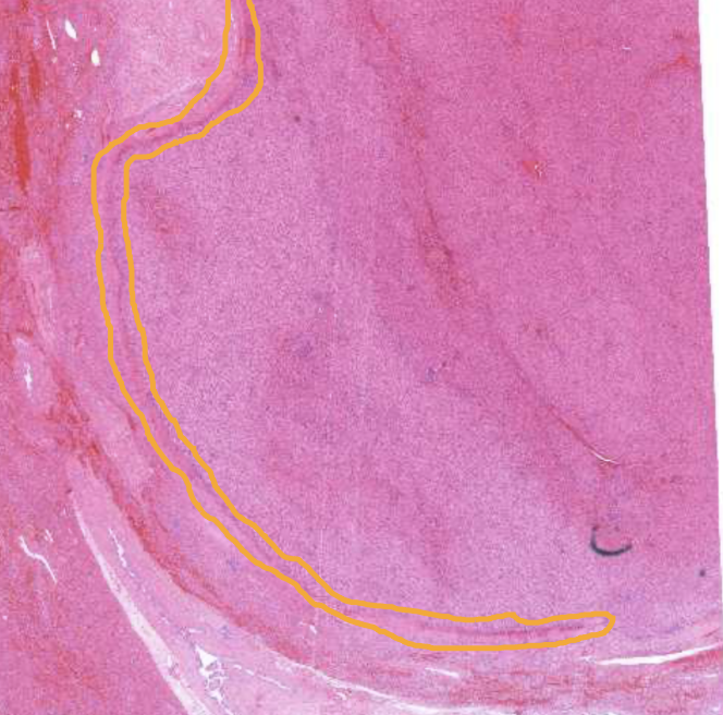
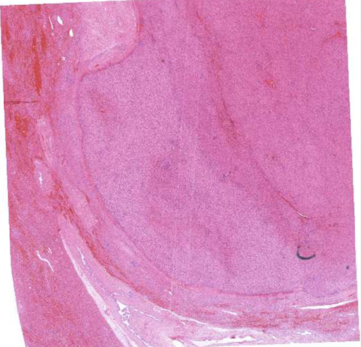
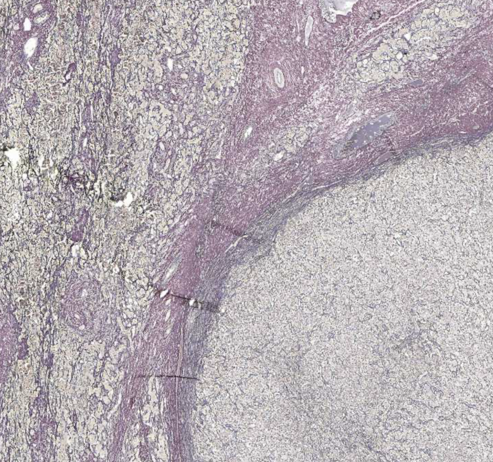
HCC - Capsule surrounding tumour area
Collagen type I
Encapsulates tumor area
Outside pretty much normal liver, inside is tumour and immune cells
Formed by the expansion of the tumour
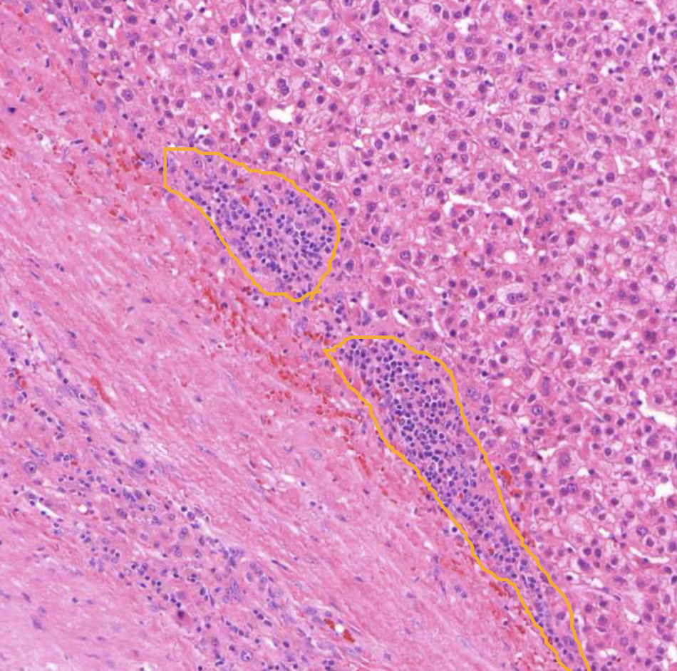
HCC - Immune cells
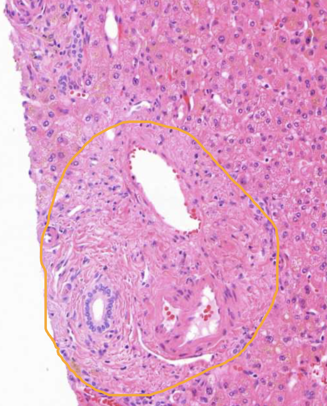
Normal portal triad
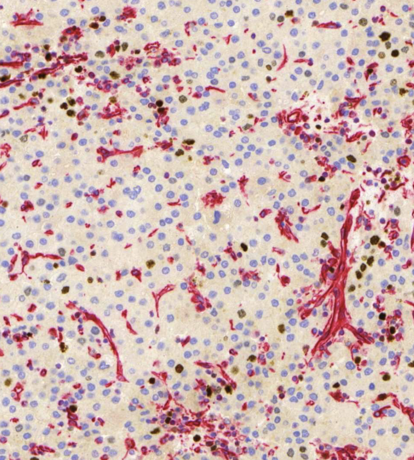
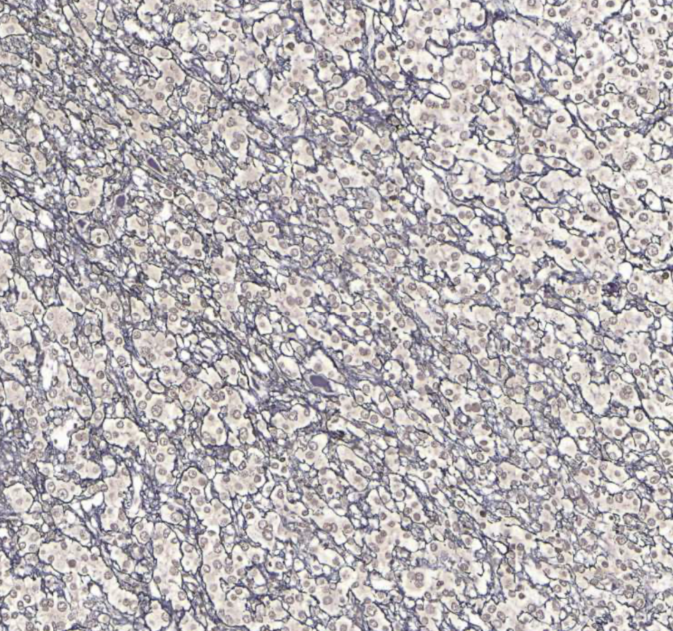
Liver with HCC stained with anti-Ki67/anti-vimetin
Brown → Ki67-positive → Proliferating cells
Red → Vim-positive → Cells of mesenchymal origin (e.g. fibroblasts, endothelial cells, smooth muscle) or showing mesenchymal differentiation
In carcinomas, vimentin positivity often indicates epithelial–mesenchymal transition (EMT), which is associated with increased invasiveness and metastatic potential - NOT seen in this case
Mesenchymal cells should not be found in liver because liver cells are from endoderm origin - Presence of them here indicates carcinoma
Roughly around 5% of nuclei are brown, rest are blue - Gives us an indication of fast the tumor is growing (in this case slow growing → better prognosis)
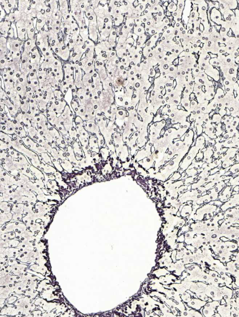
Normal liver stained with GoS
Black → Collagen type III
Purple/brown → Collagen type I
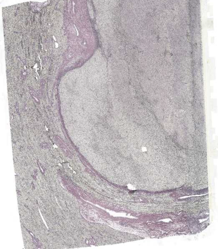
Liver with HCC stained with GoS
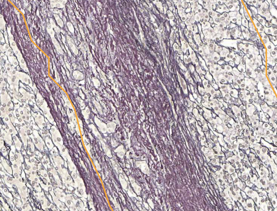
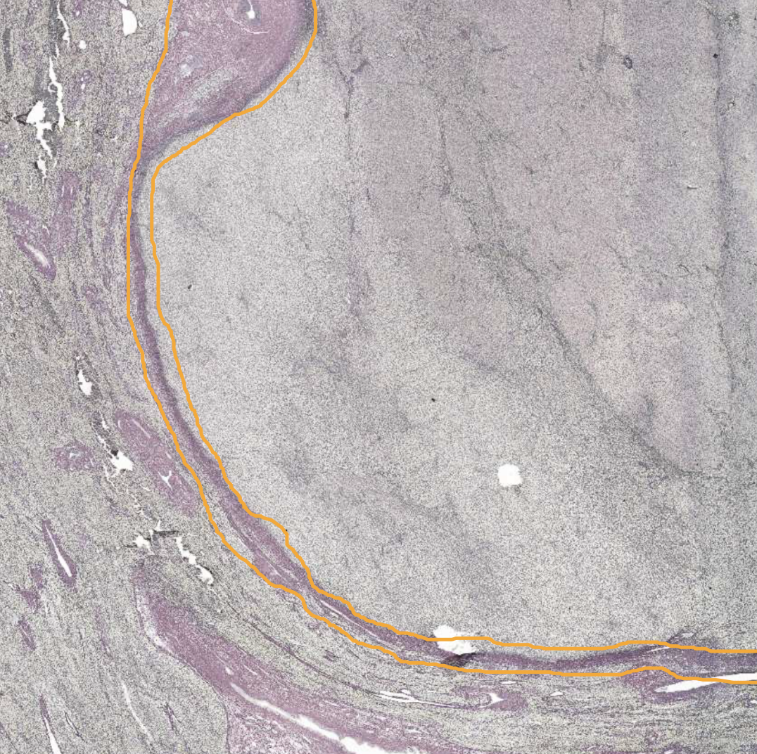
HCC stained with GoS - Capsule around tumor area
Purple → Collagen type I
HCC stained with GoS - Tumor area
Loss of a normal sinusoidal pattern (less black collagen type III)
No portal triads or similar surrounded by purple collagen type I