A&P II LAB- Intro + Endocrine
1/18
There's no tags or description
Looks like no tags are added yet.
Name | Mastery | Learn | Test | Matching | Spaced | Call with Kai |
|---|
No analytics yet
Send a link to your students to track their progress
19 Terms
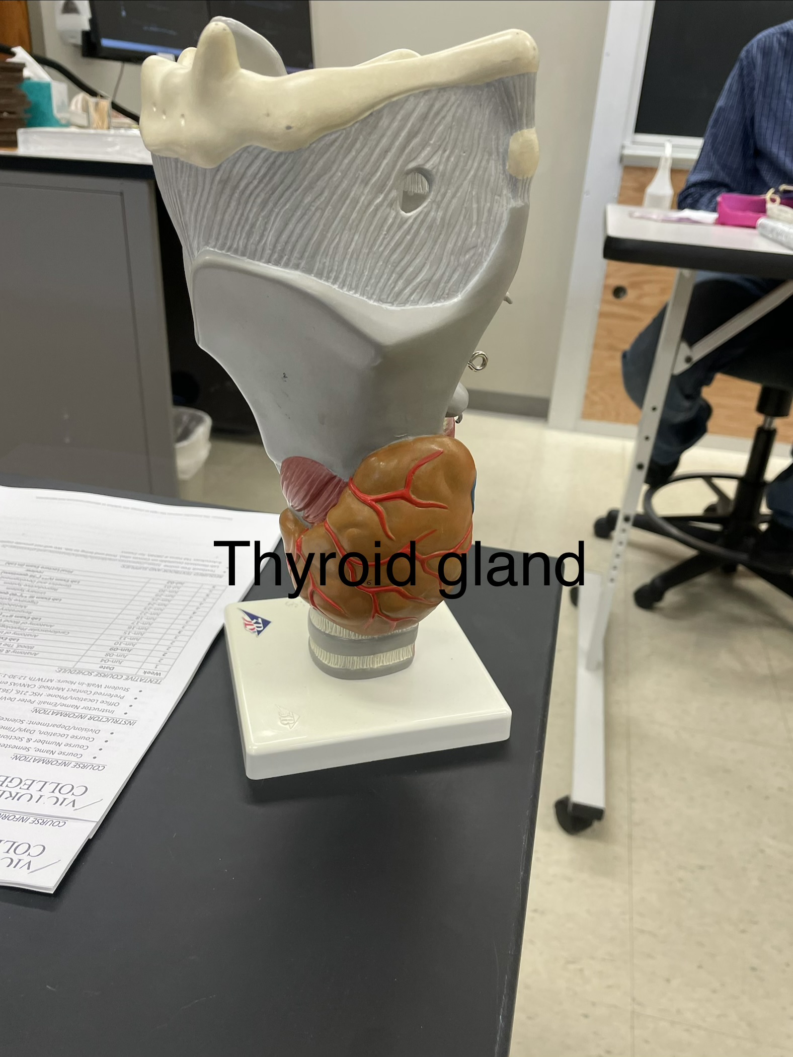
Thyroid Gland visual
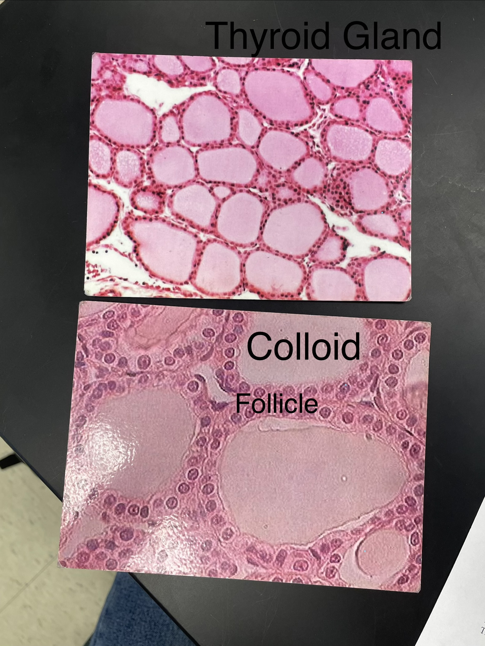
Thyroid Gland
Under larynx
thyroid hormones produced in colloid
Has distinctive “pools” of colloid surrounded by follicle cells (simple cuboidal epithelium).
Looks like cobblestone street. The “stones” are the colloid and the “mortar” is the follicle cells.
Be able to identify under the ‘scope:
Gland
Follicle
Colloid
Hormones:
Calcitonin
Thyroid Hormones (T4 & T3)
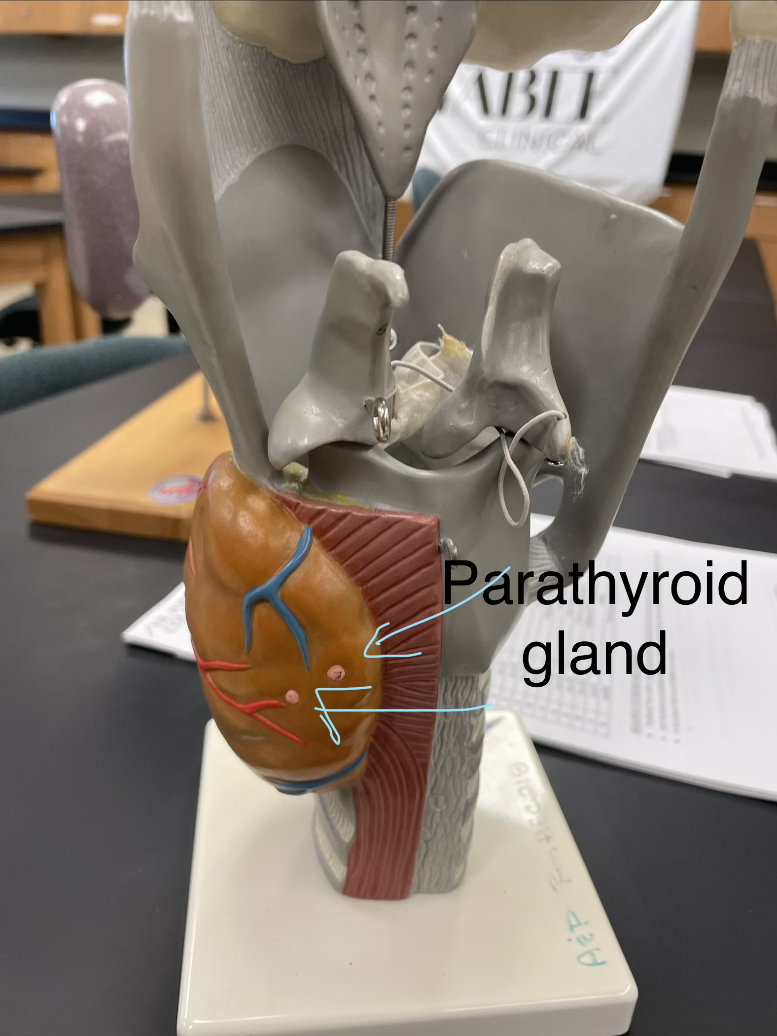
Parathyroid gland visual
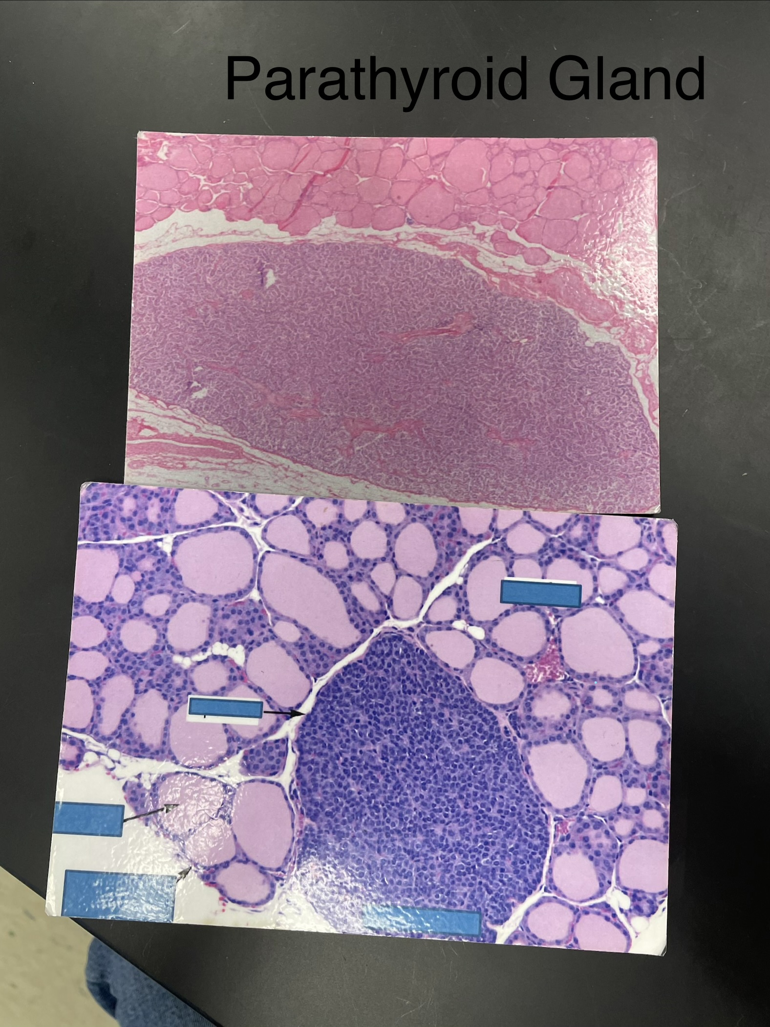
Parathyroid Gland
pink dots
seedy vacuole
Indistinct compared to the other glands.
May see the thyroid gland in the field of view on low power.
Be able to identify under the ‘scope:
gland
Hormones:
Parathyroid Hormone
Pancreas visual
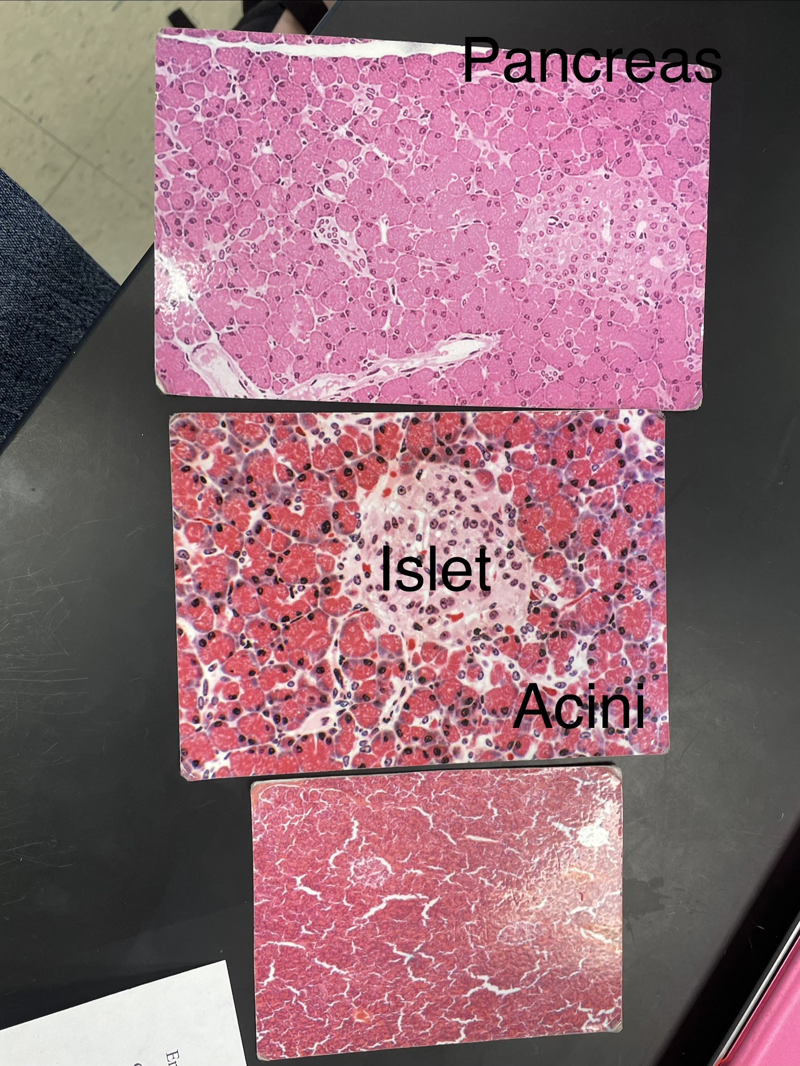
Pancreas
I want to eat your pancreas
endocrine and exocrine glands (into digestive)
The collections of lighter stained cells (Islets) surrounded by “grape like” darker stained cells (Acini)
dots are nuclei
Be able to identify under the ‘scope:
Gland
Pancreatic Islet (of Langerhans)
Acini
Hormones:
Insulin
Glucagon
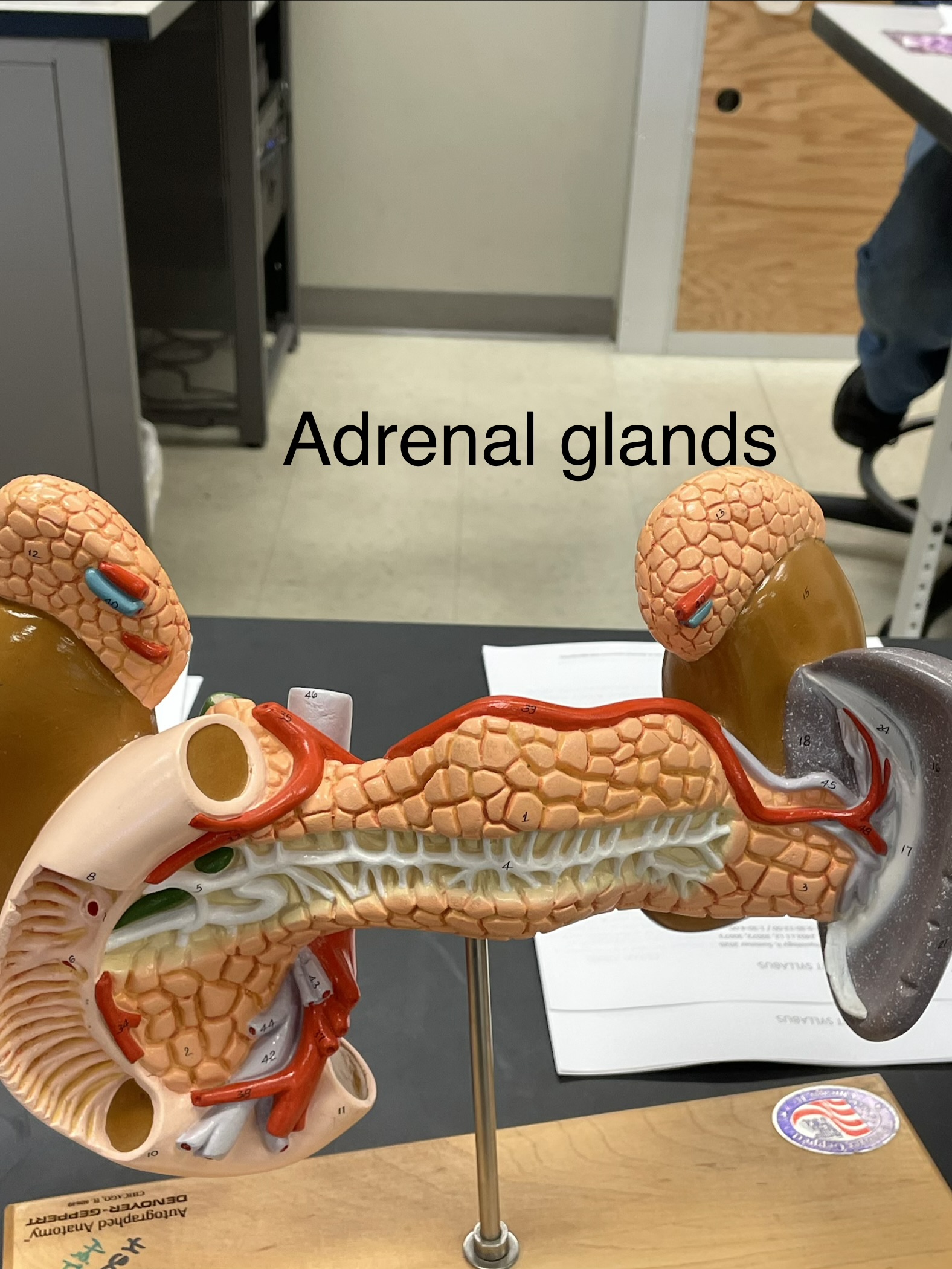
adrenal glands (suprarenal) visual
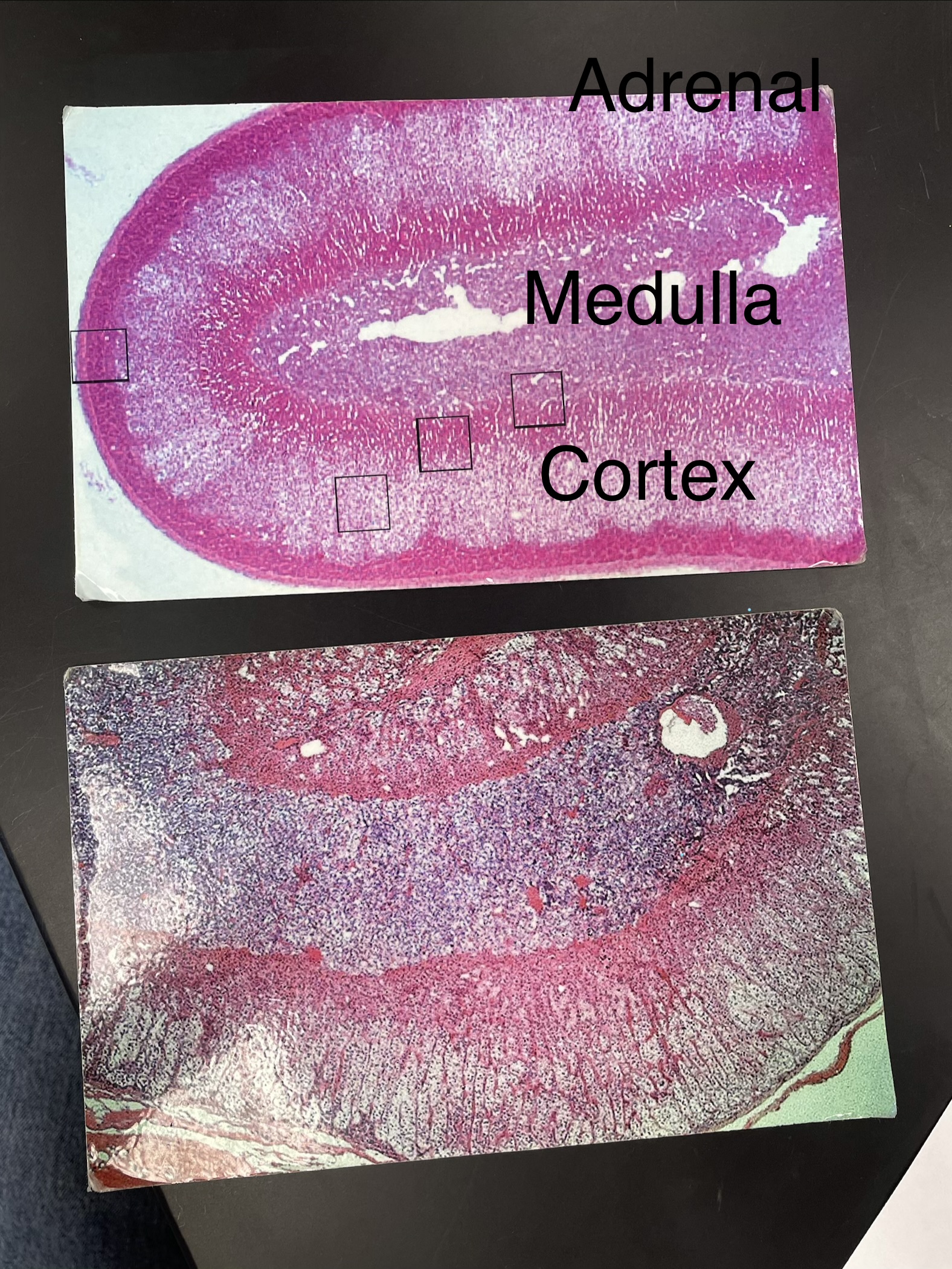
Adrenal Gland (suprarenal)
Has a medulla and a cortex.
The middle layer of the cortex has cells thatare arranged in distinctive columns
Be able to identify under the ‘scope:
Gland
Cortex
Medulla
Hormones:
Medulla
Epinephrine
Norepinephrine
Cortex
Corticosteroids
Mineralocorticoids
Aldosterone
Glucocorticoids
Androgen
Testis visual
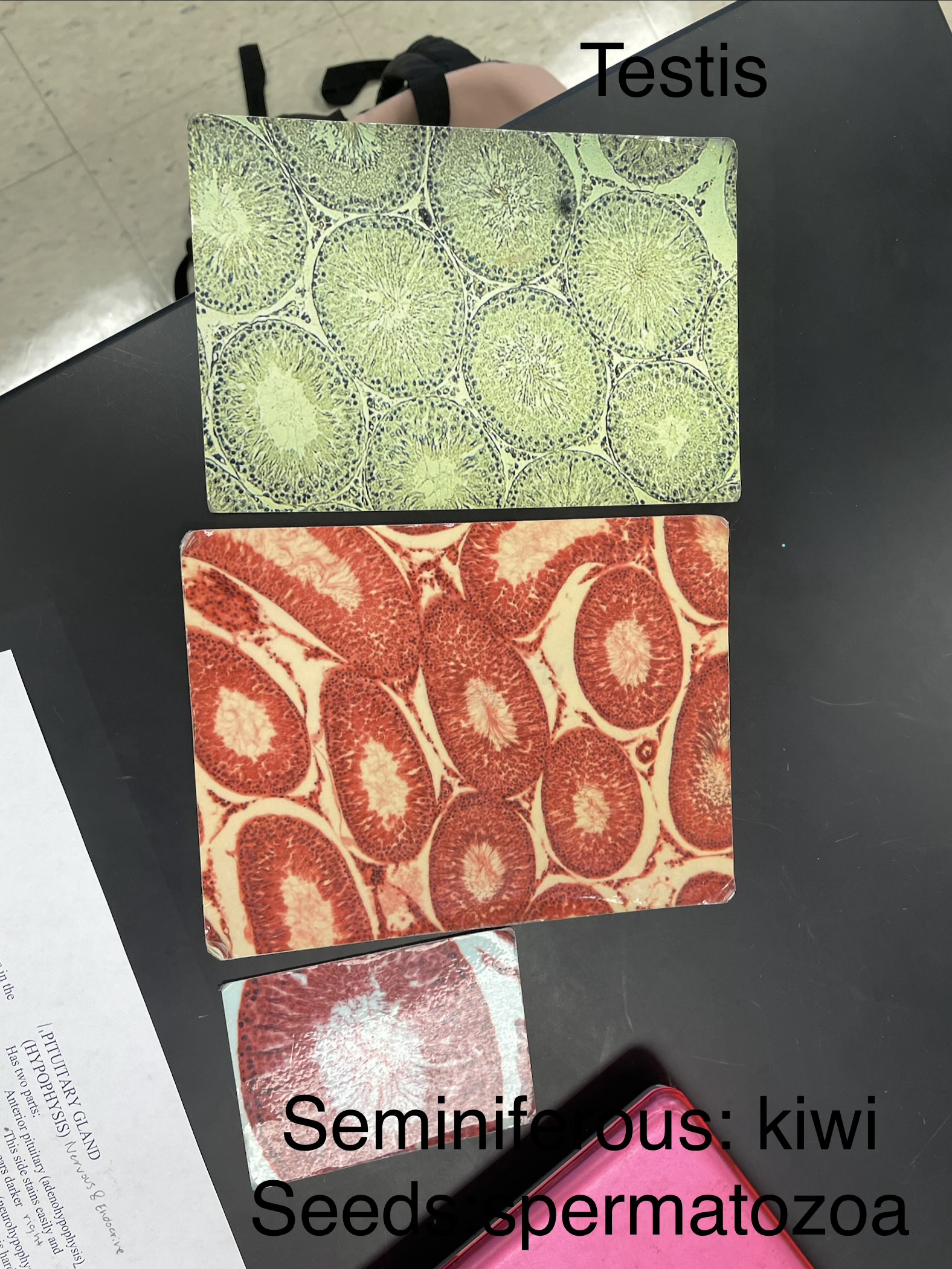
Testis
sperm in seminiferous tubules
interstitial- testosterone
The seminiferous tubules look like kiwis and the spermatozoa look like the seeds in the kiwi.
The cells between the kiwis are interstitial cells.
The entire field of view is the testis.
Be able to identify under the ‘scope:
Gland
Seminiferous Tubules
Interstitial Cells
Spermatozoa
Hormones:
Testosterone
pituitary gland (hypophysis) visual
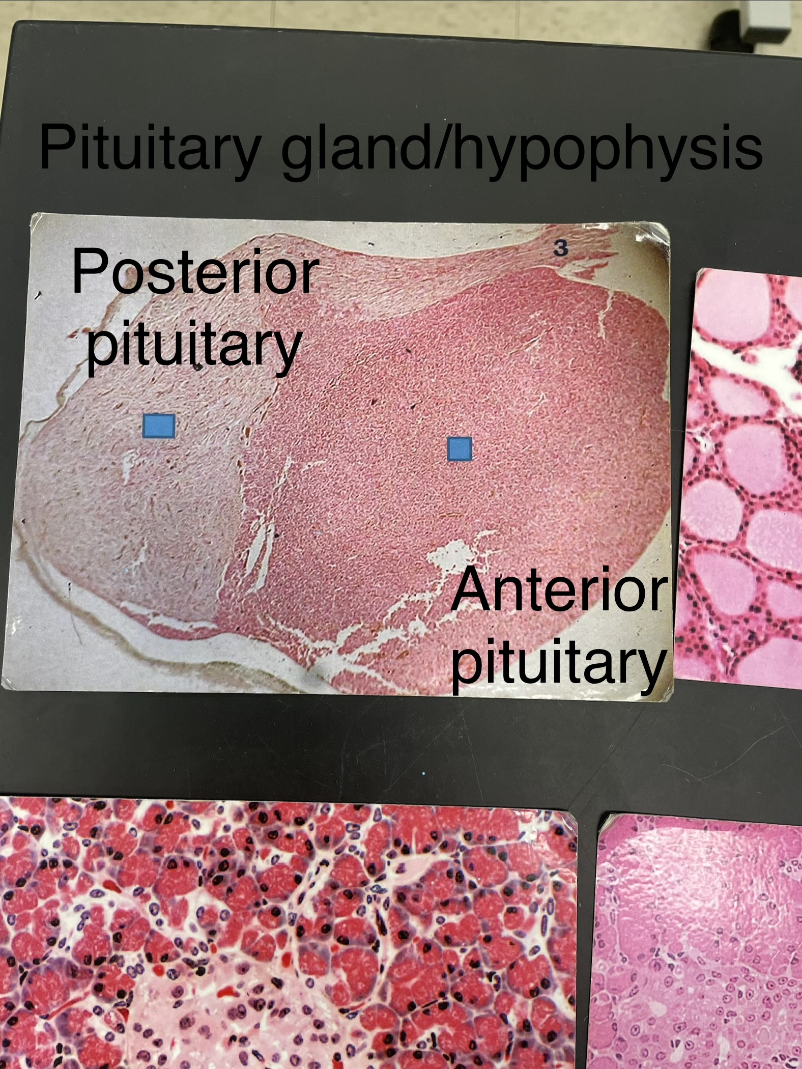
Pituitary gland (hypophysis)
Has two parts:
Anterior pituitary (adenohypophysis)
This side stains easily and appears darker
Heart → hypothalamus → anterior
(hypothalamus releases hormone for anterior to release hormone)
blood
Posterior pituitary (neurohypophysis)
The nervous tissue is hard to stain and you can almost see the hypothalamic hypophyseal tract
doesnt produce hormones
hypothalamus → posterior
move hormone from 1 organ to the other without leaving cell
myelinated axons; white matter
Be able to identify under the ‘scope:
Gland
Anterior pituitary (adenohypophysis)
Posterior pituitary (neurohypophysis)
Hormones:
Adenohypophysis
Associated releasing
hormones
FSH & LH
ACTH
TSH
GH
Prolactin
Neurohypophysis
ADH
Oxytocin
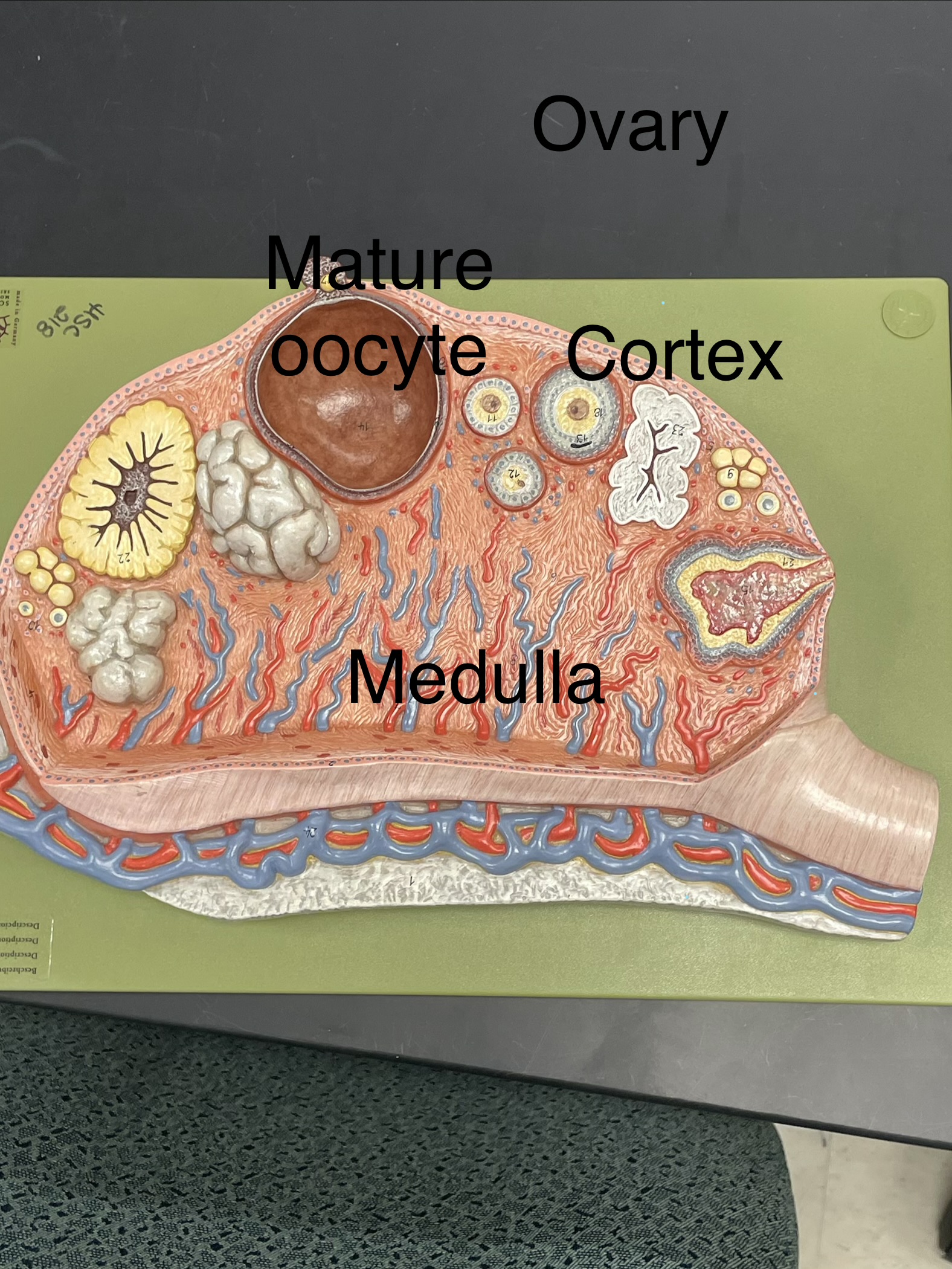
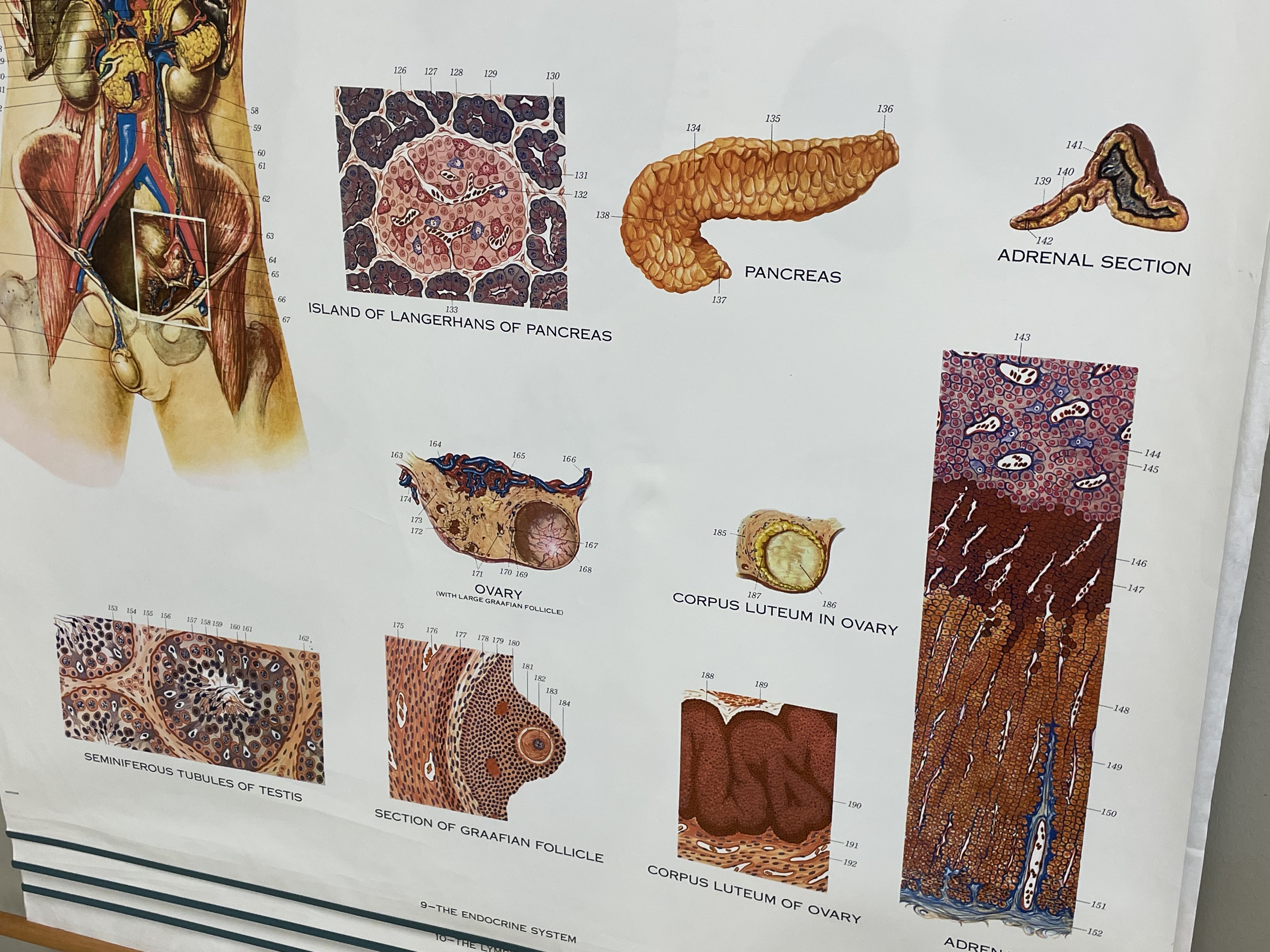
Ovary visual
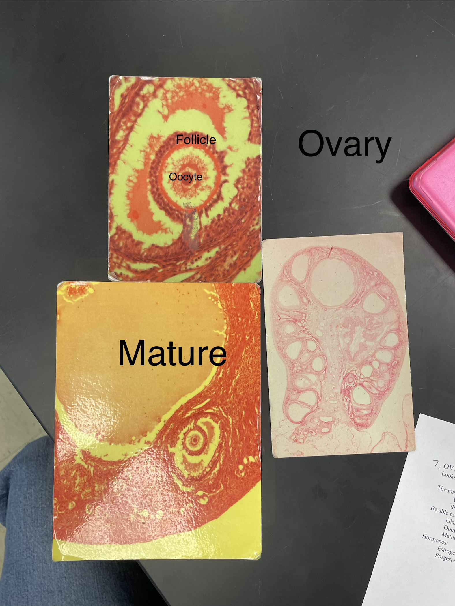
Ovary
Looks like the cartoon representations in the text.
The mature follicle is the LARGE structure.
You may or may not be able to view the ovum
big diagram: medulla “vessels” and cortex “business”
fluid space in follicle= mature
Be able to identify under the ‘scope:
Gland
Oocyte (Immature Ovum)
Mature Follicle
Hormones:
Estrogen
Progesterone
Parathyroid gland diagram
Thyroid gland diagram
Adrenal glands diagram
ovary diagram