CT Boards, Patient Care
1/33
There's no tags or description
Looks like no tags are added yet.
Name | Mastery | Learn | Test | Matching | Spaced | Call with Kai |
|---|
No analytics yet
Send a link to your students to track their progress
34 Terms
What are Mild Side Effects of IV Contrast Administrations?
Warmth
Nausea
Metallic taste
Mild itching
Anxiety symptoms
What are Moderate Side Effects of IV Contrast Administrations?
Normal INR Range
0.8–1.2
Normal GFR Levels
A normal eGFR for adults is 90 and above, although this number varies according to several factor
Layers of the Meninges
From the outside in, the layers are the dura mater, arachnoid mater, and pia mater
What are the Retroperitoneal Organs?
Ascending and Descending Colon
Adrenal Glands
Kidneys
Pancreatic Head and Body
Ureters
Aorta and Inferior Vena Cava
Severe Contrast Reactions
Anaphylactic shock
Severe bronchospasm
Cardiopulmonary arrest
Severe hypotension / cardiovascular collapse
Normal Creatine Levels
0.5 to 1.3 mg/dL
The Three Vessels that branch of Celiac Artery
common hepatic artery
splenic artery
left gastric artery
Calcium Scoring
demonstrates calcium in the left main coronary artery and circumflex coronary artery. The calcifications appear brighter than the surrounding tissue. Because calcifications appears in the left main coronary artery, this patient was likely referred for a cardiac catheterization
Congestive Heart Failure
when the heart muscle is unable to sufficiently pump blood, symptoms include irregular heartbeat, persistent cough, increased frequency of urination, and weight gain from fluid retention. Congestive heart failure requires lifelong management, usually with medication
may be the result of coronary artery disease, high blood pressure
Left Sided Heart Failure
fluid backs up in the lungs and may cause shortness of breath
Right-sided heart failure
, fluid may back up in the abdomen, causing swelling or edema of the lower extremities
Pulmonary Edema
it is known as cardiogenic pulmonary edema and results from increased pressure in the heart that pushes fluid through vessel walls and into the interstitial spaces of the lungs
Pneumonia
an infection of the lungs that causes inflammation of the air sacs. It can be classified according to where the infection occurred and the type of infectious agent. The most common type is community-acquired pneumonia
Pneumothorax
the abnormal collection of air in the pleural space
Atelectasis
the partial or complete collapse of the lung and occurs when the alveoli become deflated or filled with fluid, preventing normal expansion of the lung. Obstructive atelectasis occurs from an obstruction or blockage of the airway, such as from a foreign body, mucus plug, or mass in the airway
Bezoar
, is a collection of foreign material, such as hair, fiber, plastic, or other indigestible matter inside the digestive tract
Hernia Categories:
congenital or acquired. Congenital hernias are present at birth and are caused by embryonic developmental defects, whereas acquired hernias usually develop later in life, often due to trauma
Diverticulitis
a result of a weakness in the muscle layers of the digestive system that causes a “pocket” or outpouching. Although most common in the large intestine, diverticula can also appear in the esophagus, stomach, and small intestine
Classified as uncomplicated or complicated
Uncomplicated diverticulitis symptoms are abdominal cramping, bloating, flatulence, and irregular defecation
Complicated diverticulitis symptoms are rectal bleeding and anemia
P Wave
the first small round upward bump
Atrial Depolarization

Q Wave
the first downward deflection of QRS Complex
normal electrical activation, hearts middle wall

R Wave
first upward deflection in the QRS complex
Thick walls of the ventricles
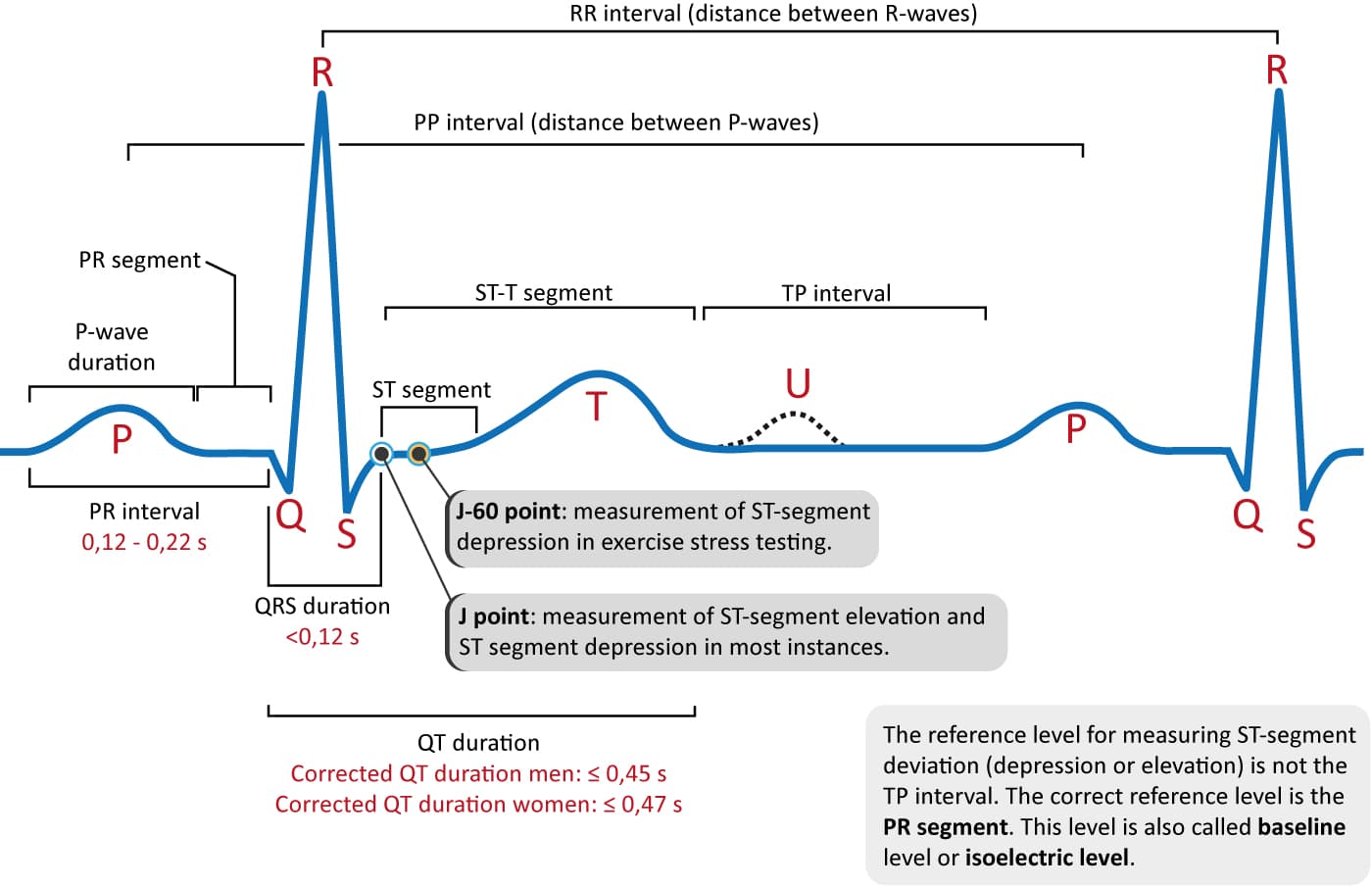
S Wave
he final phase of ventricular depolarization (electrical activation of the heart's lower chambers).

T Wave
repolarization of the heart’s ventriculus
Next bump after QRS Complex
QRS Complex
the sharp, prominent series of spikes seen on an electrocardiogram
represents the electrical depolarization (activation) of the heart's ventricles.
This electrical signal triggers the ventricles to contract and pump blood to the lungs and the rest of the body
R-R Interval
a full heartbeat represented
Lethargic
Drowsy, but easily awakened by speaking in a normal or slightly louder voice; may drift back to sleep when not stimulate
Obtunded
is a medical term used to describe a dulled or reduced level of alertness, arousal, or consciousness. An obtunded patient has decreased interest in their surroundings, appears drowsy, and exhibits slowed responses to stimulation
Stupor
A person in a stupor can only be temporarily aroused by vigorous, intense, or painful stimulation, and they quickly relapse into an unresponsive state once the stimulus stop
Cerebral Blood Flow (CBF)
how much blood is moving through brain tissue per minute
Units: mL/100g/min
Cerebral Blood Volume (CBV)
how much blood is currently in the tissue
Reflects capillary + small vessel blood content
Low CBV = infarcted (dead) tissue
Mean Transit Time (MTT)
MTT=CBF/CBV
What it means: how long blood takes to pass through tissue
High MTT = sluggish or blocked flow
Time to Peak (TTP)
Time from contrast arrival → maximum enhancement
Delayed TTP = delayed perfusion / ischemia