Thrombus and Embolic Events
1/23
There's no tags or description
Looks like no tags are added yet.
Name | Mastery | Learn | Test | Matching | Spaced | Call with Kai |
|---|
No analytics yet
Send a link to your students to track their progress
24 Terms
What is the difference between a thrombus and an embolus
thrombus is a blood clot attached to a vessel wall
embolus is a free floating clot
Conditions which increase likelihood of thrombus?
there is stasis (low-flow of blood)
there is a substrate
there is hypercoagubility of blood (more likely to clot)
What is the triad for causes of vascular thrombosis called ? What are the categories?
Virchow’s triad:
Stasis
Vessel wall damage
Hypercoagulation
Echo sign of stasis of blood
Spontaneous echo contrast
What does Spontaenous echo contrast mean in terms of risk?
increased likelihood of thrombus formation
What causes spontaneous echo contrast? What can exacerbate it
usually happens in AF, abnormal pump function of LA causes slower moving blood
exacerbated in AF with MS
List all the substrates for a thrombus?
RWMAs
Akinetic apex: apical thrombi
Aneurysmal LV region
Akinetic LV region
LAA: MS, MR, AF?
RA: L to R shunting?
Intracardiac devices: valve replacements, pacing leads (RA and RV), intravascular catheter, LV assist device, ASD occluder device (Amplatzer)
What increases chances of LA thrombus?
AF (loss of normal atrial contraction)
MS:
What in the LA / LAA can be mistaken for a thrombus?
pectinate muscles
In a patient with AF, MS and an echogenic mass in the LA, what is the likelihood this is a thrombus?
high
What can cause right heart thrombus?
conditions + pacing / defibrillator leads/ intravacular catheter
thromboembolism in peripheral veins (i.e DVT) in transit to pulmonary system
Are right heart thrombi usually formed in the right heart?
can be but this is rare
more often formed in the peripheral veins and travel back to right heart
What typically causes LV thrombus
Akinetic walls
Aneurysmal walls
Valve replacement
Name the different types of thrombus?
mural
laminated
How does thrombus usually attach to myocardium?
broad base attachment rather than via a stalk (perdunculated)
Medicines to prevent thrombus?
anticoagulants
Apixiban, rivaroxiban, dabigatran, edoxaban
Warfarin
Occasionally aspirin which is a blood thinner but not 1st line anticoagulant
Medicines to break down an existing thrombus?
thrombolytic drugs that induce fibrinolysis
What are the risks associated with a thrombus?
thrombus can dislodge/ detach from wall and become embolus
Embolic event
pulmonary embolism
brain aneurysm: Stroke

Terrible image quality but what can be seen in the PA bifucraction?
Saddle shaped embolus
Likely an acute pulmonary embolism
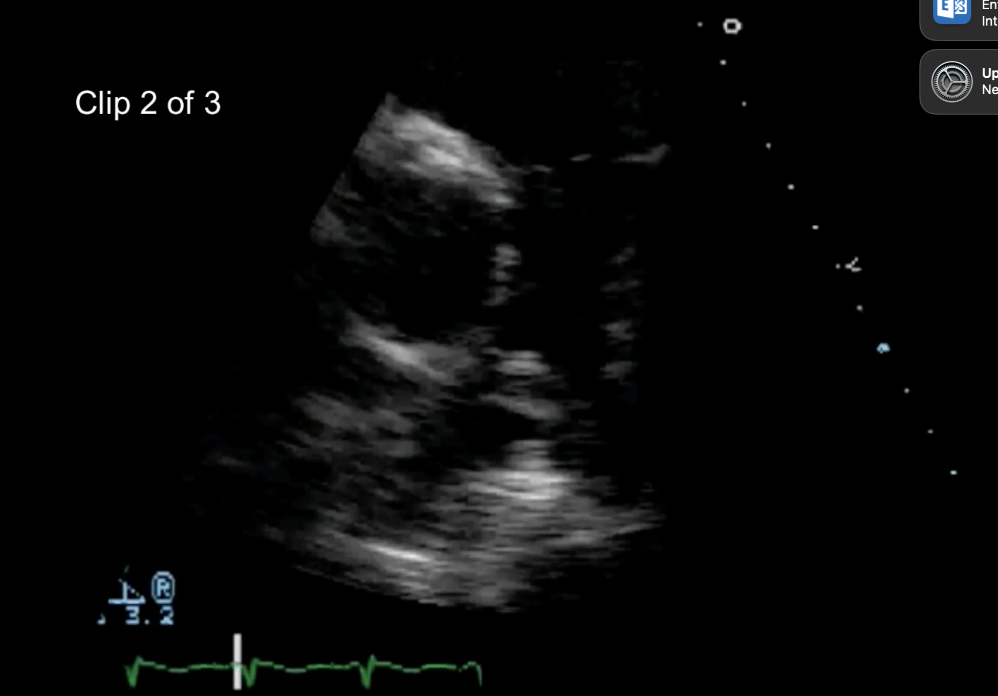
After seeing this, what other echo features to check for?
RV free wall: look for McConnell’s sign (akinesis with apical sparing) and 60/60 sign: PAT <60 ms and RVSP <60 mmHg
What is the echogenicity of thrombus like?
different to myocardium/ darker but change based on age. Older can look brighter due to calcification.
Thrombus vs a tumour: how can we tell the difference?
echogenicity: tumour should have similar echogenicity to myocardium/ liver (speckled parenchyma appearance)
attachment: perdunculated or broad base?
ask yourself: are there the conditions for a thrombus ? Stasis/ substrate/ hypercoagubility
What is the best ultrasound imaging technique for LA or LAA thrombus?
TOE due to reduced distance from the LAA (higher temporal resolution, and spatial res (sharpness) because higher frequencies can be used because penetration is less)
What is the best ultrasound imaging technique for LV thrombus?
TTE because this is closer to LV than TOE would be (particularly the apex which is a major site of thrombi)