EKG(Part 2): Lesson 17-Junctional and Ventricular Arrhythmias, Heart Blocks
1/20
There's no tags or description
Looks like no tags are added yet.
Name | Mastery | Learn | Test | Matching | Spaced | Call with Kai |
|---|
No analytics yet
Send a link to your students to track their progress
21 Terms
You are working in an ambulatory setting and collecting an EKG on a patient who complains of intermittent heart palpitations. What common abnormal heart rhythm will the EKG show?
Premature junctional contraction
Which wave may not be seen on an EKG with a premature junctional complex?
P wave
Which statement describes the heart rate for a patient with junctional bradycardia?
A heart rate below 40 beats per minute.
While performing an EKG, an inverted P wave right after the QRS complex is noted with a regular rhythm and heart rate of 92 beats per minute. Which type of rhythm is described?
Junctional escape rhythm
You are performing an EKG and notice an accelerated junctional rhythm on your patient's EKG tracing. This means that the QRS complex appears:
normal
Which atrioventricular block will show the PR interval getting longer until the QRS complex is dropped?
Second degree atrioventricular block Type I
You are attending to a patient who has a history of ventricular dysfunction. While collecting the EKG you notice abnormal P waves. How will the P wave appear if the junctional rhythm is initially depolarized?
The P wave appears after the QRS complex.
What type of condition is present on an EKG reading from a patient that shows a QRS complex is wider than normal, the T wave and the QRS complex deflect in opposite directions, and the P wave is absent.
Ventricular arrhythmia
Which rhythm is characterized by a widened and abnormal looking QRS complex every other beat?
Bigeminy premature ventricular contraction
You obtain an EKG tracing from a patient that indicates a condition of fast QRS complexes. What type of arrhythmia would be visible on the EKG?
Ventricular tachycardia
This occurs when the natural pacemaker, the SA node, stops
working or sends signals that are too slow or weak. Your heart responds by using
one of your backup pacemakers instead.
junctional rhythm
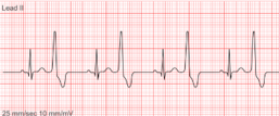
It is an early or
premature electrical impulse that originates in the atrioventricular junction also known as junctional tissue, occurring before the next
expected P wave, leading to an irregular rhythm. Has abnormal or absent P waves, and has short than normal PR intervals. Very narrow QRS complex because His-Purkinje fibers are mainly used. Can have either very slow or fast heart rate.
premature junctional complex (PJC)
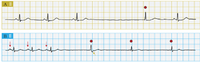
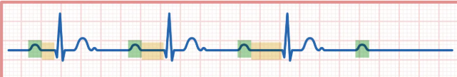
Anytime the rate of the SA node is not adequate an area at the AV junction can pace the heart. heart beats 40-60bpm. The atria are depolarized through a process known as backward conduction, resulting in inverted P waves. Any “time” this happens to a QRS complex, they are escape “beats”
junctional escape rhythm
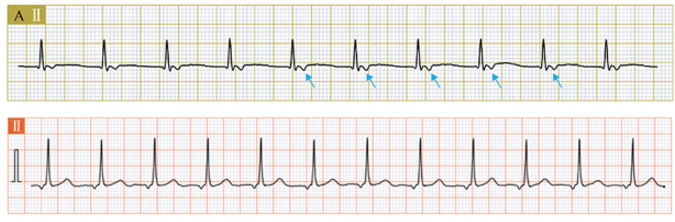
basically the 60-100bpm version of junctional escape rhythms
accelerated junctional rhythm
is a regular rhythm with the PR interval constantly prolonged. All other aspects of
the EKG may appear normal.
first degree AV block
the PR intervals stays the same but, “some P waves” don’t conduct to QRS
Second-Degree Atrioventricular Block: Mobitz type 2
PR intervals gradually lengthen, until P waves don’t conduct to QRS
Second-Degree Atrioventricular Block: Mobitz type 1
When the electrical signals that regulate the heartbeat come from the ventricles (the lower chambers of the heart), as opposed to their usual origin in the sinoatrial (SA) mode. This implies that the signals controlling the heartbeat come from a different
channel than normal.
ventricular arrhythmia
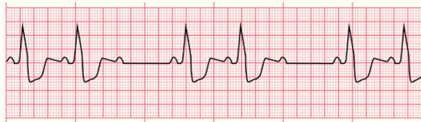
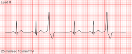
occurs when an area within either the right or left ventricle becomes irritated causing the
ventricles to contract early.
Premature Ventricular Contraction (PVC)
If every other beat is a PVC,
Bigeminy PVC
If every third beat is a PVC,
Trigeminy PVC