1751 Facial Bones Anatomy
1/41
There's no tags or description
Looks like no tags are added yet.
Name | Mastery | Learn | Test | Matching | Spaced | Call with Kai |
|---|
No analytics yet
Send a link to your students to track their progress
42 Terms
What are the 14 Facial Bones
2 Nasal
2 Maxillary (Maxilla Singular)
2 Lacrimal
2 Zygomatic
2 Palatines
2 Inferior Nasal Conchae
Vomer
Mandible
(The 2 palatines and vomer are located internally and are not visible on a skeleton from the exterior. Facial bones contribute to the shape and formation of a person’s face)
What do facial bones provide
Structure, shape and support for the face. They form a protective housing for the upper ends of the respiratory and digestive tracts
Maxilla (2)
Largest immovable bones of face
Articulated with all other facial bones, except mandible
Each maxilla assists in formation of 3 cavities of the face: roof of the mouth, nasal cavity, and part of floor of orbit
4 processes (frontal, zygomatic, alveolar, palatine)
All other facial bones are closely associated with the 2 maxillae; thus they are structurally the most important bones of the upper face
Zygomatic Bones (2)
Prominence of cheeks
Forms part of side wall and floor of orbital cavities
Temporal Process unites with zygomatic process of temporal bone to form the zygomatic arch
Zygomatic arch is the cheek bone
What bones do the zygomatic bones articulate with
Frontal bone superiorly
Temporal bone side
Maxilla anteriorly
Sphenoid posteriorly
Tripod (Trimalar) Avulsion Fracture
Fracture of lateral wall
Results in a free floating zygoma
Caused by a blow to the cheek
Fracture in 3 places
Zygomatic bone - frontal process
The maxilla - Zygomatic process
The Temporal Bone - Zygomatic Process
Lacrimal (2)
Smallest bones in the skull
About the size of a fingernail
Lie anteriorly and medially within orbit
Posterior to frontal processes of maxilla
Each bone contains a lacrimal foramen through which the tear duct passes
Both lacrimal and nasal bones articulate with 2 cranial bones and 2 facial bones
What bones do the lacrimal bones articulate with
Frontal Bone
Ethmoid Bone
Maxilla
Inferior Nasal Conchae
Nasal Bones (2)
Small and thin
Vary in size
2 join together at the MSP to form bridge of nose
Point of junction of 2 nasal bones with frontal bone - nasion
Most commonly fractured facial bone
What bones do the nasal bones articulate with
Other nasal bone
Frontal bone
Ethmoid bone
Maxillae
Inferior Nasal Conchae
Long narrow very thin bones with a lateral curl
Gives scroll like appearance
Upper 2 nasal conchae are processes of ethmoid bone. The inferior is a separate bone
The 3 nasal conchae are covered with a mucous membrane to warm, moisten and clean inhaled air
The purpose of the 3 pairs of nasal conchae are to divide the nasal cavity into various compartments. These compartment break up the flow of air coming into the nasal cavities before it reaches the lungs. The air is warmed and cleaned
Palatine Bones
2 L shaped bones
Vertical portion lies between the pterygoid process of sphenoid and maxilla
Horizontal portion fuses with palatine process of maxilla to complete bony palate
Composed of vertical and horizontal plates
Located internally and are not visible from the outside
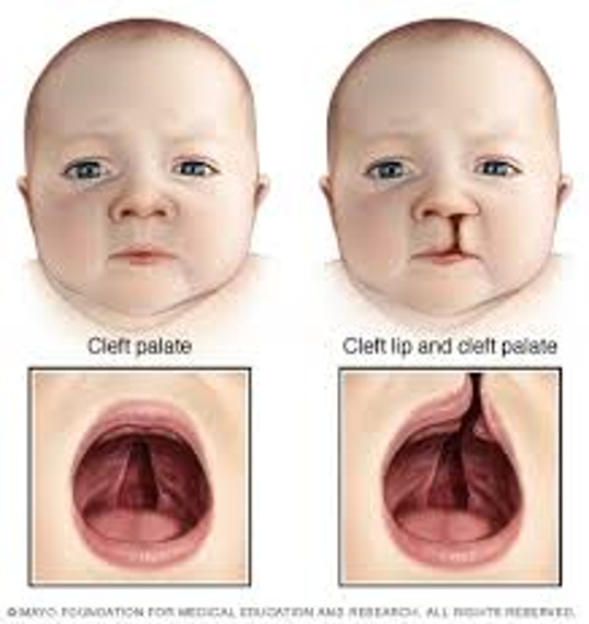
The maxilla palatine processes to form the hard palate. Separation of the 2 causes cleft palate
Vomer Definition
Plowshare due to the resemblance to the shape of a plow cutting blade
Vomer Bone
Thin plate of bone
In the MSP of the nasal cavity
Forms inferior portion of nasal septum
The surface of the vomer are marked by small furrow-like depressions for blood vessels, thus the source of nosebleed with trauma to the nasal area
Deviated septum describes the clinical condition wherein the nasal septum is deflected or displaced laterally from the midline of the nose. This deviation usually occurs between the septal cartilage and the vomer
Mandible
Largest and densest facial bone
Body of Mandible
Curved horizontal portion
Rami of Mandible
2 vertical portions on each side of body
Angle of mandible (Gonion)
Junction of body and ramus
Mental Protuberance of Mandible
Anterior, triangular prominence
Symphysis of Mandible
Most anterior and centra part where left and right halves fuse
Alveolar Portion of Mandible
Superior border of body; consists of spongy bone that supports roots of teeth
Mental Foramina of Mandible
Small openings on each side below the second premolar; transmit nerves and blood vessels
Coronoid Process of Mandible
Anterior process on top of ramus
Condylar Process of Mandible
Posterior process on top of ramus
Mandibular Notch
Concave area at top of ramus between coronoid and condylar process
Mandible and TMJ
Mandible is the only moveable bone on adult skull
TMJ only moveable joint in skull
Formed by the condyle (head) of condyloid process
Fits into the temporomandibular fossa of temporal bone
Slants posteriorly 15 degrees and inferiorly/medially 15 degrees
Just anterior to and slightly superior to the EAM
Hyoid Bone
Small U-Shaped bone situated at the base of the tongue
Accessory bone of axial skeleton - not a facial or cranial bone
Only bone in the body that does not articulate with another bone
Held in position by the ligaments extending from the styloid process of the temporal bone
Hyoid serves as an attachment for certain muscles of the larynx and tongue and is easily palpated just above the larynx
What bones compose the Orbits (7 each, 14 total)
3 Cranial Bones
Frontal
Sphenoid
Ethmoid
4 Facial Bones
Maxilla
Zygoma
Lacrimal
Palatine
Orbits
Cone shaped
Rim is base
Apex is posterior portion of cone and corresponds to the optic foramen (opening for optic nerve)
Using OML parallel to floor, orbits will project upward 30 degrees and toward MSP 37 degrees
Each orbit contains 3 openings in the posterior portion
Superior orbital fissure
Inferior orbital fissure
Optic Foramen - For transmission of optic nerve which is a continuation of the retina
Each orbit has the 3 openings for 12 pairs of cranial nerves to pass
Blow Out Fracture
Results from a direct blow to the front of the orbit that transfers the force to the orbital walls and floor (orbital floor just above maxillary sinuses)
Tissue - FIbrous
Type - Suture
Movement - Immovable
Coronal Suture
Sagittal Suture
Lambdoidal Suture
Squamosal Suture
Tissue - Synovial
Type - Hinge and Gliding
Movement - Freely Moveable
Temporomandibular
Tissue - FIbrous
Type - Gomphosis
Movement - Immovable
Alveolar Sockets
Tissue - Synovial
Type - Ellipsoidal
Movement - Freely Moveable
Atlantooccipital
Basal Fracture
Fracture located at the base of the skull
Blowout Fracture
Fracture of the floor of the orbit
Contre-Coup Fracture
Fracture to one side of a structure caused by trauma to the other side
Depressed Fracture
Fracture causing a portion of the skull to be pushed into the cranial cavity
Le Fort Fracture
Bilateral horizontal fracture of the maxillae
Linear Fracture
Irregular or jagged fracture of the skull
Tripod Fracture
Fracture of the zygomatic arch and orbital floor rim and dislocation of frontozygomatic suture