Human Body Systems
1/36
There's no tags or description
Looks like no tags are added yet.
Name | Mastery | Learn | Test | Matching | Spaced | Call with Kai |
|---|
No analytics yet
Send a link to your students to track their progress
37 Terms
Homeostasis
A body’s process of maintaining a stable balanced internal environment. It is important for organisms as it allows them to exist in a changing environment, for instance when it is hot and you are more active you sweat instead of immediately overheating.
Negative feedback loop
A negative feedback loop occurs when a stimulus sends a signal triggering a response that decreases the original stimulus. This is what allows organisms to maintain homeostasis, as they counteract a change to bring it back to normal which is essentially what homeostasis does. Example: Dehydration → Colon being allowed to absorb more water. Temperature regulation.
Positive feedback loop
A positive feedback loop occurs when a stimulus sends a signal triggering a response that increases the original stimulus. This can still be trying to stop something like stop bleeding from a cut, but instead it amplifies the stimulus. It is not used to maintain homeostasis thus as it amplifies a change rather than counteracting one. Example: platelets to repair a blood tear → more platelets. Child birth.
Endocrine system
Much slower and longer lasting than the nervous system. Glands secrete/release hormones (chemical signaling) which are carried in the circulatory system (blood) to target cells where hormones bind to them through target cell receptor proteins. The range of targets can vary from many to only a few. They change how the genes are expressed (off or on) by impacting primarily proteins. If there is a problem with production of something, like testosterone, is actually maybe due to a problem with another gland, like the pituitary not releasing FSH and LH to stimulate the testes to release testosterone.
Peptide hormones are hydrophobic, so they only reach a receptor to trigger the cellular activity. Steroid hormones are hydrophilic so they directly reach the nucleus by way of a steroid receptor in the cytoplasm to trigger the cellular response.
Major glands
Hypothalamus, Pineal Gland, Pituitary Gland, Thyroid Gland, Adrenal Gland, Pancreas, Ovaries, Testes. Pituitary secretes FS (Follicle stimulating)H and L(Luteinizing)H, ovaries secrete estrogen/progesterone, testes secrete testosterone.
How do the male and female reproductive systems (testes, ovaries) make and deliver gametes.
Eggs are produced in the ovaries, and are released to the fallopian tube where they will be fertilized and then attached to the uterus with the uterine lining if fertilized, if not it won’t attach and will be shed with the uterine lining. The sperm is taken care of in the male reproductive system as it must be able to keep it alive, nurtured, warm, protected and be able to ejaculate it to be able to reach an egg.
Spermatogenesis vs Oogenesis
Spermatogenesis
10 weeks
After puberty → death
4 functional sperm
Much greater production daily, especially with mitosis in the testes, many diploid cells created to become haploid cells.
Testes
Oogenesis
28 days
Pre-birth (Meiosis 1) →PAUSE→ puberty (Meiosis 2, only around 1 per month goes through it, technically only complete after fertilization)→ menopause
Cytoplasm does not evenly split, one gets the nucleus one gets the majority of the cytoplasm and such. Sperm kicks out the second nucleus.
1 egg and 2-3 polar bodies
Ovaries
Smaller production cyclically (monthly typically)
Genetically unique offspring how
Genetic variation through crossing over, independent assortment and random fertilization. Also environment, with the behavior of the mother and history of both parents, the same fertilized egg put into another mother or the same mother at a different time would result in a genetically different child. Fertilization occurs in the oviduct (fallopian tube).
Ovarian and menstrual cycles of a female mammal.
The pituitary gland releases LH and FSH, triggering the maturation of a follicle, which secretes estrogen. The rise in estrogen stimulates the pituitary gland to release more LH and FSH, creating a positive feedback loop. Once the follicle has fully matured the egg is released and the cells are repurposed into the corpus luteum. The corpus luteum now has a new job, and still releases estrogen but also now progesterone. Together progesterone and estrogen act as negative feedback on the pituitary gland causing a decrease in the secretion of LH and FSH and then steadying, up until the full degradation of the corpus luteum. The corpus luteum full degradation leads to a major drop in estrogen and progesterone levels, and the shedding of the uterine lining, allowing for an increase of stimulation of the LH and FSH/less negative feedback stimulating the start of the cycle again. The menstrual cycle occurs over 28 days, with the first follicular phase occurring until ovulation on day 14, which then starts the luteal phase.
Key events of embryonic & fetal development
Trimester 1
All major organ systems are established, 9 weeks after fertilization called a fetus (body structures start to appear, embryo from fertilization), and the fetus begins to be able to move its arms and legs and look distinctly human.
Trimester 2
Increase in size and refinement of human features
Some growth, roughly 7.6 inches and 1 pound at 20 weeks.
Trimester 3
Time of rapid growth, circulatory and respiratory systems mature (pulmonary surfactant), muscles thicken and skeleton hardens.
Purpose of digestion
Break down the food and liquid consumed into essential nutrients.
Chemical digestion
Breakdown of chemical bonds by way of hydrolysis, which is aided by enzymes.
Mechanical digestion
Breakdown (physical) of large food pieces into smaller particles, without altering their chemical structure.
Human digestive system
Mouth
Salivary glands
Pharynx
Esophagus
Stomach
Gallbladder, liver, and pancreas.
Small intestine
Large intestine (including colon)
Rectum
Anus
Ingestion
Food is ingested through the mouth.
Digestion (Mechanical is peristalsis)
Digestion begins in the mouth, and goes until the small intestine. Food is mechanically digested in the mouth (teeth and tongue) and through the movement muscles (contract and relax) surrounding the esophagus and stomach continue the mechanical breakdown. Chemical digestion begins in the mouth, with the salivary glands producing amylase which causes the immediate breakdown of carbs in the mouth. Food moves down the esophagus into the stomach, where the gastric juices (enzymes and acid) breakdown chemical bonds, before food moves into the small intestine where digestive enzymes (produced by the pancreas) help with digestion, as well as bile produced by the liver and stored in the gall bladder helps break down (hydrophobic) fats mechanically, so that lipase can chemically break them down further (from the pancreas), before the food moves into the large intestine.
Absorption
Absorption begins in the small intestine, where nutrients are absorbed through the thin walls of the intestine, which surface area is maximized by way of the villi (small folds in the intestinal lining) into the bloodstream (capillaries) to the liver, and lymphatic system (fats, fat soluble vitamins) absorbed by lacteals to the lymphatic system.The remaining indigestible material continue to in the large intestine, with the absorption of water, by way of the colon (section of the large intestine), into the bloodstream, leaving behind waste (fiber from cellulose) that stays in the rectum till excretion through the anus.
Digestive enzymes digesting macromolecules.
They break down large complex macromolecules into their smallest chemical building blocks so the body can absorb them (proteins, speed up breakdown by way of hydrolysis).
Digestion of carbs
Amylase, produced by salivary glands, denatured by stomach acid, gets reproduced in the small intestine. Begins in the mouth. Monosaccharides to polysaccarides.
Digestion of lipids
Lipids (fats/triglyceride) are hydrophobic, so they don’t mix with water, cling to each other, into globs, and the enzyme lipase can only interact with the surface area. So the liver creates bile (stored in gallbladder) and it ends up released to the small intestine, breaking down big globs into smaller globs, so that lipase can interact w/ more fat broken down into fatty acids and glycerol. Technically digestion begins in the mouth, with chewing.
Digestion of proteins
Proteins are broken down in the stomach, where pepsin as well as stomach acid begins breaking them down into amino acids.
Digestion of nucleic acids
Nucleic acids are broken down into nucleotides by way of nucleases, beginning in the stomach.
Nervous system
Sensory receptors detect a change, and send sensory input by way of sensory neurons to the brain, where the information is integrated and a reaction is determined, which is sent through the spinal cord (communication between the brain and PNS) to trigger a motor output, by way of motor neurons, triggering glands or muscles, or more neurons.
Neuron
Not the most abundant, but the most functional. They are essentially communication cells. Axon branches lead to the axon terminal (synaptic terminals).
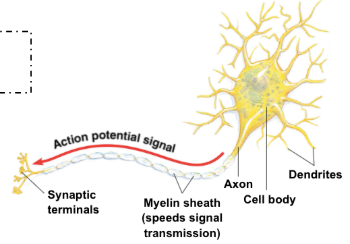
Neurotransmission.
The action potential (an electrical signal) arrives from the axon to the synaptic/axon terminal. This triggers the influx of calcium into the presynaptic terminal/axon terminal. This leads to the fusing of the synaptic vesicle (containing neurotransmitter neurons) with the plasma membrane. This releases neurotransmitters through exocytosis into the synapse. They diffuse across the synapse, and bind to the receptor proteins located on the dendrite. The binding triggers the action potential being passed along, either inhibiting or exciting the receiving neuron. Any remaining neurotransmitters are recycled through/into reuptake proteins located on the synaptic axon/terminal back into the synaptic/axon terminal, or they are broken down by enzymes.
Requirements of Respiratory Surfaces
Moist - allows gasses to dissolve, then diffuse into cells.
Thin - allows for rapid diffusion of gases
Large surface area - allows for efficient gas exchange (a higher volume of gases can pass through, think large doorways versus small doorways), allows for bulk flow.
Structures of the respiratory system
Nasal cavity
Pharynx (Esophagus)
Larynx
Trachea
Bronchus
Bronchiole
Diaphragm
Lungs
Blood capillaries
Alveoli
Nasal cavity, Pharynx (Esophagus), Larynx, Trachea, and the Bronchus.
Nasal cavity / Nose / Primary entry point for the respiratory system
Pharynx (Esophagus) / Essentially where the throat is / An open air channel, connecting the nose and mouth to the larynx and trachea, also a passageway for food.
Larynx / Right below the esophagus / Only a channel for air flow and houses your vocal cords, and it is connected with the epiglottis (which prevents food from going down the windpipe (trachea).
Trachea / Below the Larynx and Pharynx (long throat like part between lung and head) / A major passageway for air, and it also filters, warms, and moistens the air. It contains cartilage rings, which provide structure to the trachea.
Bronchus / Branch off from the trachea into the lungs / another passageway that conducts, warms, moistens, and filters the air.
Lungs, blood capillaries, alveoli, diaphragm, and bronchiole.
Lungs / perform gas exchange (transfer oxygen into the bloodstream through diffusion and remove carbon dioxide, it diffuses out of the blood). The right lung is larger (55%) vs the left lung (45%).
Blood capillaries / Connect to the alveoli, thinner than the bronchioles / where diffusion of O2 into the bloodstream occurs (thin walls) and diffusion of CO2 out of the bloodstream occurs. The capillaries also then connect to the rest of the body’s bloodstream helping distribute the nutrients.
Alveoli / Broccoli like things in the lungs / The facilitate gas exchange, oxygen diffuses across the thin walls of the alveoli into the capillary bloodstream, and vice versa with the carbon dioxide removal.
Bronchiole / connect to the alveoli (mini branches off of bronchus) / smaller versions of the bronchus that deliver oxygen-rich air to the alveoli and carry carbon dioxide out during exhalation, while regulating air flow and filtering out dust and germs before gas exchange occurs.
Diaphragm / Bottom rainbow-like thing at the lungs / Muscle separating the chest cavity from the abdominal cavity, and its contraction and relaxation are very significant.
Inhalation
During inhalation the rib muscles contract and the diaphragm contracts (lowers it,less rainbow) causing the rib cage to expand, increasing the volume of the chest cavity. This causes a decrease in pressure, as gas particles have more space to move around and so bump into each other and the barriers less, meaning a decrease in gas pressure. This decrease in pressure makes the outside pressure greater, and so the greater pressure will force air into the body.
Exhalation
During exhalation the rib muscles relax and the diaphragm relaxes (more rainbow), causing the chest cavity’s volume to decrease, leading to an increase in pressure (see above for how gas pressure changes as it is measure in how often the particles contact the container), and so now there is a greater air pressure inside instead of outside, forcing air out of the lungs.
Pathway of a red blood cell from the right atrium to the aorta, valves, and the septum.
Starting in the right atrium, the blood cell flows into the right ventricle and then into the pulmonary arteries to the lungs where the blood receives oxygen, becoming oxygenated, and then proceeds to flow through the pulmonary veins back to the heart, into the left atrium, which pumps blood into the left ventricle, when then pumps blood to the rest of the body through the aorta, where it becomes deoxygenated (distributes the nutrients and oxygen), before it returns to the heart through either the superior vena cava or inferior vena cava (higher vs lower to the heart) into the right atrium, starting the cycle again. There are valves in-between each atrium and ventricle and then between the ventricle and the arteries/aorta, to prevent back flow of the blood to maximize efficiency. The septum separates the two ventricles, also maximizing efficiency as the blood sent to one part is all the “correct“ type rather than it being a mix of both where only some would be properly used/in the right location.
Muscles
The left ventricle has much thicker muscle than the right ventricle, and the ventricles have much thicker muscles than the atrium. This is all due to pumping blood, as the distance from the atria to the ventricles is much shorter than the distance from the ventricles to the rest of the body and lungs. However, the rest of the body is a much greater distance than the lungs to the heart, so the muscles for the left ventricle must be greater. Think about squeezing a water bottle and a kindergartner vs a bodybuilder, how far will the water squirt out.
Heart, arteries, veins, capillaries, red blood cells role in transporting nutrients and oxygen to cells and removing cellular waste (oxygen depleted blood is waste heavy).
The human body is too large for just diffusion to satisfy this transportation. Instead, through the circulatory system (pulmonary circulation and systemic circulation), with our 4 chambered heart, can transport oxygen and nutrients to cells and remove cellular waste. The hemoglobin in the red blood cell attaches to oxygen and CO2, allowing it to be transported with the blood flow, which is from the heart which pumps it through arteries away from the heart to capillaries which go to more specific parts, to veins which bring the blood back to the heart.
Structure of blood vessels
An artery has a rigid wall, with a thick outer wall and small lumen, as well as a thick layer of muscles and elastic fibers, and experience high pressure as they carry blood away from the heart (pump it away).
A vein is more flexible, with a thin layer of muscles and elastic fibers, a large lumen, and a fairly thin outerwall as it just takes blood back towards the heart (low pressure). The movement of skeletal muscle, forces blood against gravity back to the heart.
A capillary has a very small lumen, and the wall is made of a single layer of cell, and is very fragile. This facilitates diffusion, and helps explain why you get bruises.
Blood pressure
The force blood exerts on walls of blood vessels. Systolic/Diastolic, systolic is the high blood pressure while the heart contracts, and diastolic is the low blood pressure while the heart relaxes. Systolic is typically between 90 and 120, and diastolic is typically between 60 and 80.
Heart attack major risk factors
High blood pressure places excess stress on artery walls, and can cause tears which result in plaques forming/collecting at the tears (harder over time), causing the passage to narrow, and stiffen the artery to no longer regulate blood flow easily, so the heart must do more work to pump the blood further, and a plague fully building up prevents the flow of blood back to the heart, from a clot forming from a plaque rupture. High salt intake, as due to osmosis this creates a hypertonic environment, so more water flows in, in response, expanding the volume increasing blood pressure. Also smoking damages the blood vessels and heart, and thickens blood and increases blood pressure.