Endocrine System
1/103
There's no tags or description
Looks like no tags are added yet.
Name | Mastery | Learn | Test | Matching | Spaced | Call with Kai |
|---|
No analytics yet
Send a link to your students to track their progress
104 Terms
What are hormones?
They are chemical messengers
Travel throughout the body
Coordinate activity between cells, tissues, and organs
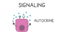
Ways to secrete hormones
Endocrine
Paracrine
Autcrine
Endocrine
Hormones secrete hormones directly into the bloodstream
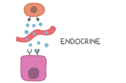
Paracrine
Hormones secrete into neighboring cells
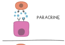
Autocrine
Hormones secrete into the same cell that is secreting it.
Three main hormone types
Peptide hormones
Steroid Hormones
Amino Acid Derived hormones
Peptide Hormones
What are they made of
How do we make them?
Hormones based on proteins
Synthesis: They are produced in the rough endoplasmic reticulum by linking amino acids through peptide bonds.
What do peptide hormones do? How do they act?
Bind to cell surface receptors rather than freely passing through cellular membranes
Proteins are water soluble, not lipid soluble
Indirect Stimulation
Peptide hormone interaction with cell surface receptors kicks of a signal transduction pathway in order to carry out the signaling
Ligand gated receptors
Receptor proteins that are capable of inducing intracellular signal transduction pathways once an extracellular ligand is bound
Second Messengers
Plus all 4 examples of them
Signaling molecules independent of the original hormone which propagate signals within the cell (intracellular effect)
cAMP (cyclic AMP),
IP3 (inositol triphosphate)
DAG (diacylglycerol),
Ca2+ (calcium ions)
GCPRs
GPCRs (G Protein Coupled Receptors): Associated with a G Protein on the intracellular domain that is responsible for conveying intracellular signals
Ex Pathway: IP3/DAG
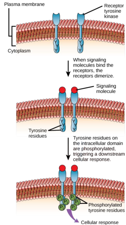
Receptor Tyrosine Kinases
Receptor Tyrosine Kinases (RTKs) Twin components dimerize to and cross phosphorylate in order to influence intracellular signals
Ligand Gated Ion channels
Ligand-Gated Ion Channels: Channel proteins which change shape once a ligand is bound in order to allow the flow of ions across membranes
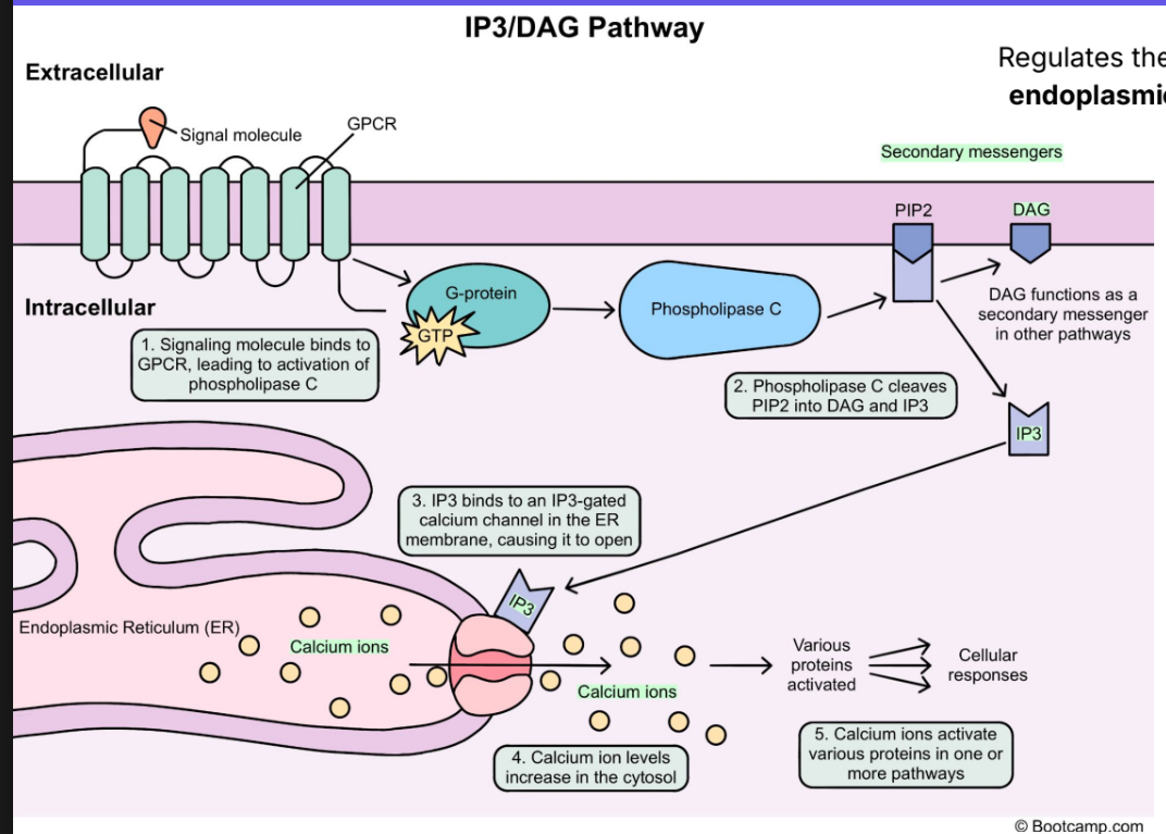
IP3/DAG Pathway Purpose
Regulates the release of stored Ca2+ from the endoplasmic reticulum into the cell cytosol
IP3/ DAG Pathway
1. The pathway is activated when an appropriate peptide hormone binds the GPCR
GPCR activates the associated G protein via the binding of one GTP molecule
3. G protein activates the enzyme Phospholipase C
Activated Phospholipase C cleaves the lipid PIP2 into two separate second messengers (IP3 and DAG)
5. IP3 binds ligand gated Ca2 channels on the surface of the ER
6. ER calcium channels open, releasing Ca2+ into the cytosol
7. Free Ca2+ goes on to affect multiple other pathways
G Proteins —> goes from GDP to GTP to get activated.
Receptor Tyrosine Kinases
Peptide hormone receptor with an independent mechanism from the GPCR
Made of two mirror components that dimerize once a peptide hormone is bound
Intracellular RTK domains cross-phosphorylate to kick of a second messenger response
RTK components phosphorylate each other and affect other proteins
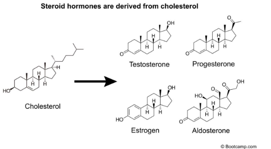
Steroid Hormones
What are they made of
Hormones made from modified cholesterol molecules
Synthesis: Production occurs in the smooth endoplasmic reticulum
Structure: Basic fused 4-ring structures with differentiating functional groups
Where are steroid hormones made?
Synthesis: Production occurs Smooth Endoplasmic Reticulum. (same as fats, remember that)
Structure of steroid hormones
Basic fused 4-ring structures with differentiating functional groups
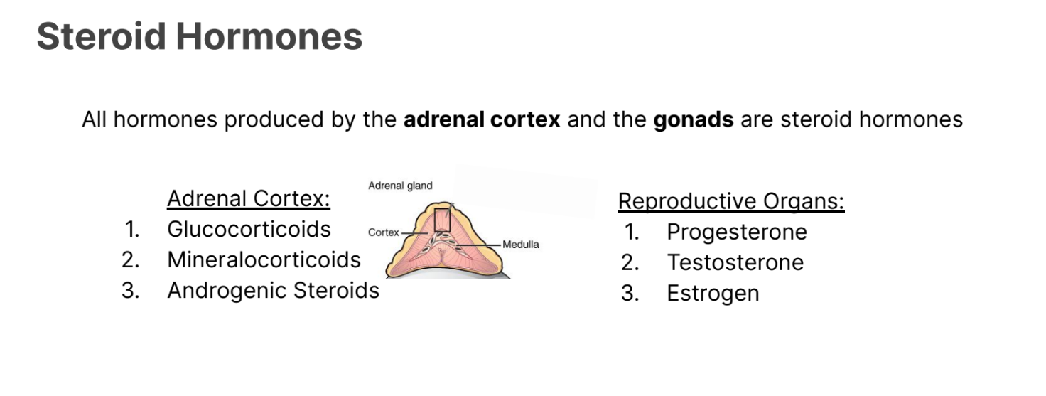
Where are hormones produced by the Adrenal Cortex and gonads stored?
They are stored as steroids.
Adrenal Cortex:
Glucocorticoids
Mineralocorticoids
Androgenic Steroids
Reproductive Organs
Progesterone
Testosterone
Estrogen
What do steroid Hormones do?
How do they move around
How do they activate things?
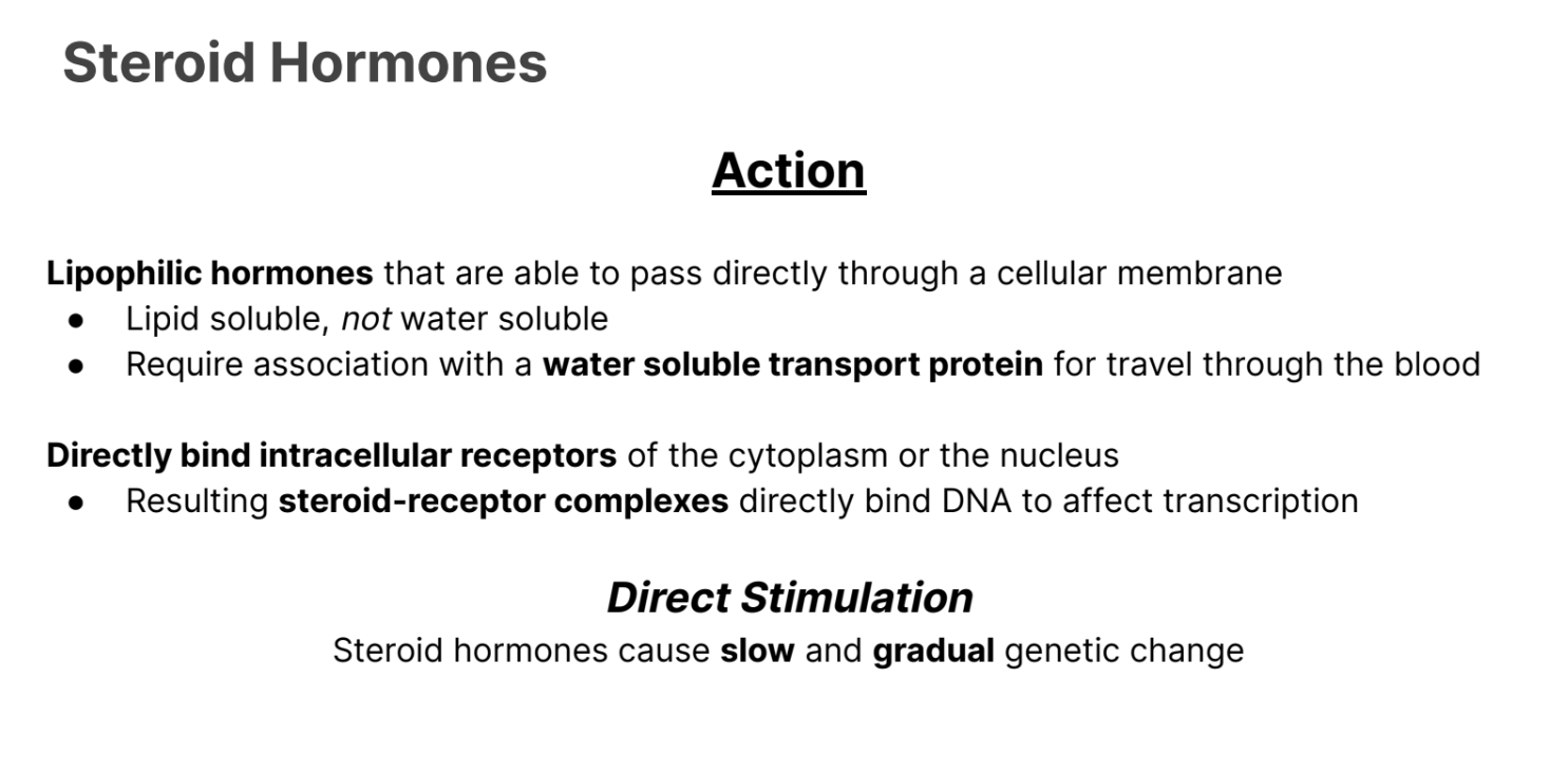
They are lipophillic hormones and can pass directly through a cellular membrane.
Lipid Soluble NOT water soluble.
Require association with water soluble transport protein to travel through the blood.
Directly bind intracellular receptors of the cytoplasm or the nucleus
Results in steroid receptor complexes that directly bind to NDA to affect transcription
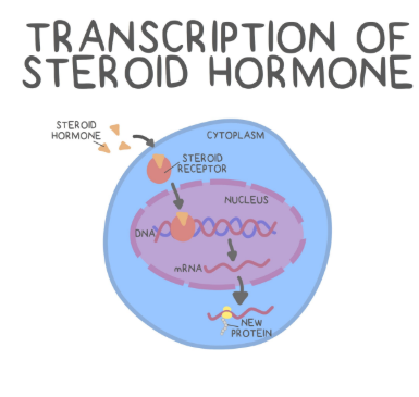
What is direct stimulation in hormones, and what type undergoes it?
Steroid Hormones undergo slow and gradual genetic change.
Amino Acid Derived Hormone Properties
How are they made
What are some example hormones.
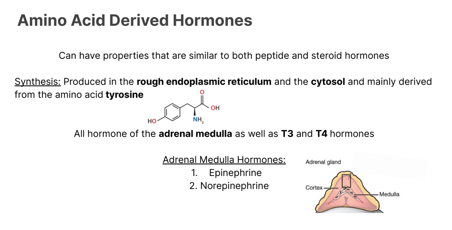
Can have properties that are similar to both peptide and steroid hormones.
Synthesis: Produced in rough endoplasmic reticulum, and the cytosol and mainly derived from the amino acid tyrosine.
All hormones of the adrenal medulla as well as T3 and T4 hormones.
Adrenal Medulla Hormones
Norepinephrine
Epinephrine
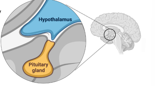
Hypothalamus
Purpose
The main thing respinsible for hormone secretion in the body.
Coordinates the body’s internal environment and maintains homeostasis.
Pituitary Gland
a.k.a (hypophysis)
Crucial gland for hormone production, storage, and release
Rests just below the hypothalamus
Anterior pituitary and posterior pituitary
Posterior Pituitary gland
Posterior Pituitary (Neurohypophysis): The direct neuronal extension of the hypothalamus constructed of neuronal tissue
Stores and releases hormones produced by the hypothalamus
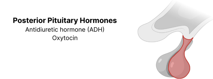
Posterior Pituitary Gland Hormones (ADH)
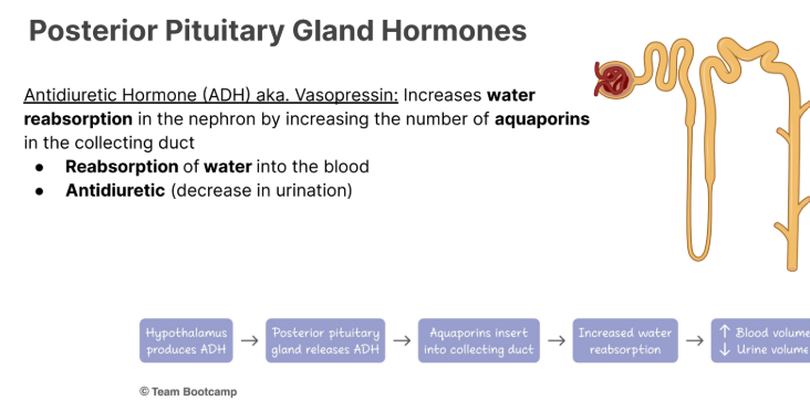
Antidiuretic Hormone (ADH) aka. Vasopressin: Increases water reabsorption in the nephron by increasing the number of aquaporins in the collecting duct
Reabsorption of water into the blood
Antidiuretic (decrease in urination)
Posterior Pituitary Gland Hormones (Oxytocin)
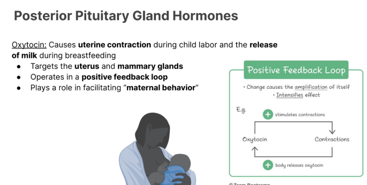
Causes uterine contraction during child labor and the release of milk during breastfeeding
Targets the uterus and mammary glands
Operates in a positive feedback loop
Plays a role in facilitating “maternal behavior”
Anterior Pituitary Gland
Anterior Pituitary (Adenohypophysis):
Produces it own hormones and is composed of glandular tissue rather than neural tissue
Hypothalamic Releasing Hormones
Hormones released by the hypothalamus in order to stimulate the release of hormones generated by the anterior pituitary gland
Hypothalamic Inhibiting Hormones
Hormones released by the hypothalamus to inhibit the release of other hormones by the anterior pituitary
Hypophyseal Portal System,
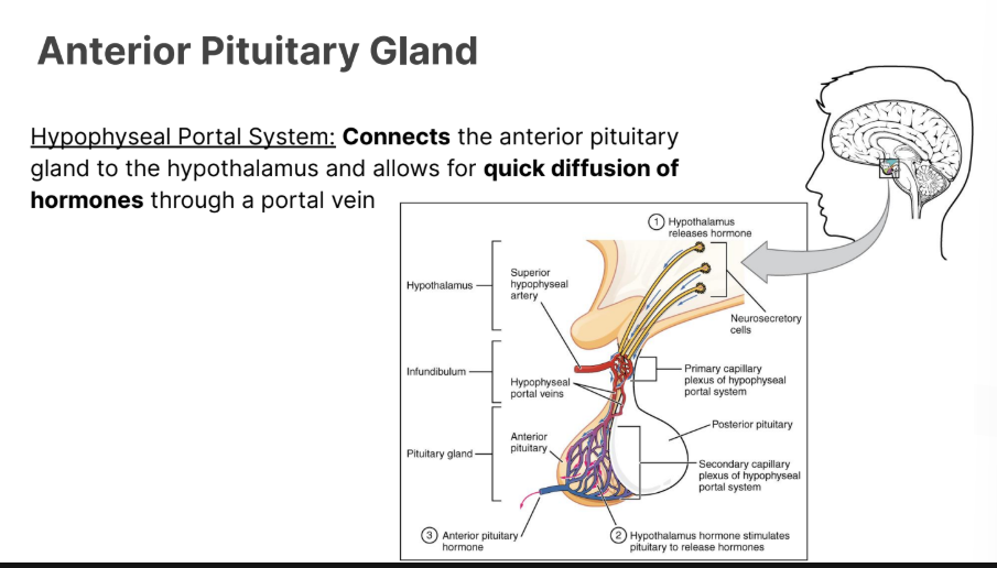
Hypophyseal Portal System:
Connects the anterior pituitary gland to the hypothalamus and allows for quick diffusion of hormones through a portal vein
Connects hypothalamus and the anterior pituitary.
Anterior Pituitary Gland Stimulating Hormones (4)
GnRH
TRH
CRH
GRH
GnRH
(Gonadotropin Releasing Hormone):
Causes the release of luteinizing hormone (LH) and follicle stimulating hormone (FSH)
TRH
TRH (Thyrotropin Releasing Hormone):
Causes the release of thyroid stimulating hormone (TSH)
CRH
(Corticotropin Releasing Hormone):
Causes the release of adrenocorticotropic hormone (ACTH)
GRH
(Growth Hormone Releasing Hormone):
Causes the release of growth hormone (GH)
Anterior Pituitary Gland Hormones
Once stimulated, the anterior pituitary can release its own hormones
Tropic and Direct Hormones
Tropic Hormones
Target other endocrine glands for further hormone release
ACTH
TSH
LH
FSH
Direct Hormones
Directly Targets organs to cause effects
TROPIC
FSH
Purpose
Follicle growth in females and sperm maturation in male gonads
(follicle stimulating hormone)
TROPIC
LH
Lutenizing Hormone
Stimulates ovulation, the formation of the corpus luteum in females (keep in mind this eventually produces estrogen), and the production of testosterone in male gonads
TROPIC
ACTH
(Adrenocorticotropic Hormone):
Stimulates the release of glucocorticoids from the adrenal gland to fight stress
Increases the body’s glucose levels
TROPIC
TSH
TSH (Thyroid Stimulating Hormone): Stimulates the thyroid gland to produce T3 and T4 in order to increase metabolism
LIPOPHILLIC
TROPIC
Direct Hormones of the Anterior Pituitary
Prolactin
Growth Hormone (somatotropin)
Prolactin
Stimulates the development of the mammary gland and increases milk production following childbirth
After childbirth, prolactin also increases milk production by stimulating lactation in the female.
Growth Hormone
Stimulates the growth and division of body cells
Anterior Pituitary Gland Hormones (pneumonic)
(tropic)
F Follicle Stimulating Hormone (FSH)
L. Luteinizing Hormone (LH)
A. Adrenocorticotropic Hormone (ACTH)
T. Thyroid Stimulating Hormone (TH)
(direct)
P. Prolactin
i.
G. Growth Hormone (Somatotropin)
Pineal Gland
Gland in the brain responsible for the production of melatonin
Hormone involved in regulating the circadian rhythm
Thyroid Gland
The largest of the endocrine organs.
In front of the trachea.
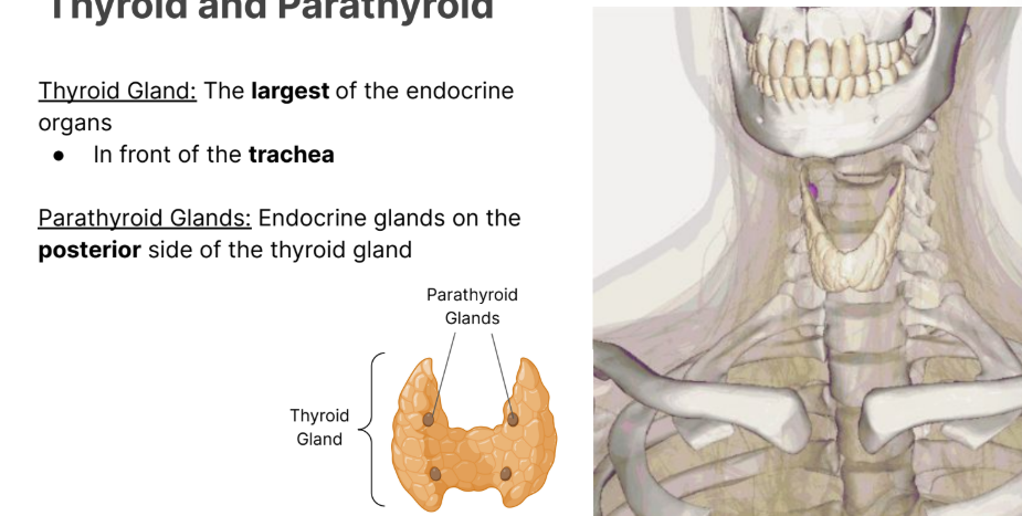
Parathyroid Gland
Endocrine glands on the posterior side of the thyroid gland.

Thyroid Gland Hormones
T3: triiodothyronine
T4: thyroxine
Calcitonin
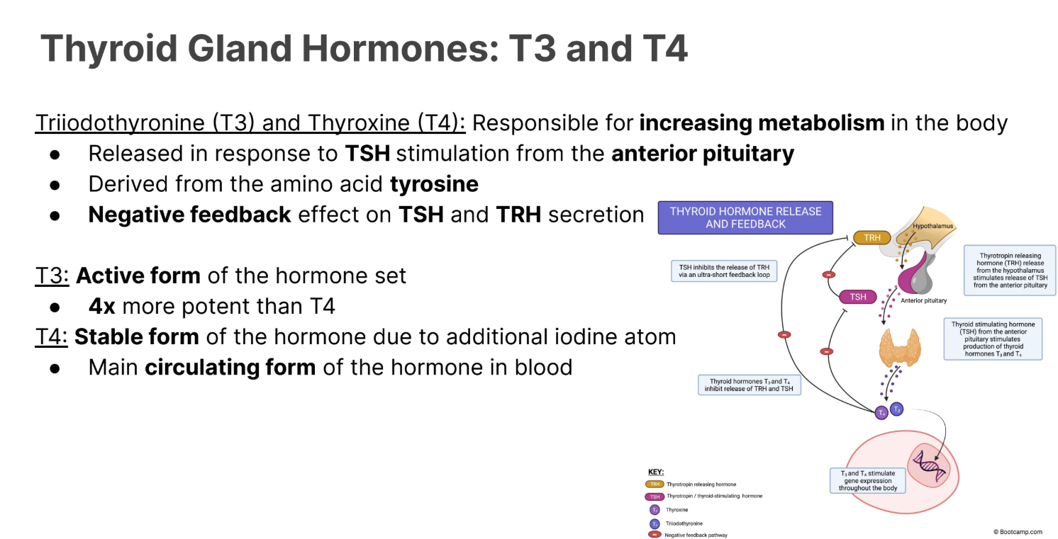
T3 and T4
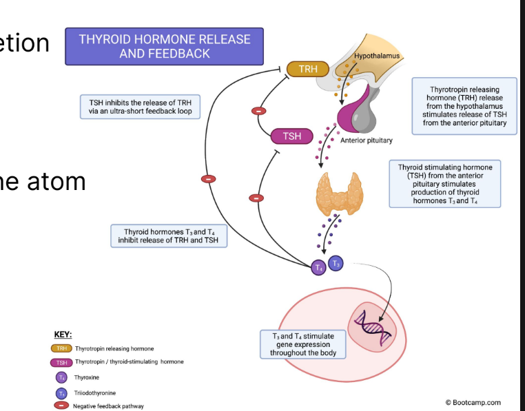
Triiodothyronine (T3) and Thyroxine (T4): Responsible for increasing metabolism in the body
Released in response to TSH stimulation from the anterior pituitary
Derived from the amino acid tyrosine
Negative feedback effect on TSH (thyroid stimulating) and TRH (thyroid releasing hormone) secretion
LIPOPHILLIC
T3
T3: Active form of the hormone set
4x more potent than T4
LIPOPHILLIC
T4
Stable form of the hormone due to additional iodine atom
Main circulating form of the hormone in blood
Lipophillic

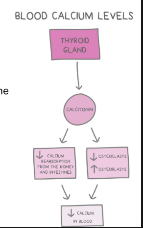
Calcitonin
Released by parafollicular thyroid cells (c cells)
Stimulates osteoblast use of calcium to generate new bone.
Inhibits osteoclasts from freeing calcium by breaking down bone.
Decreases Calcium Reabsorption in kidneys and intestines.
Hypothyroidism
Under secretion of T3 and T4, which causes reduced basal metabolic rates
Hyperthyroidism
Over secretion of T3 and T4, which causes increased basal metabolic rates.
Goiter in Hypothyroidism
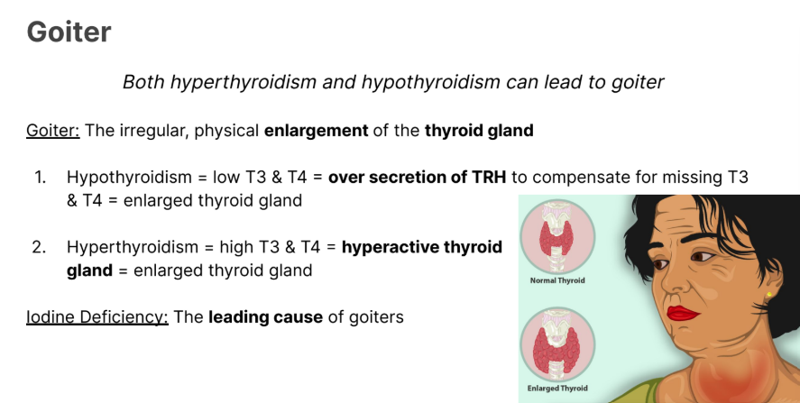
The irregular, physical enlargement of the thyroid gland
Hypothyroidism = low T3 & T4 = over secretion of TRH (thyroid releasing hormone) to compensate for missing T3 & T4 = enlarged thyroid gland

Goiters: Hyperthyroidsism.
Hyperthyroidism = high T3 & T4 = hyperactive thyroid gland = enlarged thyroid gland

What is the main cause of goiters?
Iodine Deficiency
Parathyroid Gland:
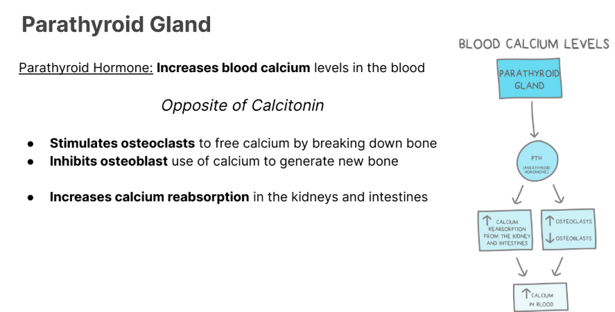
Parathyroid Hormone: Increases blood calcium levels in the blood
Opposite of Calcitonin
Stimulates osteoclasts to free calcium by breaking down bone
Inhibits osteoblast use of calcium to generate new bone
Increases calcium reabsorption in the kidneys and intestines
Calcitonin vs PTH

Pancreas purpose
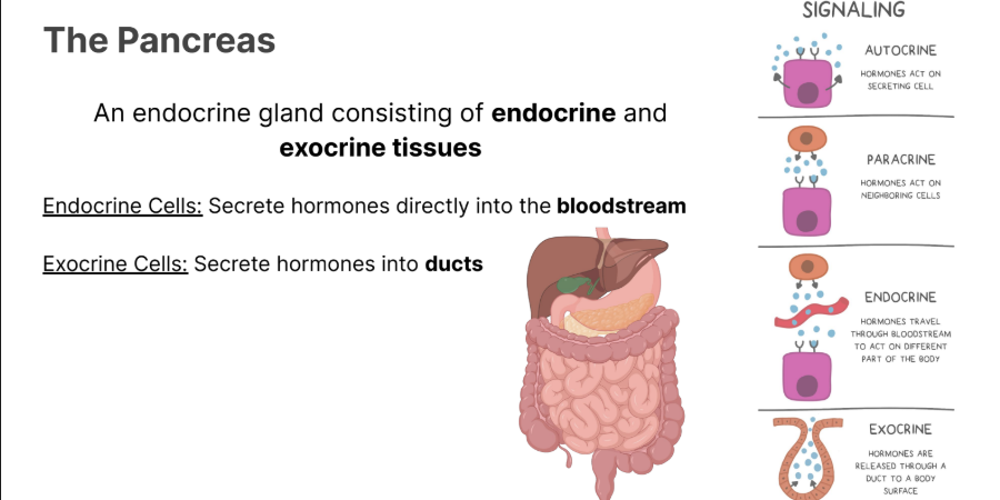
An endocrine gland consisting of endocrine and exocrine tissues
Endocrine cells
Secrete hormones into the blood stream

Exocrine
Secrete hormones into ducts
What do the islets of langerhans secrete?
What are they to an organ?
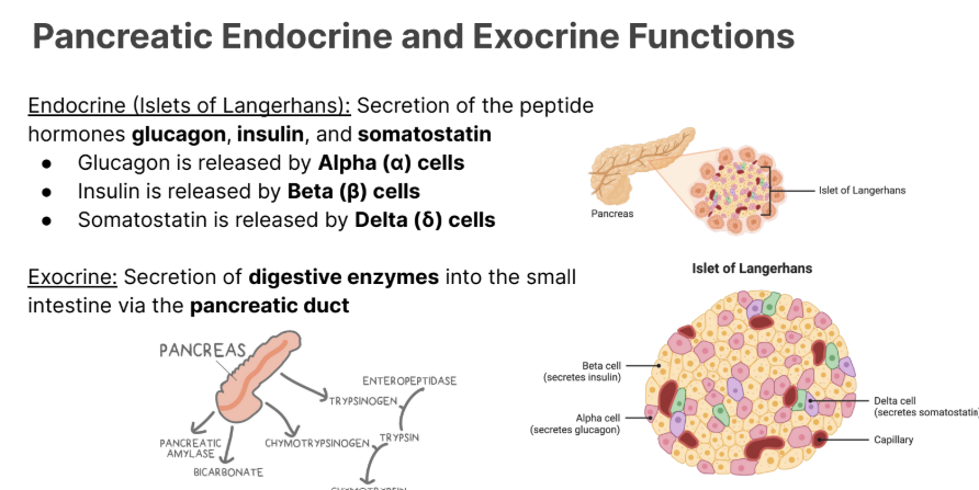
Endocrine (Islets of Langerhans): Secretion of the peptide hormones glucagon, insulin, and somatostatin
Glucagon is released by Alpha (α) cells
Insulin is released by Beta (β) cells
Somatostatin is released by Delta (δ) cells
What do Alpha cells release (pancreas)
Glucagon
What do beta cells release? (pancreas)
Insulin
What do delta cells release?
Somatostatin INHIBITS
This inhibits the growth hormone from the anterior pituitary
glucagon from alpha cells
Insulin from beta cells
Pancreatic Exocrine Functions
Exocrine: Secretion of digestive enzymes into the small intestine via the pancreatic duct
Glucagon
Released by alpha cells in times of low blood glucose.
Stimulates Liver and adipose (fat) tissues to release their stored glucose
Insulin
Released by beta cells in times of high blood glucose
Stimulates the liver, adipose (fat) and muscle tissues to store glucose.
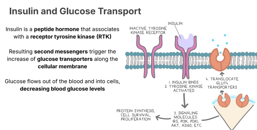
Insulin and Glucose Transport
Insulin is a peptide hormone that associates
with a receptor tyrosine kinase (RTK)
Resulting second messengers trigger the increase of glucose transporters along the cellular membrane
Glucose flows out of the blood and into cells, decreasing blood glucose levels
Somatostatin
Somatostatin from the pancreatic delta cells is an inhibitory hormone
Somatostatin is responsible for inhibiting the release of:
Insulin from the beta cells
Glucagon from the alpha cells
Growth hormone from the anterior pituitary
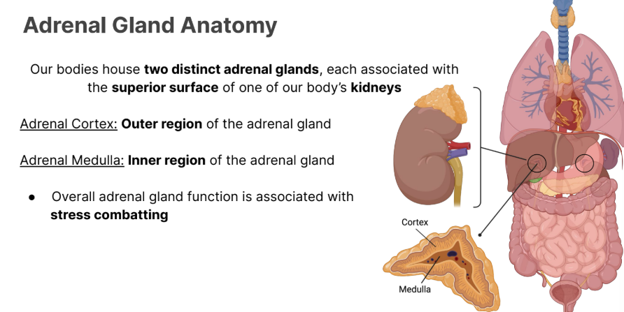
Adrenal Gland Anatomy
Our bodies house two distinct adrenal glands, each associated with the superior surface of one of our body’s kidneys
Adrenal Medulla
Adrenal Cortex

Adrenal Cortex → Location
Adrenal Cortex: Outer region of the adrenal gland
Adrenal Medulla: → location, and main function
Adrenal Medulla: region of the adrenal gland
Overall adrenal gland function is associated with stress combatting

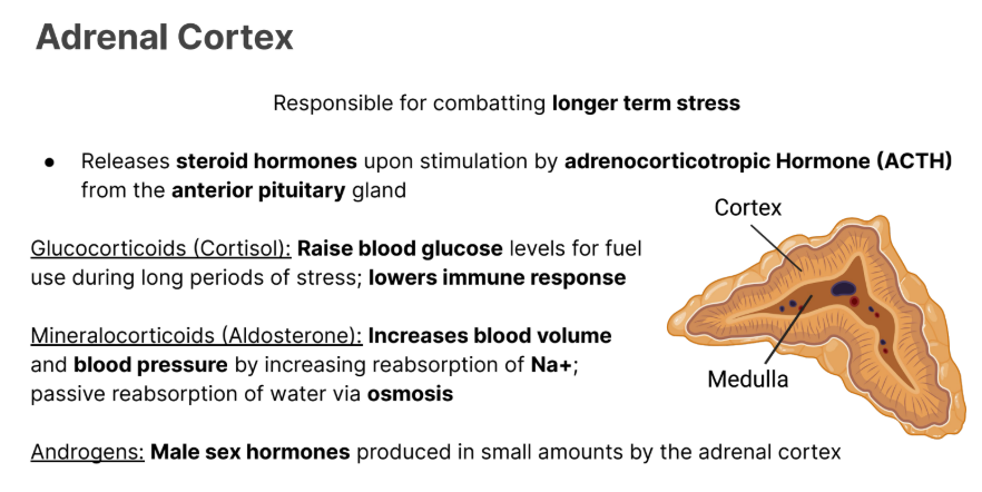
Adrenal Cortex
Purpose
What does it release
Glucocorticoids (Cortisol)
Mineralocorticoids
Androgens:
Deals with long term stress
Releases steroid hormones upon stimulation by Adrenocorticotropic hormone (ACTH) from the anterior pituitary gland.

Glucocorticoids
Glucocorticoids (Cortisol): Raise blood glucose levels for fuel use during long periods of stress; lowers immune response
Released by the adrenal cortex

Mineralocrticoids
Mineralocorticoids (Aldosterone): Increases blood volume and blood pressure by increasing reabsorption of Na+; passive reabsorption of water via osmosis
Released by the adrenal cortex

Androgens
Androgens: Male sex hormones produced in small amounts by the adrenal cortex
Released by the adrenal cortex
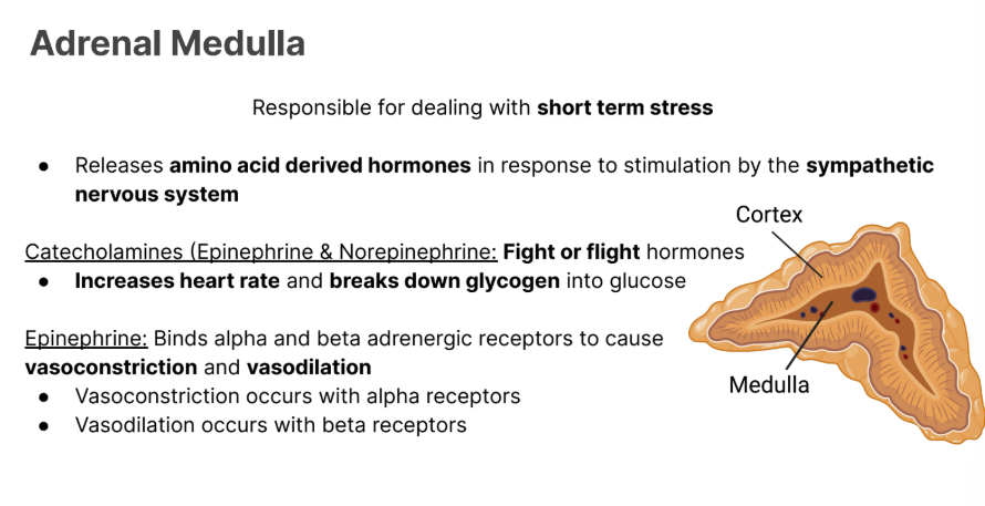
Adrenal Medulla
Responsible for dealing with short term stress
Releases amino acid derived hormones in response to stimulation by the sympathetic nervous system

Adrenal Medulla
Releases amino acid derived hormones in response to stimulation by the sympathetic nervous system
Releases:
Catecholamines
Epinephrine and Norepinephrine

Calecholamine
Epinephrine and Norepinephrine
Fight or flight hormones that increase heart rate and breaks down glycogen into glucose

Epinephrine
Epinephrine: Binds alpha and beta adrenergic receptors to cause vasoconstriction and vasodilation
Vasoconstriction occurs with alpha receptors
Vasodilation occurs with beta receptors
Long term stress
What manages it
examples
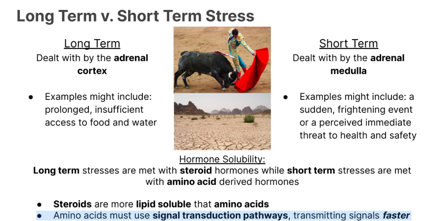
Long Term
Dealt with by the adrenal cortex
● Examples might include: prolonged, insufficient access to food and water
Short term stress
What manages it
examples
Adrenal Medulla
● Examples might include: a sudden, frightening event or a perceived immediate threat to health and safety

Long term vs short term stress
molecules
Which one is faster?
Long term stresses are met with steroid hormones while short term stresses are met with amino acid derived hormones
Amino acids must use signal transduction pathways, transmitting signals faster

Stimulation of the testes and ovaries.
What are they stimulated by?
Where are those stimulations send from
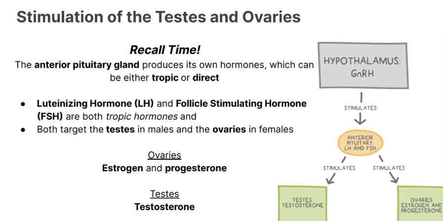
The anterior pituitary gland produces its own hormones, which can be either tropic or direct
Luteinizing Hormone (LH) and Follicle Stimulating Hormone (FSH) are both tropic hormones and
Both target the testes in males and the ovaries in females
Ovaries
Estrogen and progesterone
Testes
Testosterone
Hormones that stimulate the Testes
Testosterone
Hormones that stimulate the Ovaries
Estrogen and Progesterone.
LH in the ovaries.
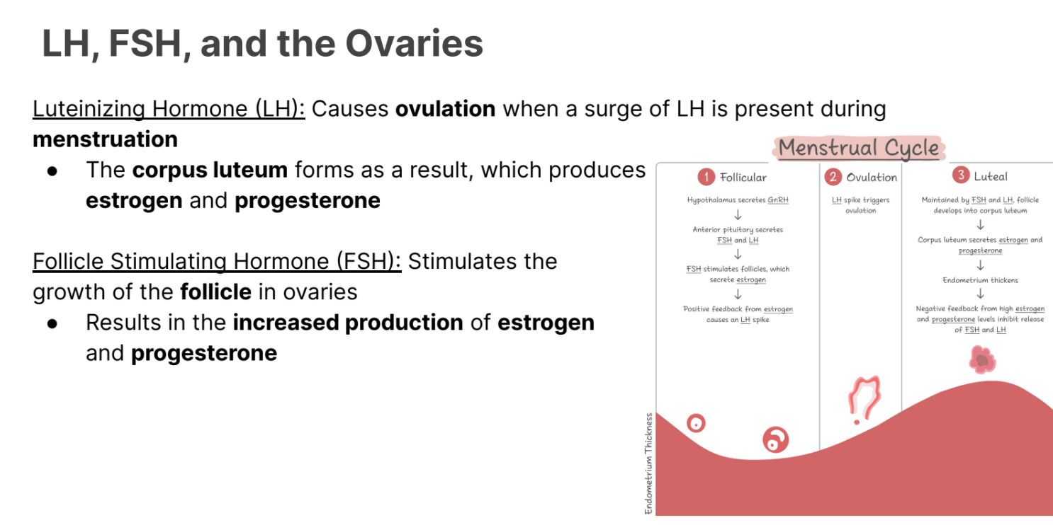
Luteinizing Hormone (LH): Causes ovulation when a surge of LH is present during menstruation
● The corpus luteum forms as a result, which produces
estrogen and progesterone
Follicle Stimulating Hormone (FSH) in the ovaries
Stimulates the growth of the follicle in ovaries
Results in the increased production of estrogen and progesterone.
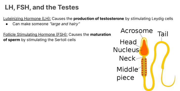
LH in the testes
Causes the production of testosterone by stimulating Leydig cells
can make someone large and hairy.

Follcile Stimulating Hormone (FSH)
Causes the maturation of sperm by stimulating Sertoli cells.
Positive Feedback Loops
The product increases the production of the stimulus
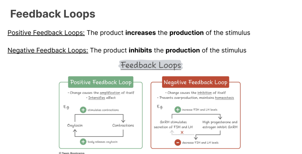
Negative Feedback Loops
The product inhibits the production of the stimulus

Catecholamines
Short term, fast acting (amines) hormones that are in charge of fight or flight. They bind to adrenergic receptors
EX
Norepinephrine
Epinephrine
Mineralocorticoids
What do they do
Example of the most famous one?
Increase blood volime and pressure
Passivley allow reabsorption of water into blood
Increase reabsorption of sodium into the excretory system
INCRESE secretion of potassium into the execratory system
ALDOSTERONE