Imaging - Radiologic Evaluation of the Spine Part I: Normal Anatomy
1/56
There's no tags or description
Looks like no tags are added yet.
Name | Mastery | Learn | Test | Matching | Spaced | Call with Kai |
|---|
No analytics yet
Send a link to your students to track their progress
57 Terms
post trauma
Pain
Pre-operatively
Suspected malignancy
Suspected anomaly or abnormality
Suspected instability
Why are C-Spine images ordered?
> 65
Dangerous mechanism
Paresthesia in extremities
What are the high risk factors when determining to take a C-spine radiograph?
Simple mechanism
Delayed onset of pain
Can sit upright
Ambulatory
Absence of midline C-spine tenderness
What are the low risk factors when determining to take a C-spine radiograph?
Yes
Do you take a radiograph of the C-spine if any high risk factors are present?
No
Do you take a radiograph of the C-spine if any low risk factors are present?
Yes
Do you take a radiography if a person can not rotate their C-spine?
No
Do you take a radiography if a person can rotate their C-spine?
Right and left Oblique
In what radiograph view do you see the intervertebral foramina of the cervical spine?
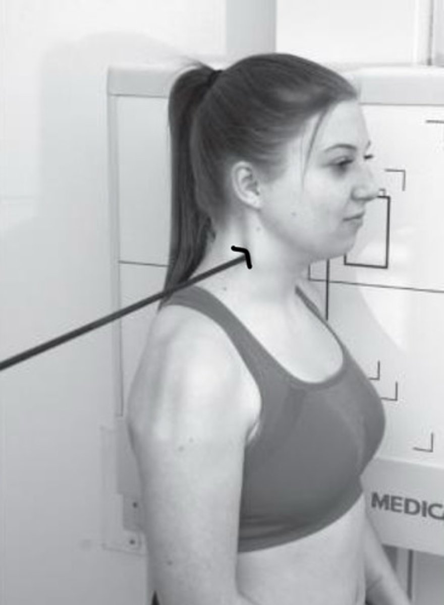
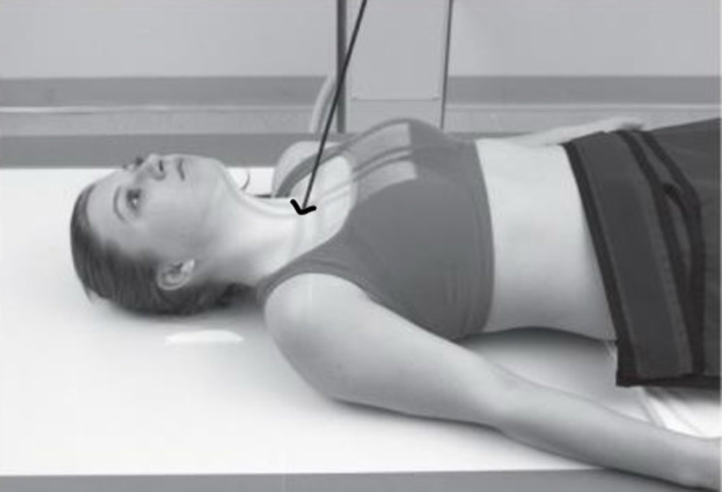
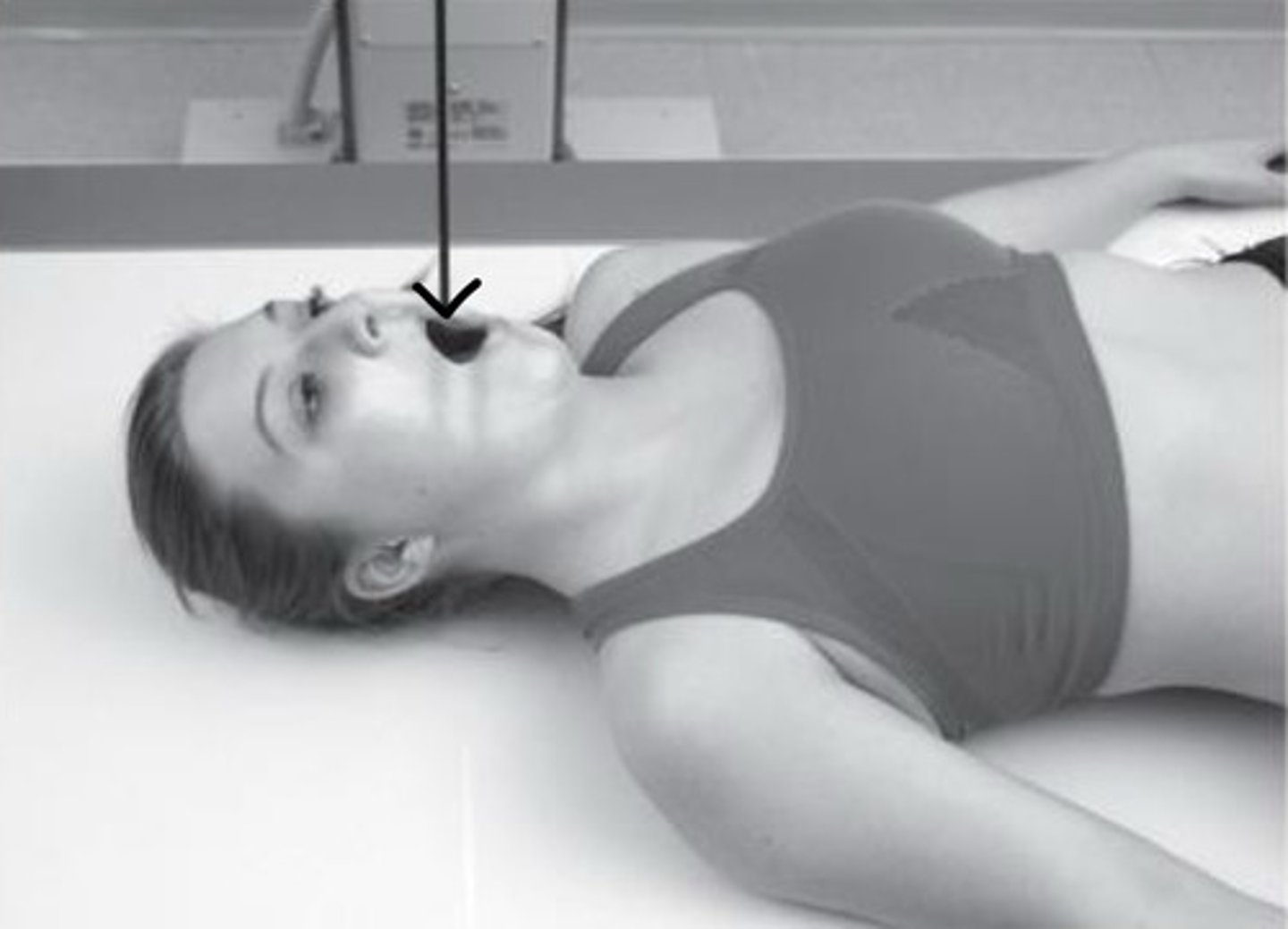
Lateral view with stress
What type of view is this?
Lateral view with stress
What type of view is this?
Oblique view
What type of view is this?
Oblique view
What type of view is this?
Lateral view
What type of view is this?
AP lower C-spine
What type of view is this?
A-P open mouth / odontoid view
What type of view is this?
5 lower cervical vertebrae
Upper thoracic vertebrae and associated ribs
Medial 1/3 of the clavicles
Trachea
What structures do you see in a AP lower C-spine radiograph?
C1-C7
intervertebral disc spaces
articular pillars and facet joints
spinous processes
What structures do you see in a lateral C-spine radiograph?
C1 and C2
What structures do you see in a AP Open-mouth (Odontoid) C-spine radiograph?
intervertebral foramina
facet joints
pedicles
What structures do you see in a R/L Oblique C-spine radiograph?
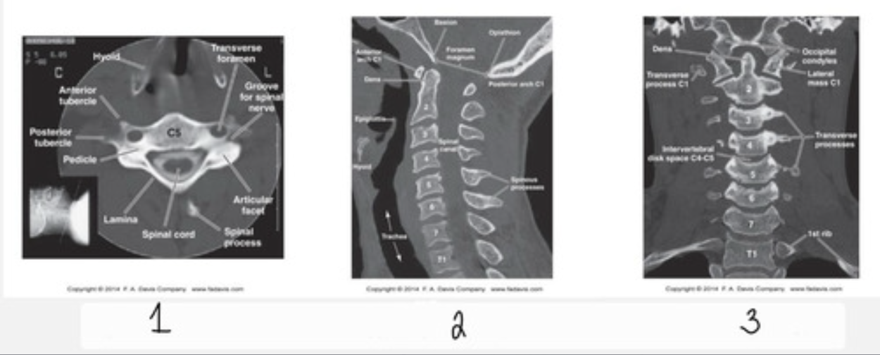
Axial plane
What type of CT view does 1 show?
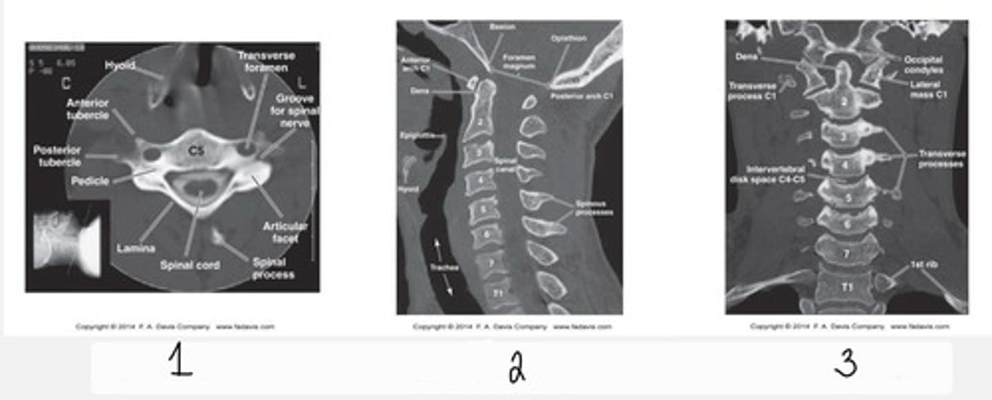
Sagittal plane
What type of CT view does 2 show?

Coronal plane
What type of CT view does 3 show?
Alignment
Bone density
Canal space
Disc integrity
Soft tissue
How do you interpret a CT?
axial and sagittal
What are the typical planes an MRI of the spine is taken in?
clarify anatomy
What is a T1 weighted MRI used for?
abnormalities in fluid
What is a T2 weighted MRI used for?
Alignment
Bone signal
Canal space
Disc integrity
How do you interpret a MRI?
AP Thoracic Spine
Lateral Thoracic Spine
What are the standard T-spine projections?
Swimmer's lateral view
Posterior Oblique view
Thoracolumbar
Coned views
PA or AP ribs
What are radiologic evaluations of the T-spine and ribs that are not as common?
Intermediate
What is 1?

Low
What is 2?

Intermediate
What is 3?

Low
What is 4?

Intermediate
What is 5?

Intermediate
What is 6?

High
What is 7?

High
What is 8?

Low
What is 9?
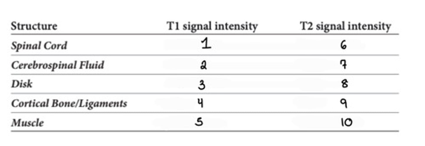
Intermediate
What is 10?

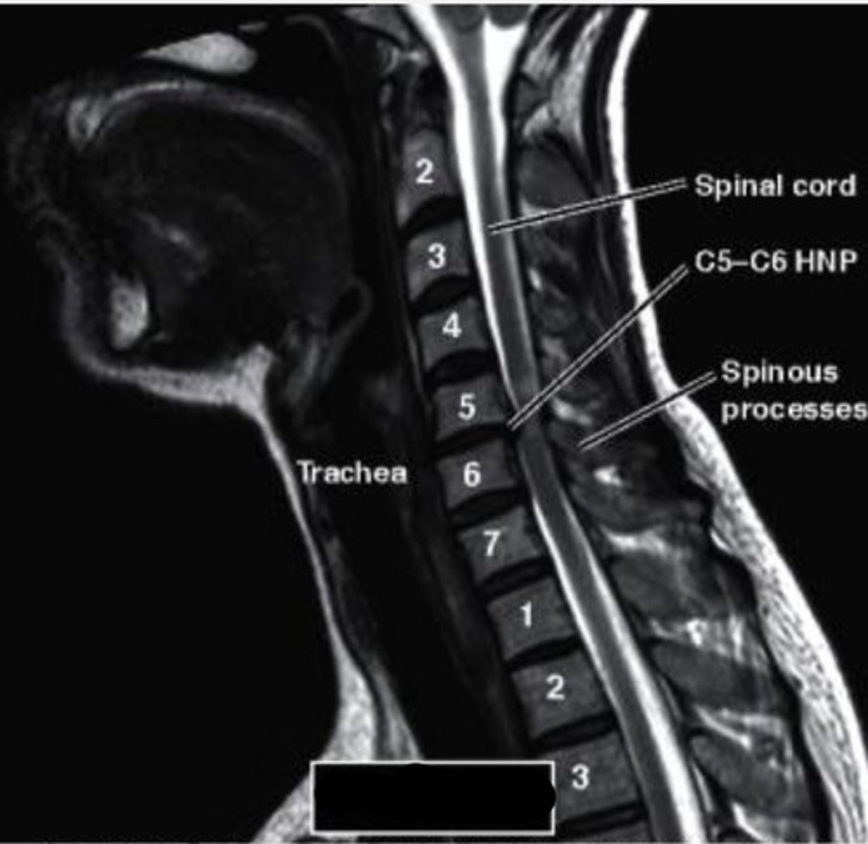
Sagittal
What type of MRI view does this show?
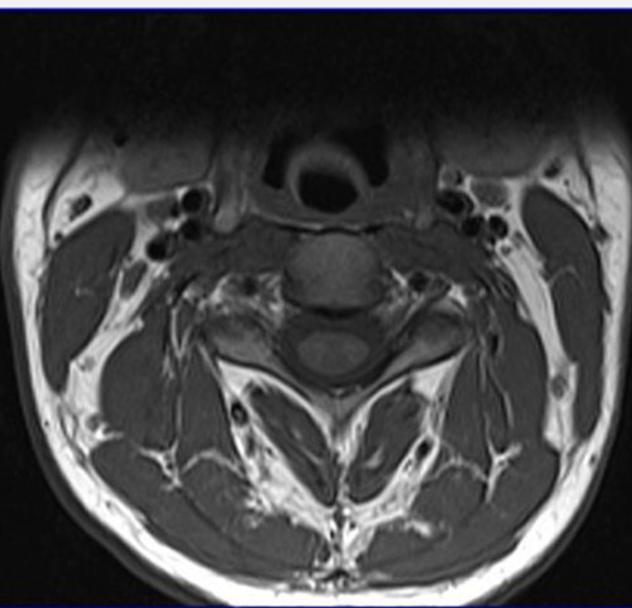
Axial
What type of MRI view does this show?
AP thoracic spine
What type of view is this?
Lateral thoracic spine
What type of view is this?
AP
Lateral
R and L Oblique
Lateral L5-S1
Sacroiliac (AP or oblique)
What are the standard radiologic projections of the lumbar spine?
facet joints
What do you see in a oblique projection of the lumbar spine?
AP lumbar spine
What type of view is this?
Lateral lumbar spine
What type of view is this?
Oblique lumbar spine
What type of view is this?
Lateral L5-S1
What type of view is this?
AP axial/sacroiliac
What type of view is this?
Oblique Sacroiliac
What type of view is this?
lumbar vertebral bodies
SP and TP
Intervertebral disk spaces
What structures do you see in an AP L-spine radiograph?
lumbar vertebral bodies
intervertebral disk spaces
SP
intervertebral foramina
lumbosacral articulation
What structures do you see in an lateral L-spine radiograph?
facet joints
superior and inferior articular process
pars inticularis
pedicles
What structures do you see in an R/L posterior oblique lumbar spine radiograph?
close up or coned view of lumbosacral junction
What structures do you see in an lateral L5-S1 radiograph?
bilateral SI joints
What structures do you see in an AP axial sacroiliac radiograph?
Individual SI joint
What structures do you see in a R/L oblique sacroiliac radiograph?