Biomed 1 - Fracture Repair Stages
1/14
There's no tags or description
Looks like no tags are added yet.
Name | Mastery | Learn | Test | Matching | Spaced | Call with Kai |
|---|
No analytics yet
Send a link to your students to track their progress
15 Terms
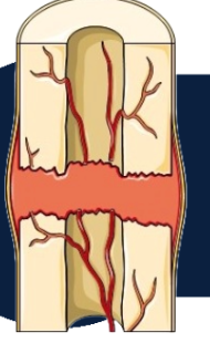
Hematoma Formation
Blood vessels that are ruptured during the break swell to form a blood clot. This mass forms between the broken bones. This clotting reduces the blood supply to many of the cells in the area of injury, and as a result, these cells die.
Hematom
Blood clot
Hematoma Formation
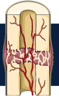
Fibrocartilage Callus Formation
New capillaries begin to form into the clotted blood in the damaged area. Connective tissues cells form a mass of repair tissue. This contains some cartilage, some bone, and collagen fibers. The combined mass closes the gap between the broken bones.
Fibrocartilage Callus Formation
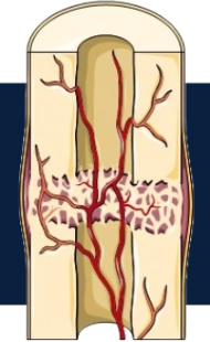
Bony Callus Formation
The callus is gradually replaced by one made of spongy bone. Osteoclasts and osteoblasts move to the area and multiply.
Bony callus formation
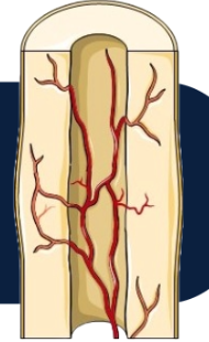
Bone Remodeling
Over the weeks and months to come, the callus is remodeled with the help of osteoclasts and osteoblasts. The shape of the bones will gradually return to normal, and there will eventually be little evidence of the fracture.
Bone Remodeling
Osteoblasts
Cell that deposits bone tissue
Osteoclasts
Cells with areas of bone resorption that helps with breaking down old or damage tissue
Osteoclasts
Cells that reabsorb bone tissue
Paget’s disease
Increased osteoblast activity compared to osteoclast activity leads to rapidly built bone tissue and weak bones easily fractured
Osteogenesis Imperfecta (OI)
Another name for paget’s disease
Hematoma Formation, Fibrocartilage Callus Formation, Bony Callus Formation, Bone Remodeling
Order of bone remodeling stages