A&P 1 Lab midterm
1/201
There's no tags or description
Looks like no tags are added yet.
Name | Mastery | Learn | Test | Matching | Spaced | Call with Kai |
|---|
No analytics yet
Send a link to your students to track their progress
202 Terms
Anatomy defintion
studies the form and structure of the body
examine the relationships among parts of the body as well as the structure of individual organs
Physiology definition
examines how the body functions
examine how organs and body systems function under normal circumstances and abnormal ones too
microscopy definition
Examines structures that cannot be observed by the unaided eye
specimen under the microscope
cytology and histology
cytology definition
the study of body cells and their internal structure
histology
study of tissues
tissues are groups of…
cells
gross anatomy
aka macroscopic anatomy
Investigates structures visible to the unaided eye
dissected for examination
systemic anatomy
studies anatomy of each functional body system
regional anatomy
examines all of the structures in a particular region of the body
what counts as the arm?
shoulder to elbow
what counts as the forearm?
elbow to wrist
what counts as the lower limb/extremity?
hip and below
what counts as the thigh?
hip to knee
what counts as the leg?
knee to foot
surface anatomy
focuses in superficial anatomic markings and internal body structures
comparative anatomy
examines similarities and differences in anatomy of different species
embryology
studies developmental changes from conception to birth
pathologic anatomy
examines anatomic changes resulting of disease (patho is abnormal)
Considers anatomic and microscopic changes
radiographic anatomy
investigates internal structures visualized by scanning procedures
cardiovascular physiology
functioning of the heart, blood vessels, and blood
neurophysiology
functioning of the nerves and nervous system organs
respiratory physiology
functioning of respiratory organs (ex: lungs)
reproductive physiology
functioning of reproductive hormones and reproductive cycle
pathophysiology
relationship between the function of an organ system and disease or injury to the system
properties common to organisms
exhibit complex organization and order
engage in metabolism
all grow and develop
all exhibit responsiveness
all exhibit regulation
all reproduce
metabolism
the sum of all chemical reactions that occur within the body
composed of anabolism and catabolism
anabolism
small molecules joined to form larger ones-build
catabolism
-large molecules broken down into smaller ones-destroy
homeostasis
ability of an organism to maintain a consistent internal environment or “steady state”
simplest to most complex level
Chemical level (most simple)
Cellular level
Tissue level
Organ level
Organ system level
Organismal level (most complex)
chemical level
involves in atoms and molecules
macromolecules
organelles
atoms definition
smallest unit of matter
molecules definition
one or more combined atoms (sugar, vitamins)
macromolecules
more complex molecules
ex: proteins and DNA
organelles
Microscopic subunits in cells cmpised of macromolecules
cellular level
consists of cells
Basic units of structure and function in organisms
Vary widely in structure, reflecting specializations needed
formed from atoms and molecules from chemical level
cells
smallest living structures
tissue level
consists of tissue
4 types of tissues
tissues defintion
groups of similar cells performing common functions
what are the 4 types of tissues?
epithelial, connective, muscle, nervous
epithelial tissue
covers exposed surfaces and lines body cavities
connective tissue
protects supports, and binds structures and organs
muscle tissue
produces movement
nervous tissue
conducts nerve impulses
organs
two or more tissue types performing specific functions
ex:the small intestine composed of all four tissue types, working to process and absorb digested nutrients
organ system level
Contained related organs that work together to achieve a common function
Eg: organs of the digestive system working together to digest food, absorb nutrients, and expel waste products
organismal level
Highest level of structural organization
All body functions working interdependently in an organism, the living being
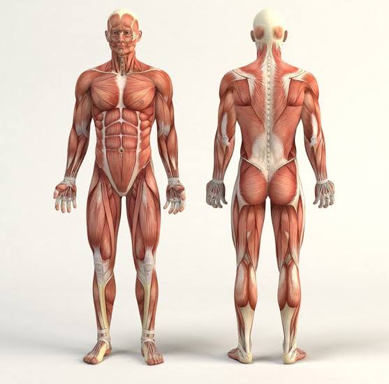
anatomic position
Anatomists use a specific position of the body as a point of common reference
Termed the anatomic position
Upright stance
Feet parallel and flat on the floor
Upper limbs at the sides of the body
Palms face anteriorly (toward the front)
Head is level
Eyes look forward
section definition
slice or cut to expose internal anatomy
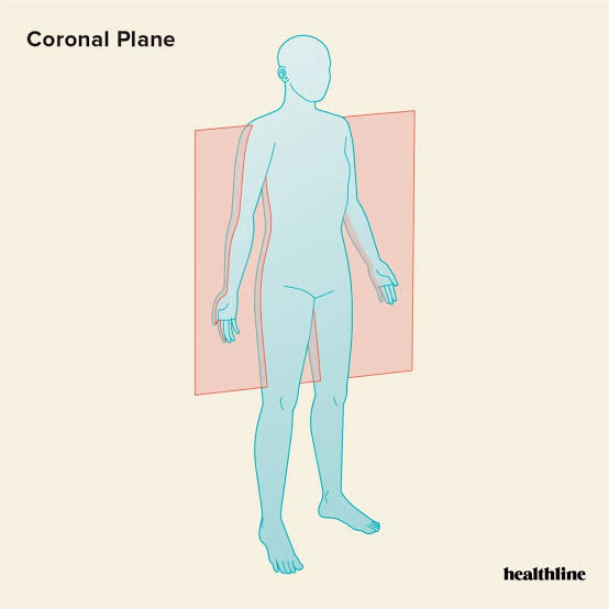
plane definition
imaginary flat surface passing through the body
what are the 3 major planes?
coronal, midsagital, transverse
anterior
front of the body
aka ventral
posterior
back of the body
aka dorsal
coronal plane
Vertical plane dividing the body into anterior (front) and posterior (back)
Also called frontal plane
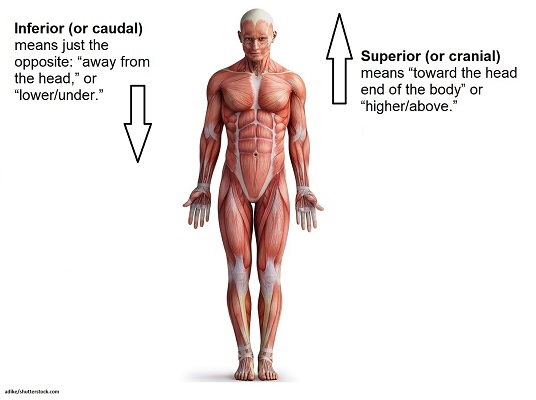
superior
above/toward the head

inferior
away from head
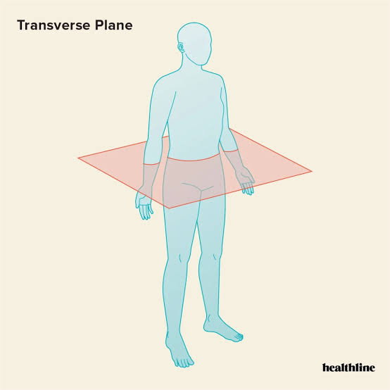
transverse plane
Horizontal plane dividing the body into superior (top) and inferior (bottom)
Aka cross-sectional plane
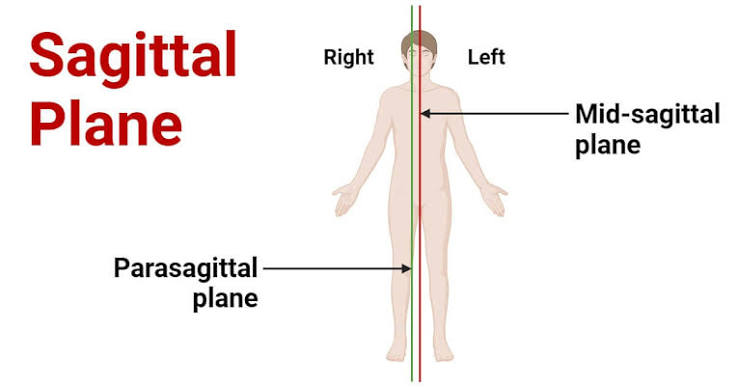
midsagittal plane
Vertical plane dividing the body into EQUAL left and right halves
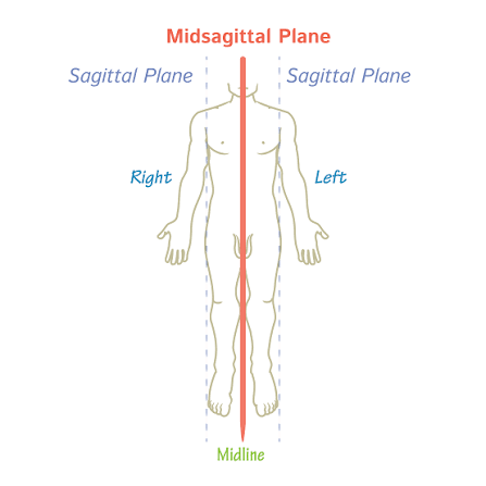
sagittal plane
Divides a structure into left and right at any number of sites (doesn’t matter if its not equal)
Parallel to midsagittal plane
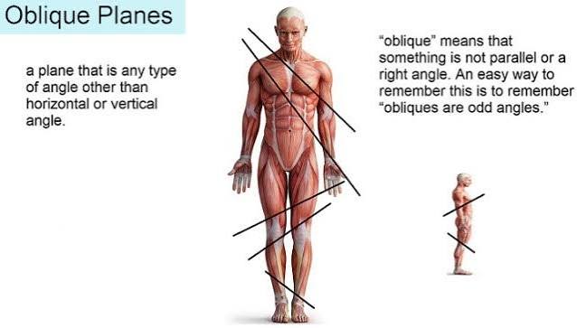
oblique plane
Pass through structures at an angle
proximal
nearer to the trunk
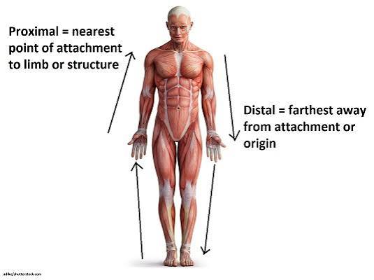
distal
farther from the trunk

posterior aspect
Contains cavities completely encased in bone
Physically and developmentally distinct from the ventral cavity
Subdivided into the cranial cavity and the vertebral cavity
cranial cavity
(endocranium) formed by bones of the cranium
Houses the brain
vertebral cavity
formed by the bones of the vertebral column
Houses the spinal cord
ventral cavity
Larger, anteriorly placed
Does not completely encase organs in bone
Partitioned into a superior thoracic cavity and an inferior abdominopelvic cavity (separated by the diaphragm)
Linked with serous membranes,
serous membranes
continuous layer of cells
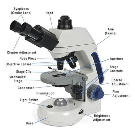
parts of the microscope
integumentary system
consists of the skin and its derivatives
Nairs, hair, sweat glands, sebaceous glands
the integument
is the skin covering the body
Aka the cutaneous membrane
Barrier to the outside world
Visual indicator of our physiology and health
Its scientific study and treatment termed dermatology
integument facts
Body’s largest organ
Protects internal body structures
Accounts for 7-8% of body weight
Area ranges between 1.5-2m^2
Thickness ranges between q.5 mm and 4.0 mm
underlying connective tissue
Provides strength and resilience
Contains smooth muscle associated with hair follicles
nervous tissue
provides information about touch, pressure, temperature, and pain
epidermis
Stratified squamous epithelium
Keratinized
contains strata
Layers from deep (bottom) to superficial (top)
Statum basale
Stratum spinosum
Stratum granulosum
Stratum lucidum
Stratum corneum
dermis
Deeper layer
Primarily dense irregular connective tissue
subcutaneous layer
Deep to dermis
Layer of alveolar and connective tissue
Termed subcutaneous layer or hypodermis
Not part of the integumentary system
Plays a role but beneath the skin
strata
specific layers
3 layers that have living keratinocytes
Statum basale
Stratum spinosum
Stratum granulosum
Most superficial 2 layers with dead keratinocytes
Stratum lucidum
Stratum corneum
stratum basale
Deepest epidermal layer
Aka stratum germinativum or basal layer
Single layer of cuboidal to low columnar cells
Attached to underlying basement membrane membrane
Separates the epidermis from the dermis
the stratum basale is occupied by these 3 cell types:
Keratinocytes
Melanocytes
Tactile cells
keratinocytes
Most abundant cells in epidermis
Found in all layers
Many kertinocytes stem cells present in this layer
Divide to regenerate new cells
Replace old cells shed at the surface
Name derived from the synthesis of keratin
keratin definition
Protein that strengthens the epidermis
melanocytes
Scattered among keratinocytes
Produce and store pigment (melanin) in response to UV light
Transfer pigment granules (melanosomes) into keratinocytes
melanin
pigment
melanosomes
pigment granules
Accumulate around nucleus of keratinocytes
Shield nuclear DNA from UV radiation
Responsible for the darker tones of skin
tactile cells
Aka Merkel cells
Few in number
Sensitive to touch
When compressed, release chemicals
Stimulate sensory nerve endings
stratum spinosum
Several layers of polygonal keratinocytes
Aka spiny layer
Daughter cells from stratum basale pushed into this layer
Begin to develop into specialized, non-dividing keratinocytes
Some in deepest level still dividing cells
desmosomes

desmosomes
Nondividing keratinocytes attached by intercellular junctions
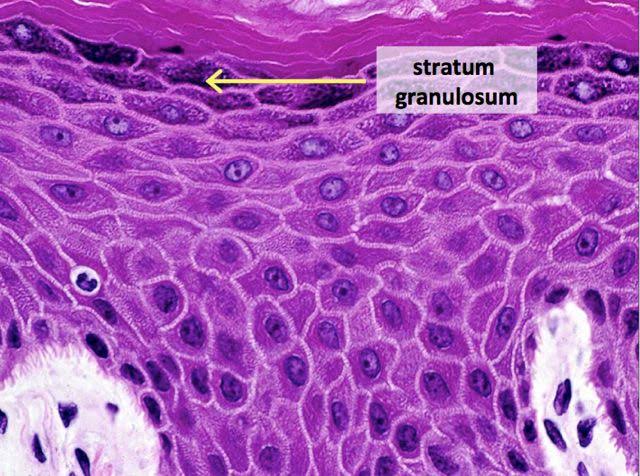
stratum granulosum
3 to 5 layers of keratinoocytes
Aka granular layer
Superficial to the stratum spinosum
First layer of keratinization
stratum lucidum
2 to 3 layers of keratinocytes
Translucent layer aka clear layer
Superficial to the stratum granulosum
Found only on thick skin within the palms and soles
Filled with a translucent protein, eledin
keratinization
Process where keratinocytes fill with keratin
Causes nucleus and organelles to disintegrate
Fully keratinized cell dead but structurally sound
Process not complete until more superficial layers
eledin
translucent protein
Intermediate product in keratin maturation
stratum corneum
20 to 30 layers of dead, interlocking keratinized cells
Cells anucleate (without a nucleus) and tightly packed
Plasma membrane enclosing keratin protein
Aka hornlike layer
Most superficial layer of epidermis
Surface unsuitable for the growth of many microorganisms
Secretions of exocrine glands also helping prevent growth
thick skin
On the palms of hands, soles of the feet, and surfaces of fingers and toes
Has all 5 layers of epidermal strata
Has sweat glands
Has no hair follicles or sebaceous glands
From 0.4 to 0.6 mm thick
thin skin
Covers most of the body
Lacks a stratum lucidum
Has sweat glands, hair follicles, and sebaceous glands
From 0.075 to 0.150 mm thick
hemoglobin
an oxygen binding compound (present in RBCs)
Bright red color upon binding oxygen
Gives blood vessels in dermis a reddish tint
Seen most easily in fair skinned individuals
More visible if blood vessels dilate
color from melanin
Pigment produced and stored in melanocytes
Occurs in black, brown, tan , yellow-brown shades
Transferred to keratinocytes in stratum basale
Amt in skin varies according to heredity and light exposure
UV stimulates melanin production’
All people with same number of melanocytes
color from carotene
Yellow-orange pigment
Acquired from yellow-orange veggies
Accumulates inside subcutaneous fat and keratinocytes of stratum corneum
Converted to vitamin A within the body
roles of carotene
In vision
In reducing free radicals
In immune function