how to do nottigam grading and one stane
1/12
There's no tags or description
Looks like no tags are added yet.
Name | Mastery | Learn | Test | Matching | Spaced | Call with Kai |
|---|
No analytics yet
Send a link to your students to track their progress
13 Terms
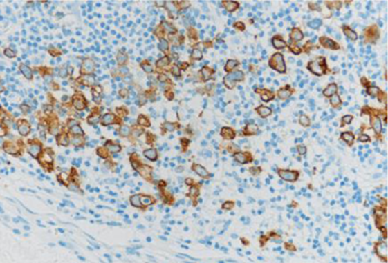
What does a positive pan-cytokeratin stain in an axillary lymph node indicate, and why is it important?
A positive pan-cytokeratin stain in an axillary lymph node indicates the presence of metastatic epithelial tumour cells, as lymph nodes do not normally contain epithelial cells. This shows that the disease has progressed from node-negative to node-positive status. This is important because it worsens prognosis by reducing 5-year survival and increasing the Nottingham Prognostic Index, and it leads to more aggressive adjuvant therapy.
A biopsy shows malignant cells confined within ducts with an intact basement membrane. No invasion seen.
Tis (DCIS)
Non-invasive
Basement membrane intact
A patient presents with a 1.4 cm invasive breast tumour, no skin or chest wall involvement.
It is a T1c equal or higer than 2 cm and is invasive
A 3 cm invasive breast mass is identified. No skin changes or chest wall invasion.
it is a T2 because it is between 2-≤ 5 cm
Scenario: A patient has a 6.2 cm breast tumour, confined to breast tissue only.
This is a T3 tumour because it is larger than 5 cm in its greatest dimension. Since it is confined strictly to the breast tissue, there is no nodal involvement or metastasis, classifying it clinically as Stage IIBt
A 2 cm tumour presents with peau d’orange and skin ulceration.
This is a T4 and is skin involvement and size irrelevant
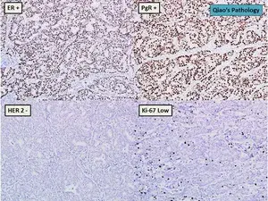
A breast tumour is ER+, PR+, HER2–, with low Ki-67.
What is the likely Nottingham grade and score?
Nottingham scoring (inferred from Ki-67 + phenotype)
Tubule formation: Likely good → 1
Nuclear pleomorphism: Likely mild → 1
Mitotic count: Low Ki-67 → 1
well differentiated, low profeliferation and better prognosis.
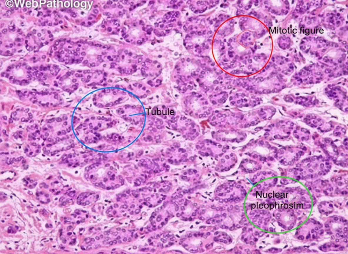
H&E shows well-formed tubules, uniform nuclei, and rare mitoses.
What is the Nottingham grade and score?
tubular differentiation, nuclear pleomorphism, and mitotic count. A Grade I (well-differentiated) tumour is defined by a total score of 3, with each category receiving 1 point for well-formed tubules, small, uniform nuclei, and rare mitoses. This cumulative score indicates a low-grade malignancy with a relatively favourable prognosis compared to poorly differentiated tumours.
H&E shows solid sheets of pleomorphic cells with poor tubule formation.
ER and PR are positive. What is the Nottingham grade?
Tubules: 3
Pleomorphism: 3
Mitoses: 3
Grade based on morphology, not receptors
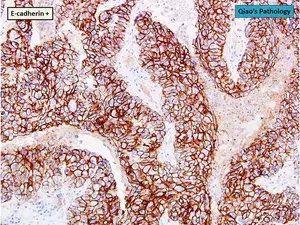
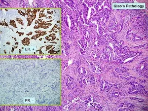
A breast tumour shows strong E-cadherin positivity with disorganised tumour architecture.
What does this suggest?
IGH grade ductal carcinoma (supporting evidence)
Why (exam features)
Strong E-cadherin positivity
Confirms ductal origin
Architecture appears disorganised and dense
Often associated with higher-grade invasive ductal carcinoma

A breast tumour shows strong E-cadherin positivity with disorganised architecture.
What is the Nottingham grade?
Nottingham scoring (from architecture)
Tubule formation: Poor, disorganised → 3
Nuclear pleomorphism: Moderate–marked → 2–3
Mitotic count: Likely elevated → 2–3
Total score: 8–9 → Grade III
What to write when explaining a high-grade cancer. EVERY TIME
The tumour demonstrates poor or absent tubule formation, marked nuclear pleomorphism, and a high mitotic rate, consistent with a poorly differentiated, high-grade (Nottingham Grade III) carcinoma.
what do you have to say for a low grade cancer
The tumour shows good tubule formation, mild nuclear pleomorphism, and a low mitotic rate, consistent with a well-differentiated, low-grade (Nottingham Grade I) carcinoma