3. Absorption, distribution and excretion
1/20
There's no tags or description
Looks like no tags are added yet.
Name | Mastery | Learn | Test | Matching | Spaced | Call with Kai |
|---|
No analytics yet
Send a link to your students to track their progress
21 Terms
What are the five ways Xenobiotics pass through cell membranes?
Passive diffusion through the membrane phospholipids
Passive filtration through aqueous pores
Active transport
Facilitated diffusion
Endocytosis: phagocytosis and pinocytosis
What are xenobiotics?
Xenobiotics are chemical substances foreign to a biological system, meaning they do not naturally occur or are not produced within an organism
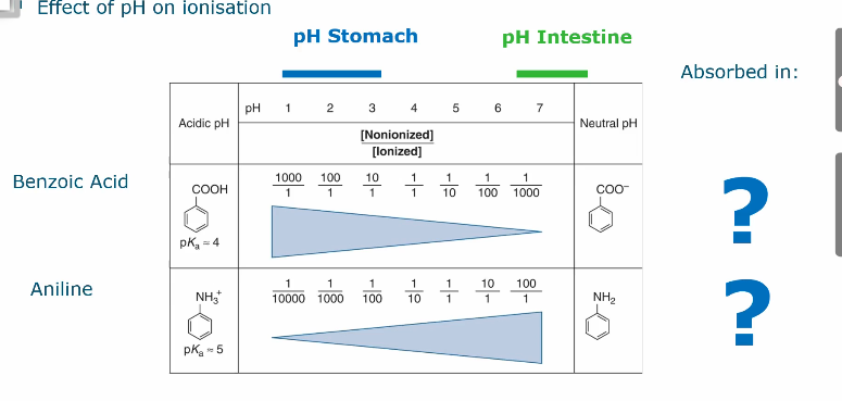
Effect of pH on ionization
At a low pH, the non ionized version of benzoic acid is more present and therefore passes the membrane better as it doesn’t have a charge'
For example in the stomach the pH is very low, weak acids like aspirin remain non-ionized and are absorbed easily here. Whereas in the small intestine where the pH is higher they are absorbed much less efficiently.
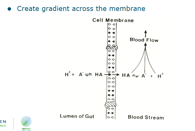
Passive diffusion and filtration
Small molecules up to MW 100-200 (ethanol, ureum)
Down a conc. gradient
Influenced by:
Lipophilicity
Ionization (different pH’s cause different charges)
Blood flow
Role of blood flow in passive diffusion and filtration
The blood flow creates a gradient across the membrane
Absorption: active transport
Chemicals are moved UP a concentration gradient
The transport system is selective and has the potential for competitive inhibition
Requires energy (ATP): sensitive to inhibition by metabolic inhibitors
The transport system is saturated at high substrate concentrations (Tmax)
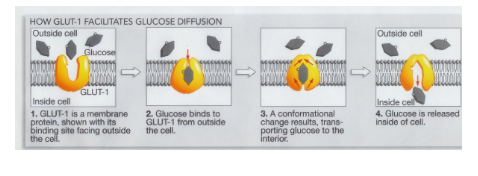
Facilitated diffusion
Similar to simple diffusion in the sense that;
It does not require energy and
transport down a concentration gradient
Two groups integral membrane proteins involved:
carrier proteins (hexose/glucose transporters)
Ion channels (Cl-, Na+)
e.g. flavonoid-glycosides can be absorbed via the glucose transporters in the small intestine
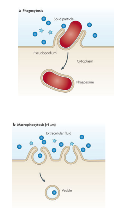
Absorption: phagocytosis and pinocytosis
Mainly used by the immune system
Phagocytosis: Cell eating
ingestion particles
Specialized cells (neutrophils, macrophages)
Pinocytosis: Cell drinking
ingestion of drops or small particles (<1 micrometer)
Almost all cells
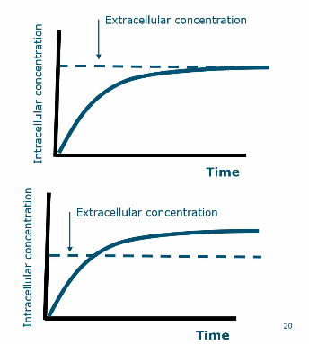
Uptake kinetics
Red = passive
High concentration difference = high rate of transport
blue = active
Is not dependent on the concentration difference as it is driven by pumps
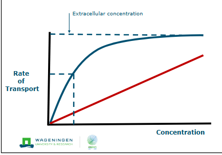
facilitated diffusion vs active transport graph
To find the differences in these graphs you need to know the concentration gradient
Bottom graph = active because it exceeds concentration gradient
Distribution: oral exposure
Gastrointestinal tract: mouth, stomach (acidic), duodenum, ileum, colon, rectum
Dependent on:
Concentration
Duration + level exposure
Area of exposure (vili)
epithelial layer
Sub epidermal blood flow
Physico-chemical properties
First pass effect
Everything entering our body will end up in the portal vein and into the liver.
Therefore everything we are exposed to does not enter in the rest of our body
Therefore not all medicines can be swallowed because the liver will break it down.
ADME
Absorption
Distribution
Metabolization
Excretion
distribution in the body
Blood flow
diffusion out of capillary bed into the cells
Active transport into cells
Volume of distribution
Binding to plasma and tissue proteins
Storage in tissue (fat, liver, bone)
Specific barriers (Blood-brain barrier)
Distribution can be uneven, this can contribute to toxicity in specific tissue
Examples of toxins being stored in the body
Toxicants and fatty acids reversibly bind to albumin which prevents them from being filtered by the kidney
Fluorine being stored in bones
Dioxins being stored in fat
Blood brain barrier
No fenestrae
While standard capillaries throughout the body contain small openings called fenestrae to allow for easy exchange of substances, BBB-forming endothelial cells are tightly sealed to restrict passive diffusion
Less permeable due to tighter junctions
Low protein content of interstitial fluid
Mainly active transport
Not fully developed at birth: toxicity for newborn
Placenta
Thought of as a barrier but is not
An organ separating the mother from the infant
Toxic agents: pass by passive diffusion
e.g. alcohol easily crosses the placental barrier
Excretion
Kidney: urinary excretion
Liver: via bile, fecal excretion
lung: exhalation
Other routes:
Mother’s milk
Sweat and saliva
Definition toxicokinetics
The study of how a substance gets into the body and what happens to it while it is there.
Focus is on what the body does to the chemical
Toxicodynamics definition
Refers to the dynamic phase of a toxicological interaction. It is defined as the phase in which the toxic substance interacts with its corresponding primary receptors or target molecules.
Focus is on what the chemical does to the body
Role of ionization in absorption
Weak acids and bases are generally lipophilic enough to pass through lipid membranes only when they are in their non-ionized (undissociated) form. Highly dissociated or ionized compounds are typically hydrophilic and are absorbed poorly, if at all, unless the epithelial barrier is damaged