Parasitiology Exam 2
 Call Kai
 Learn
Learn Practice Test
 Spaced Repetition
 Match
Match Flashcards
 Knowt Play
1/46

There's no tags or description
Looks like no tags are added yet.
Name | Mastery | Learn | Test | Matching | Spaced | Call with Kai |
|---|
No analytics yet
Send a link to your students to track their progress
47 Terms
Stool O&P looks for
Ova (EGGS)
Parasites (not eggs)
Entamoeba histolytica trophozoite
- erythrophagocytosis
- Single nucleus: Central karyosome and Peripheral chromatin
- 15-20 um
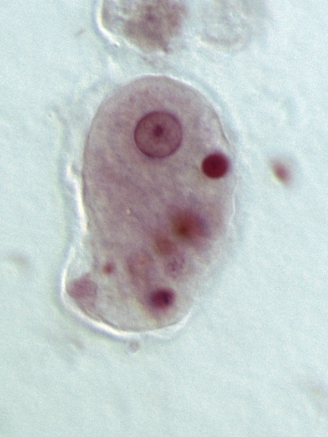
Entamoeba histolytica cyst
- Chromatid body with blunt rounded ends
- 4 nuclei
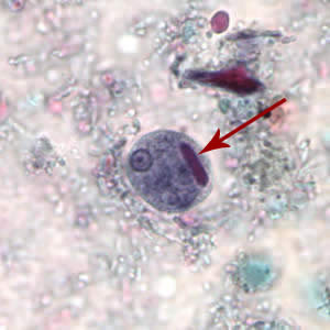
Entamoeba coli trophozite
1 nucleus w/eccentric/large karyosome
Dirty/vacuolated cytoplasm
15-50 um
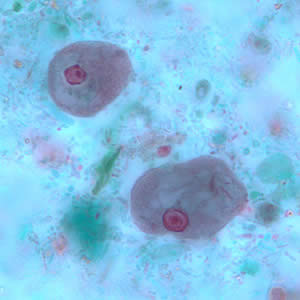
Entamoeba coli cyst
8+ nuclei (the only one w/>4)
Eccentric karyosome
10-35 um
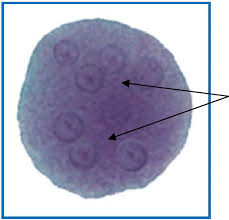
Entamoeba hartmanni trophozoite
5-15 um, small/compact karyosome
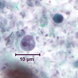
Entamoeba hartmanni cyst
Smaller (5-10 um) but like E. histolytica
Small chromatid bodies
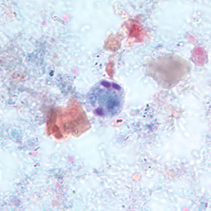
Endolimax nana trophozites
6-12 um, single nucleus
Irregular “blot-like” karyosome
Cytoplasma granular & may be vacuolated
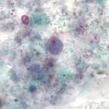
Endolimax nana cyst
Spherical (5-10 um), 4 nuclei,
Lacks peripheral chromatin
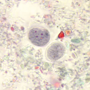
Iodamoeba buetschlii trophozoites
- Sluggish, 8-20 um, large central karyosome
- Vacuolated, achromatic vacuoles
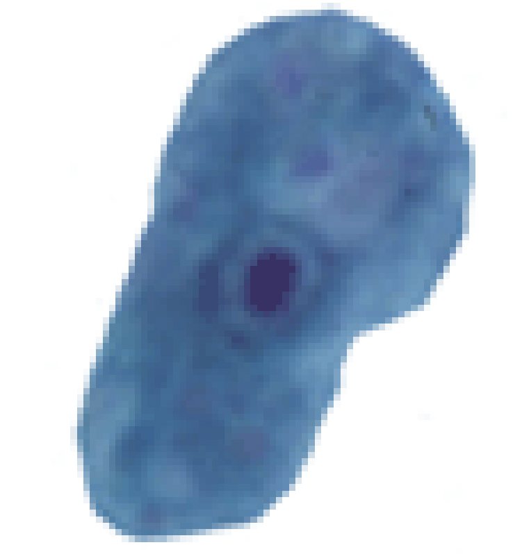
Iodamoeba buetschlii cyst
- 5-20 um, single nucleus
- One large glycogen vacuole
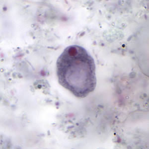
Entamoeba polecki trophozoites
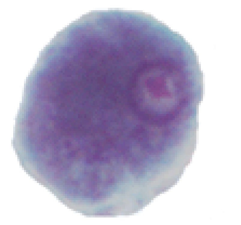
Entamoeba polecki cyst
Many small chromatid bodies with angular/pointed ends
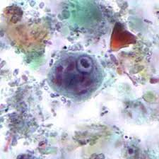
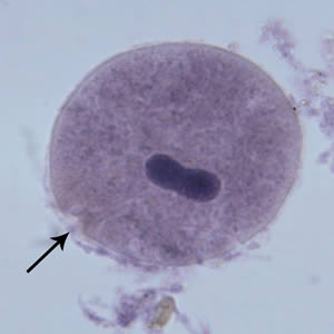
Balantidium coli
40-200 um
Cilia on surface
Cytostome (arrow)
Bean-shaped macronucleus
Cysts not often seen
Non-pathogenic amebae should ALWAYS be reported becuase
a sign of fecal-oral contamination.
Giardia duodenalis trophozoite
- Pear-shaped organisms
- 12-15 μm (range: 10-20 μm)
- 2 anteriorly-placed nuclei and 8 flagella
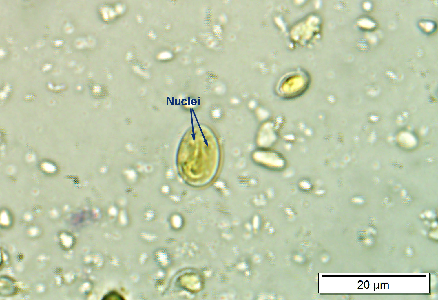
Giardia duodenalis cyst
- Ovoid to ellipsoid
- 11-14 μm (range: 8-19 μm).
- Immature cysts = 2 nuclei vs. mature cysts with 4 nuclei
- Intracytoplasmic fibrils visible
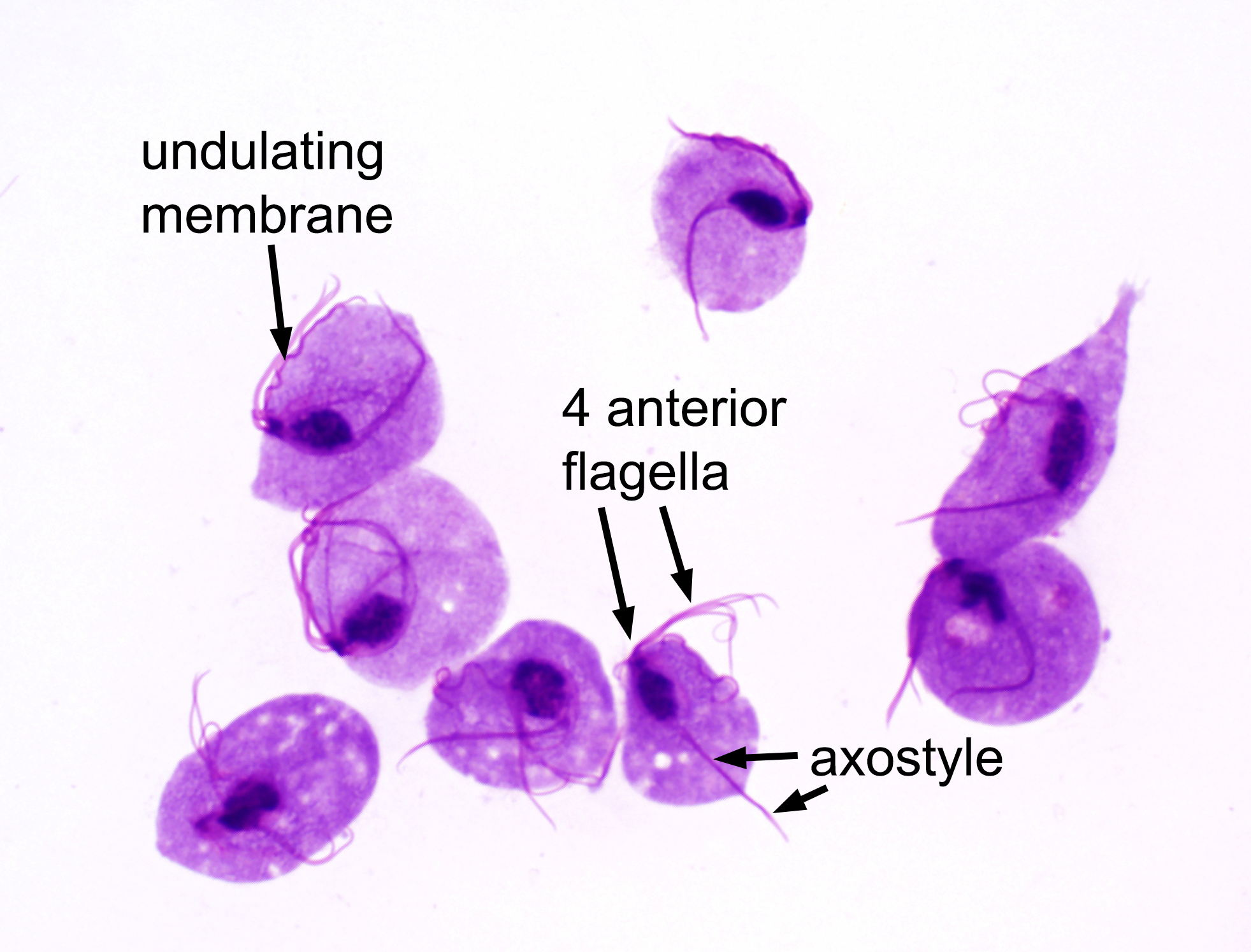
Trichomonas vaginalis trophozoite (NO Cyst stage)
- 7-30 μm long
- 5 flagella (4 anterior + 1 posterior)
- Large nucleus at wider/anterior end (contains chromatin granules and small karyosome)
- Cytoplasm with many granules
- pear shaped
Taenia’s proglottid function
absorb nutrients following past by
Taenia’s scolex function
Attaches to the intestines of the definitive host
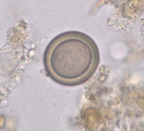
Taenia spp. egg
characteristic radiating shell
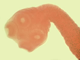
Taenia saginata
four large suckers and lack of rostellum or rostellar hooks
proglottid: >14 branches (range 12-30);motile
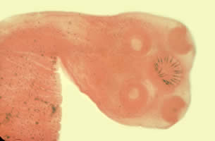
taenia solium
four large suckers and rostellum containing two rows of hooks.
proglottid: 7-13 branches
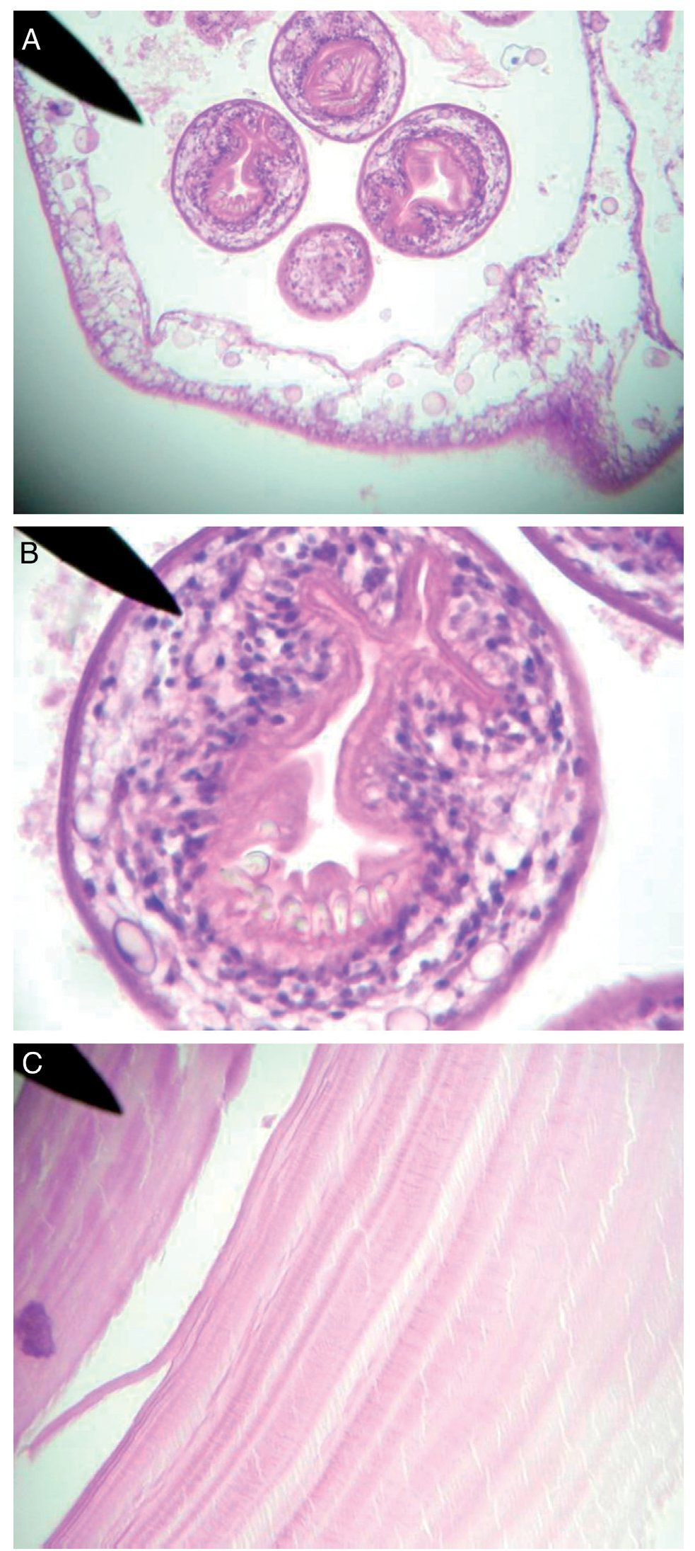
Echinococcus spp.
Hydatid brood capsule with protoscolices
‘Hydatid sand’
Refractile hooklets
Diphyllobothriid worm proglottids
Width > length compared to Taenia
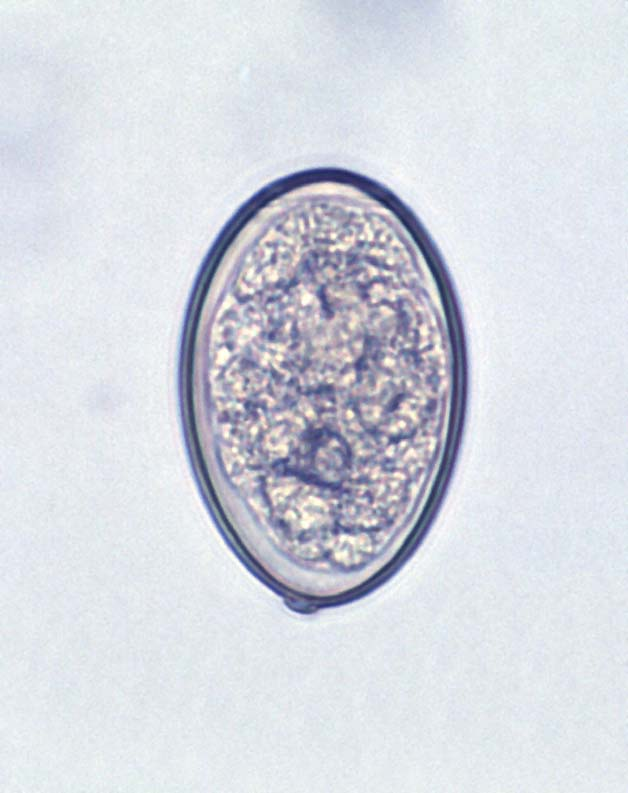
Diphyllobothriid worm eggs
55-65 x 38-42 micrometer
Smaller than Fasciola & Paragonimus eggs
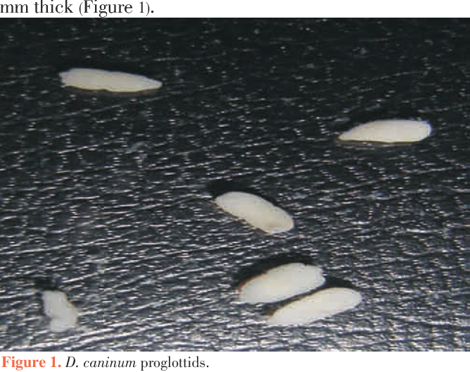
Dipylidium caninum proglottid
10-70cm
“cucumber seed” when wet or “dried grain of rice”
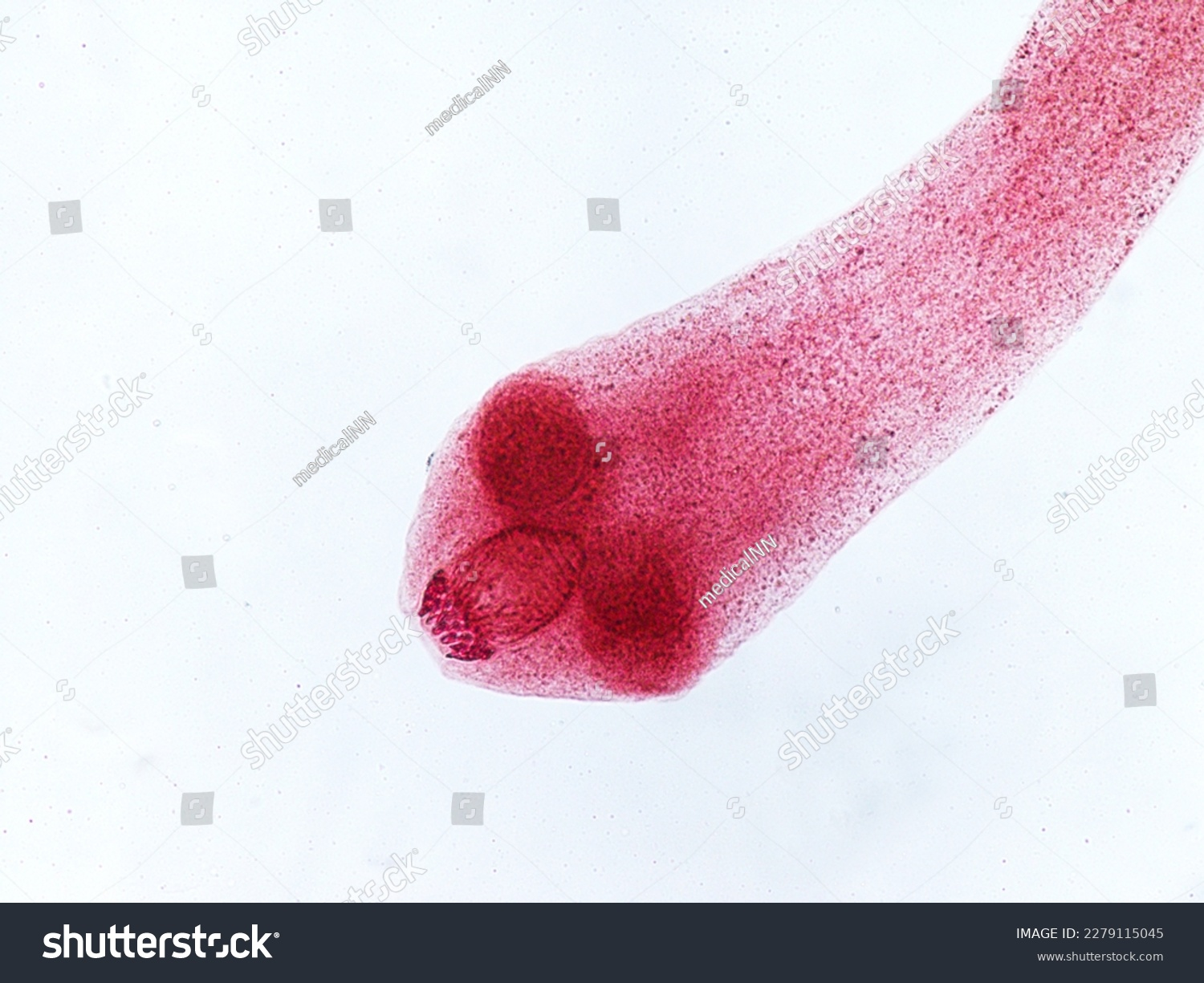
Dipylidium caninum
Armed rostellum
Four suckers
Dipylidium caninum egg pocket
– Round
– Thin shelled
– Oncosphere with six hooklets
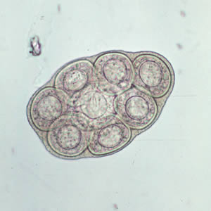
Enterobius vermicularis (pinworm)
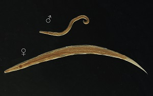
Adults:
2.5 x 0.1-0.2 mm (male)
8-13 x 0.3-0.5 mm (female)
Males: blunt posterior end with a single spicule vs. Females: long pointed tail
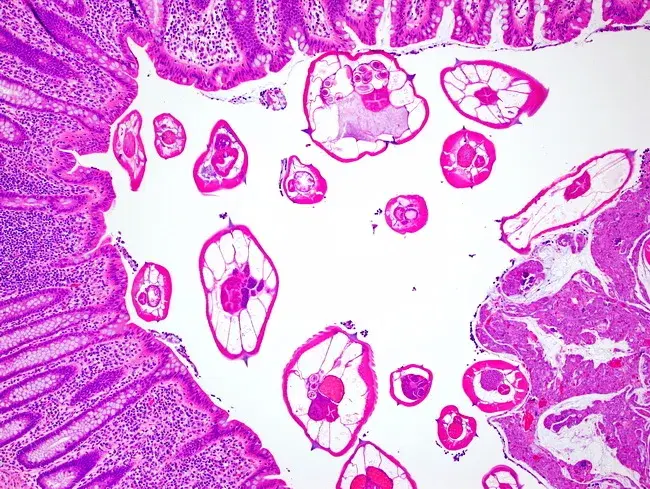
Enterobius vermicularis (pinworm) microscopic
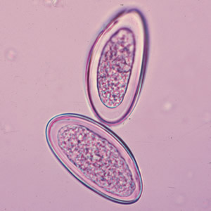
Enterobius vermicularis eggs
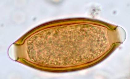
Trichuris trichiura (whipworm) Unembryonated egg
thick wall, with hyaline polar plugs
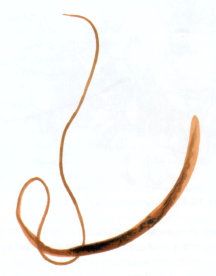
adult Trichuris trichiura (whipworm)
30-50mm long
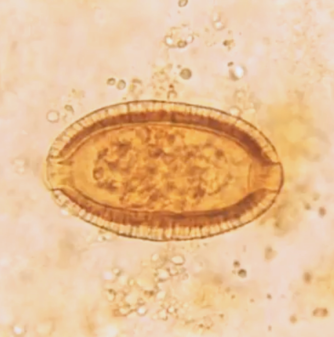
Capillaria philippinensis
resemble Trichuris trichiura (whipworm)
smaller with thick/striated shell, less prominent polar plugs
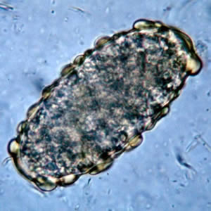
Unfertilized Ascaris lumbricoides egg
Charateristic mammalated (nodulated呈结节状的)outer shell
fertilized Ascaris lumbricoides egg
Charateristics mammalated (nodulated呈结节状的)outer shell
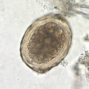
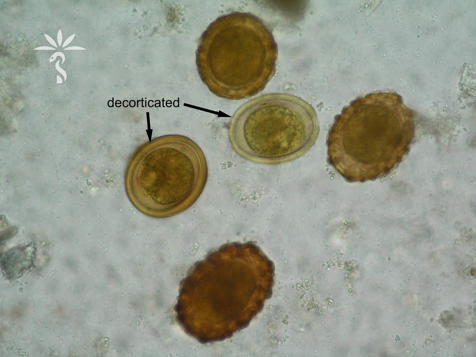
Decorticated (losing outer bumpy layer) egg of Ascaris lumbricoides
still fertilized egg
potentially infective if embryonated
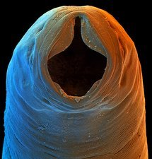
Necator americanus
cutting plate
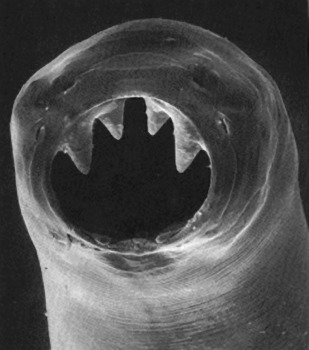
Ancylostoma duodenale
teeth
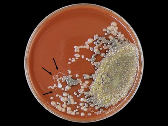
Stool culture with Larvae glide around of Strongyloides sterocolis
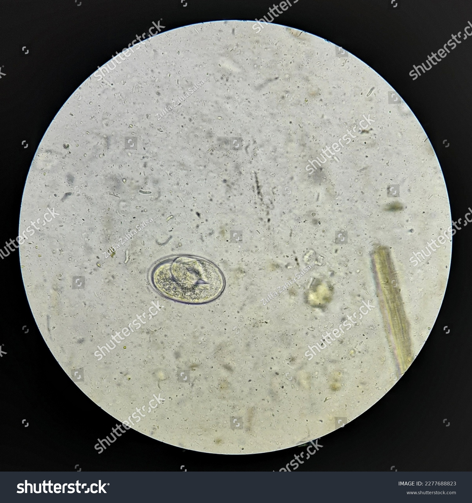
Strongyloides eggs
folded inside
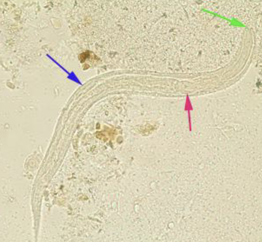
Strongyloides L1 rhabditiform larvae
Prominent genital primordium (blue arrow)
Rhabditoid esophagus (red arrow)
Short buccal canal (green arrow)
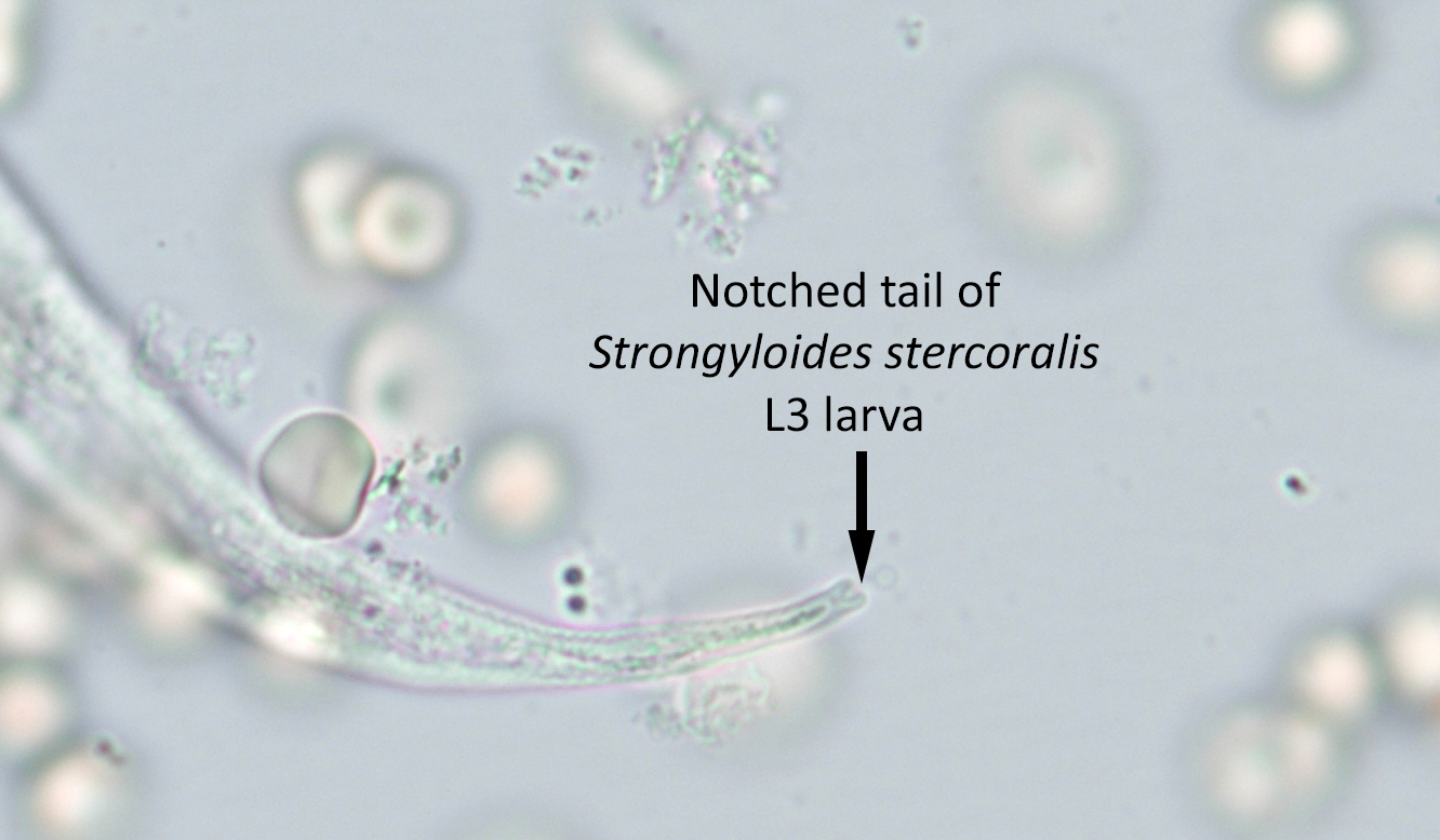
Strongyloides L3 FILARIFORM larvae
notched tail
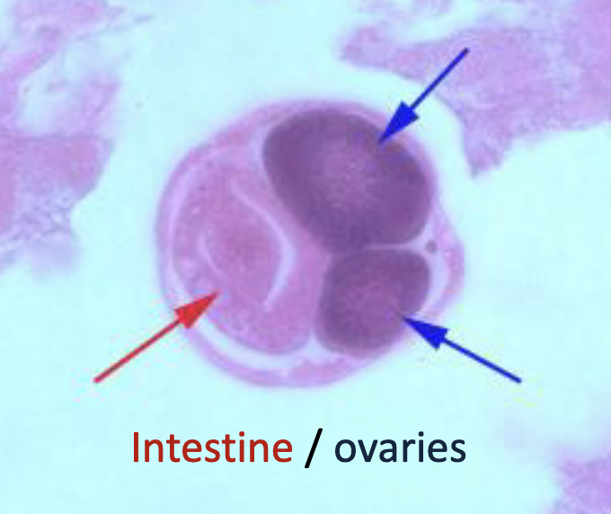
Adult female Strongyloides in Biopsy
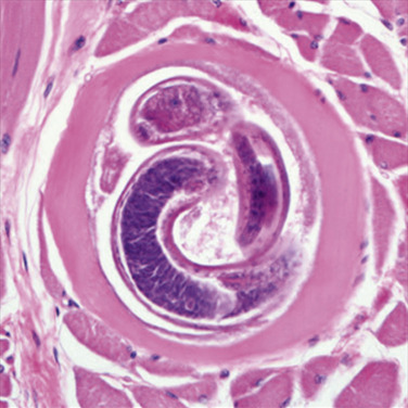
Encysted Trichinella Larvae in Muscle Tissue
Anisakis in fish tissue