AHS 131 Lab Exam 2
1/99
There's no tags or description
Looks like no tags are added yet.
Name | Mastery | Learn | Test | Matching | Spaced | Call with Kai |
|---|
No analytics yet
Send a link to your students to track their progress
100 Terms
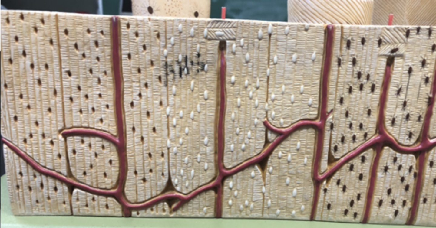
Osteon - structural unit of compact bone
What is circled in red
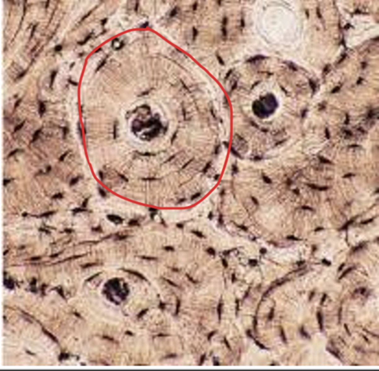
Osteon - structural unit of compact bone
What is circled in Red
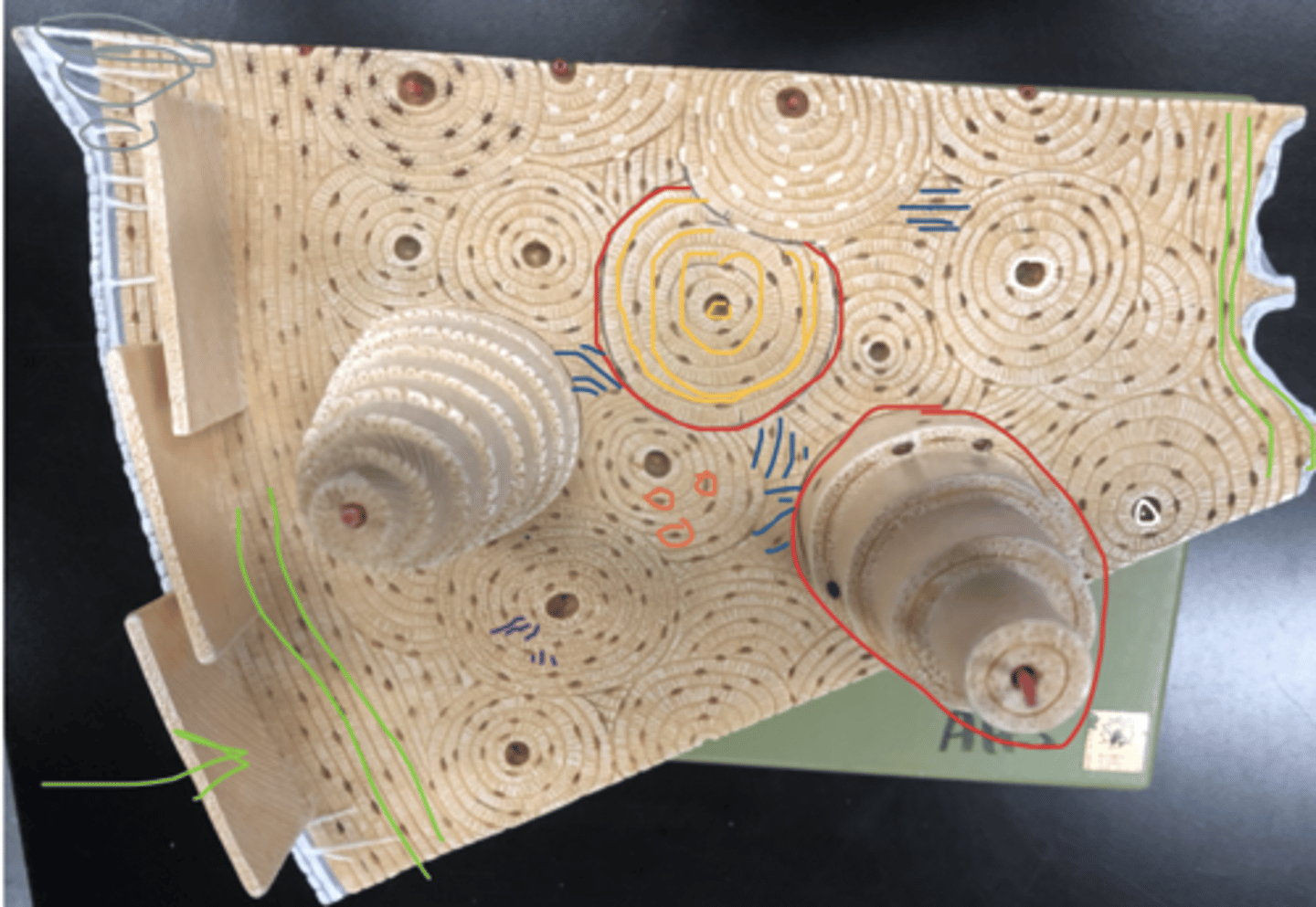
concentric lamellae - layers of bony matrix around a central canal
What is circled in yellow
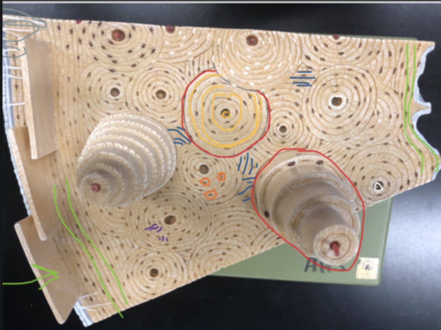
circumferential lamellae - a bony lamella that encircles the outer or inner surface of a bone
What are the green lines
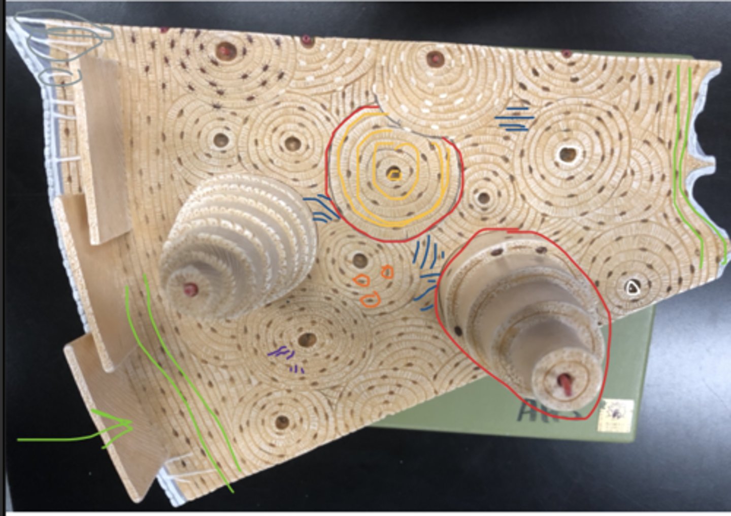
interstitial lamellae - fill spaces between osteons
What are the blue lines
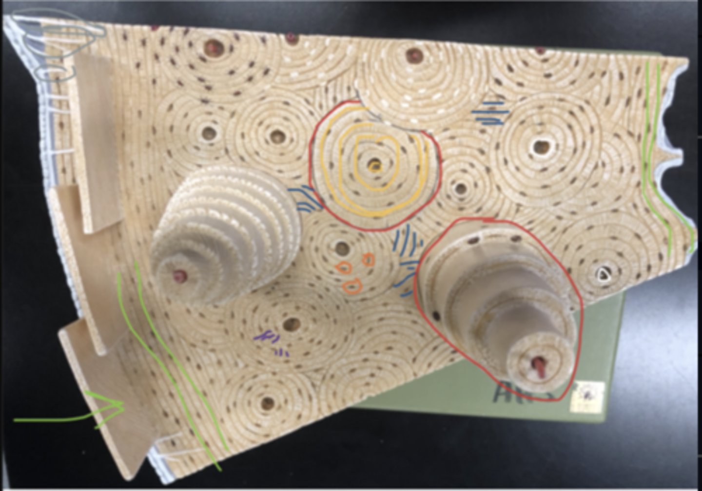
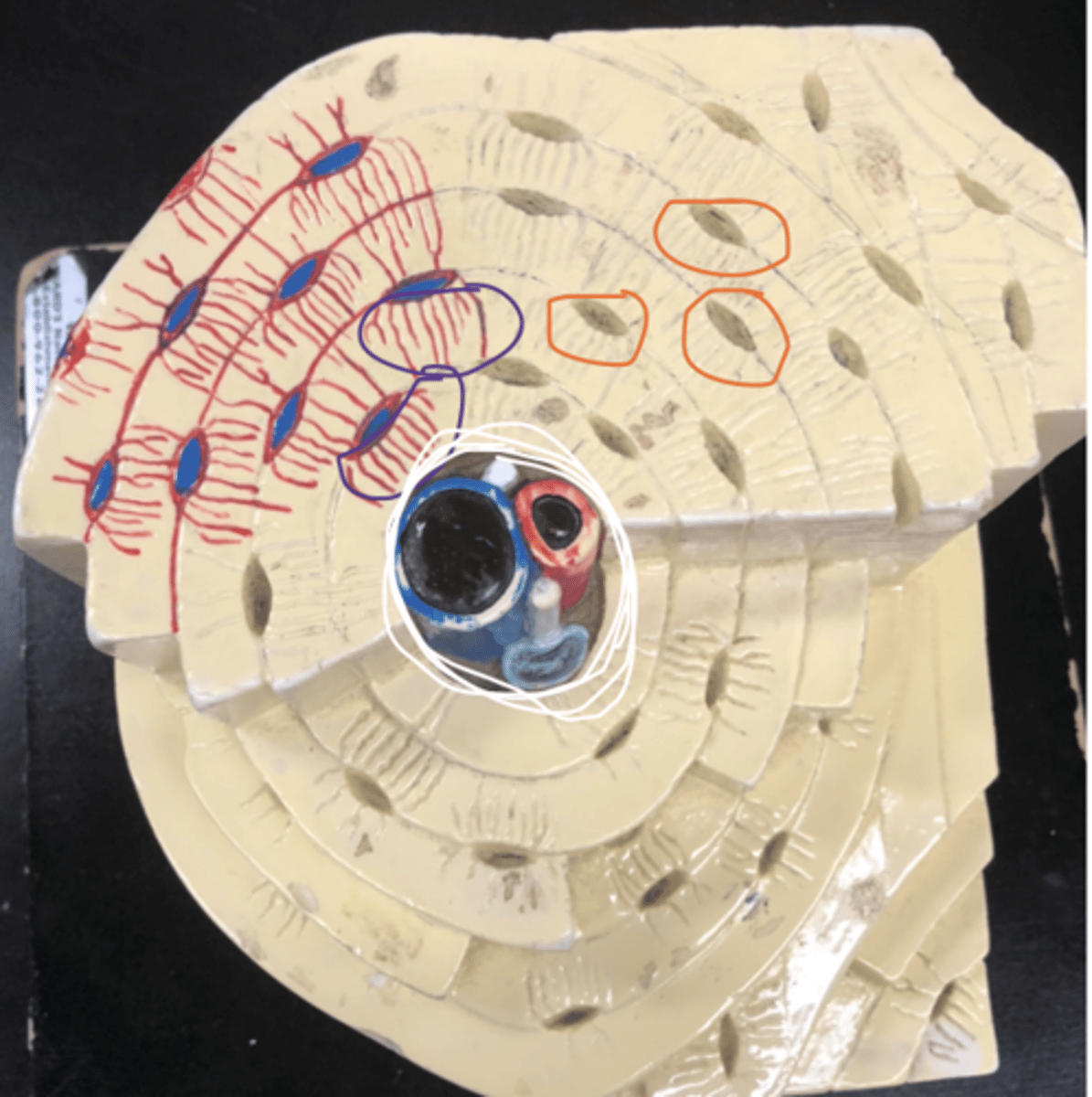
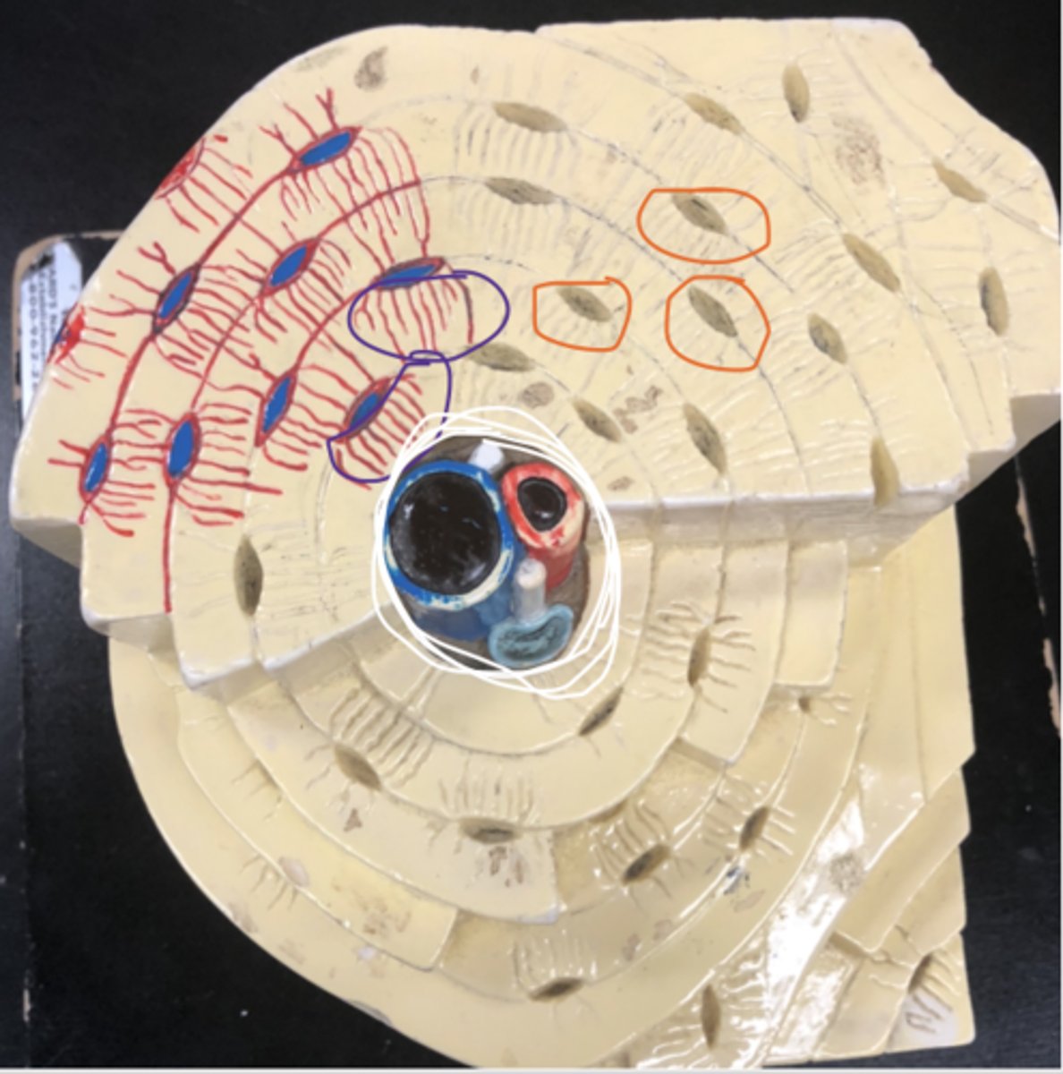
Central Canal - contains blood vessels and nerves
What is circled in white
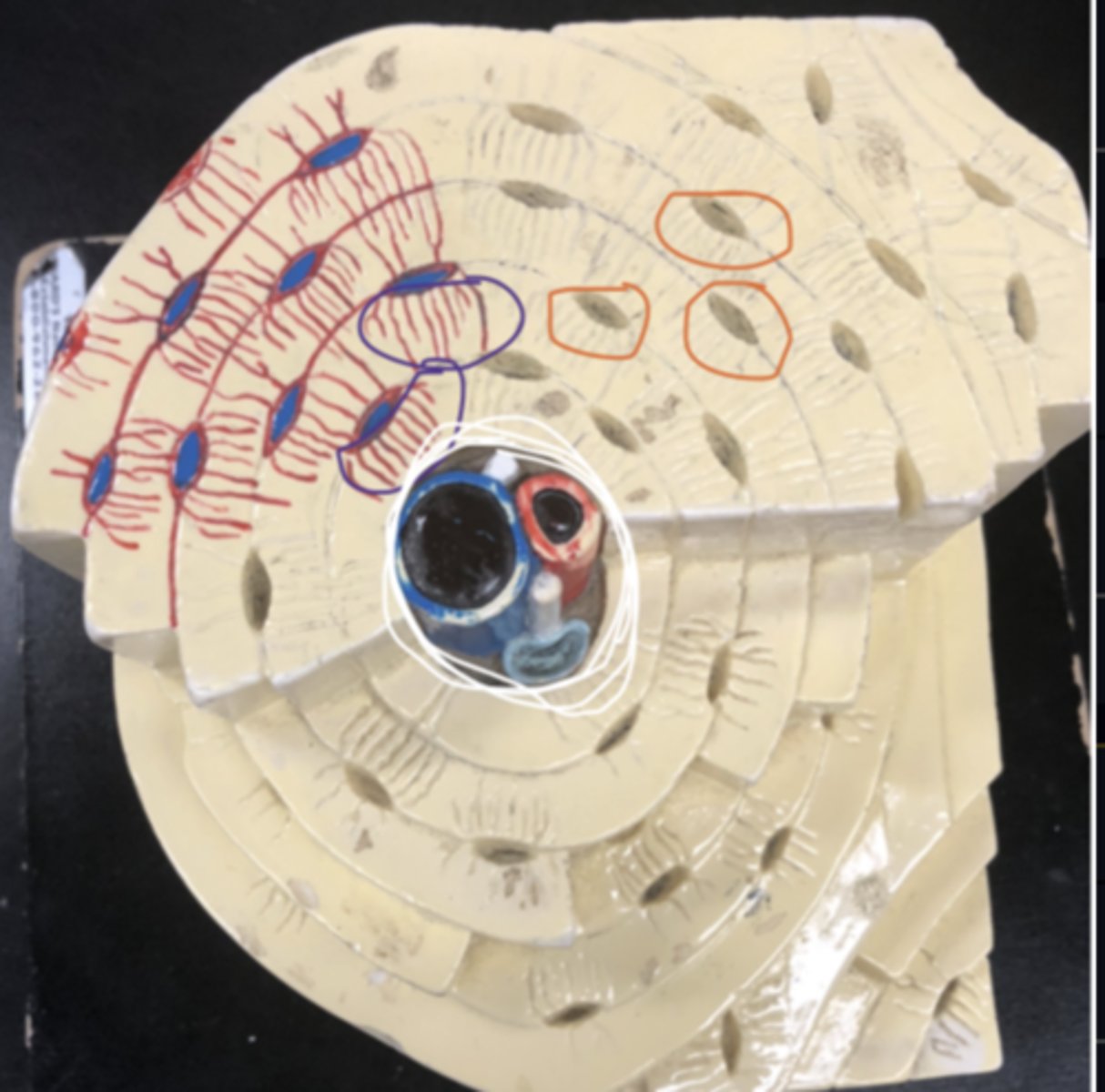
Lacuna - small cavities in bone that contain osteocytes
What is circled in orange
Lacuna - small cavities in bone that contain osteocytes
What is circled in orange
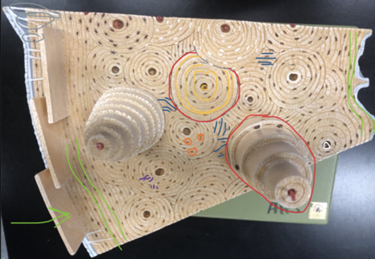
Canaliculi - Hairlike canals that connect lacunae to each other and the central canal
What is circled in purple/What are the red lines
Periosteum - A dense fibrous membrane covering the surface of bones (except at their extremities) and serving as an attachment for tendons and muscles.
What is circled in pink
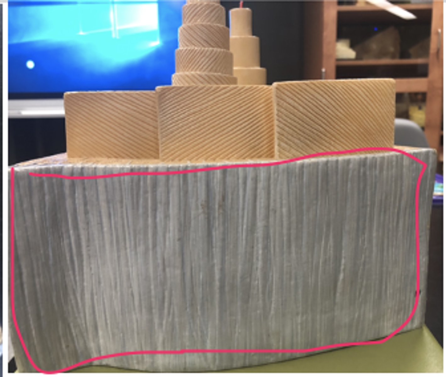
Sharpey's fibers - connect periosteum to compact bone
What is circled in grey
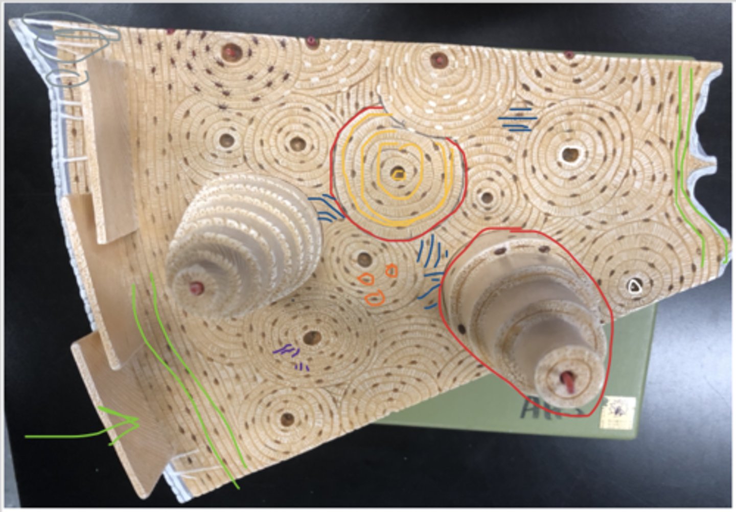
Perforating canal - canal perpendicular to the central canal, carries blood vessels and nerves
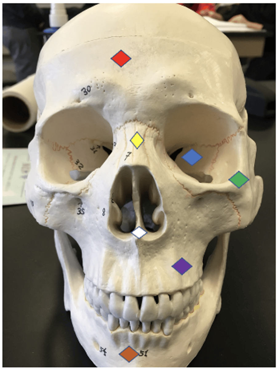
Maxillae - upper jaw
Purple
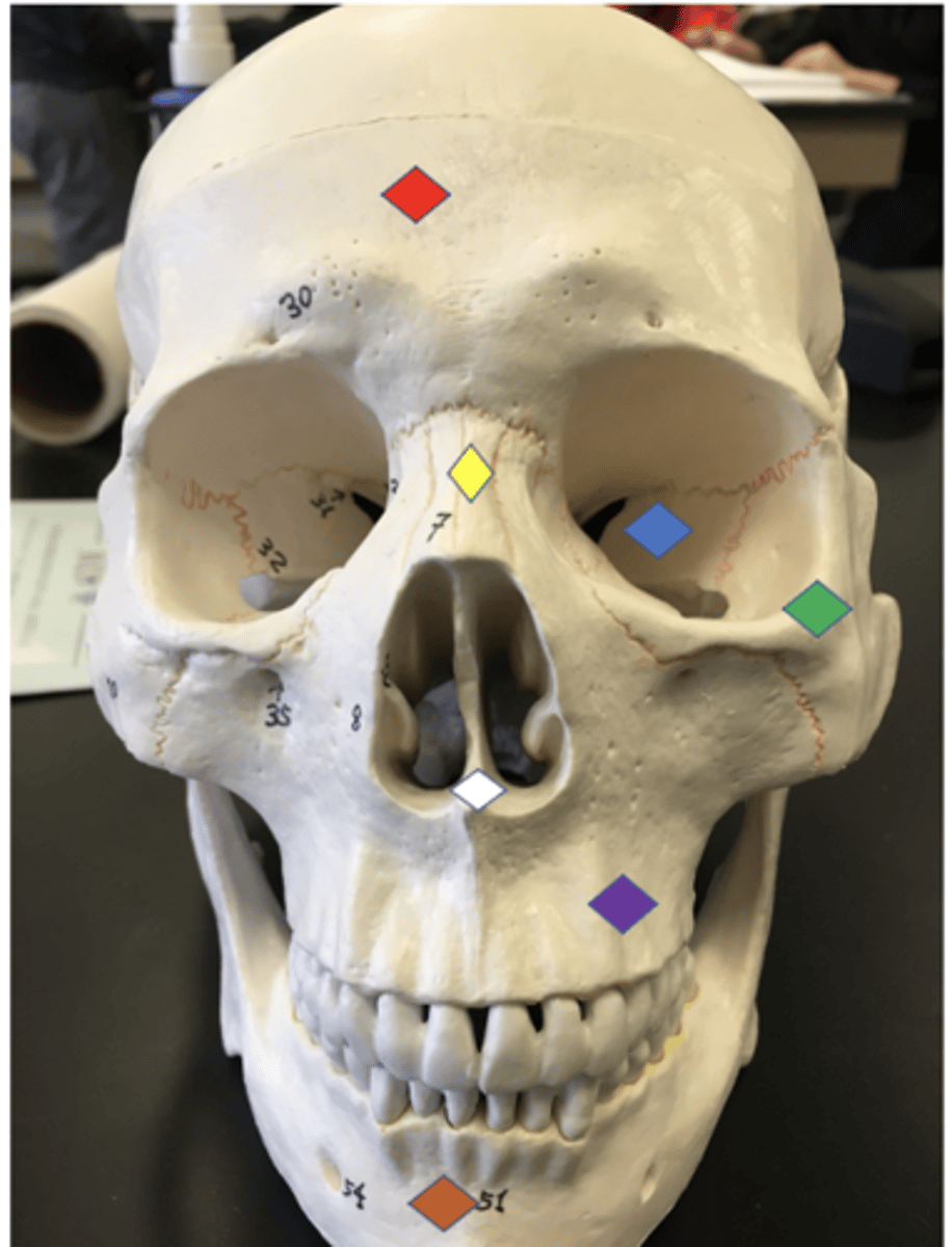
Alveolar Processes - bony points between teeth
Solid yellow lines

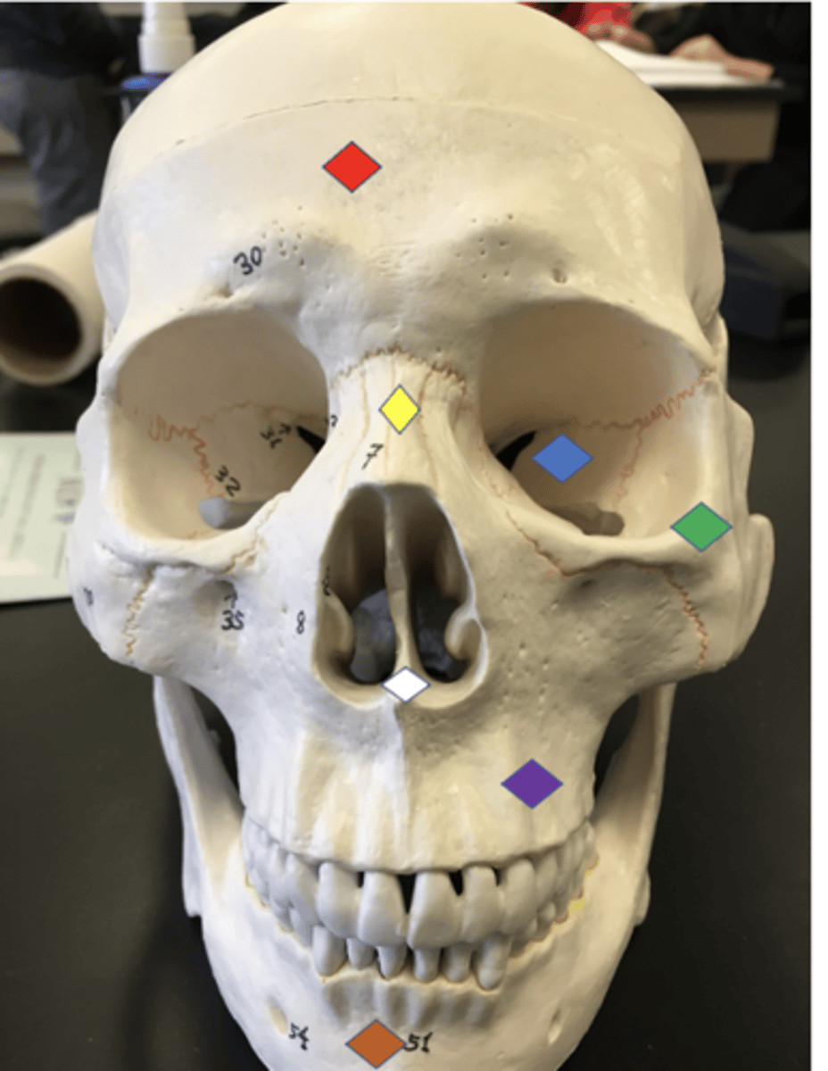
Infraorbital foramen - Hole on the cheeks/opening under the orbit carrying the infraorbital nerves and blood vessels the the nasal region
#35
Palatine Process - forms the anterior portion of the hard palate (roof) of the mouth also forms parts of the nasal cavity and eye orbits
Bright pink
Palatine - bone that forms the hard palate and parts of the nose and orbits/ above the sphenoid
Purple
Nasal - Nose bridge region
Yellow
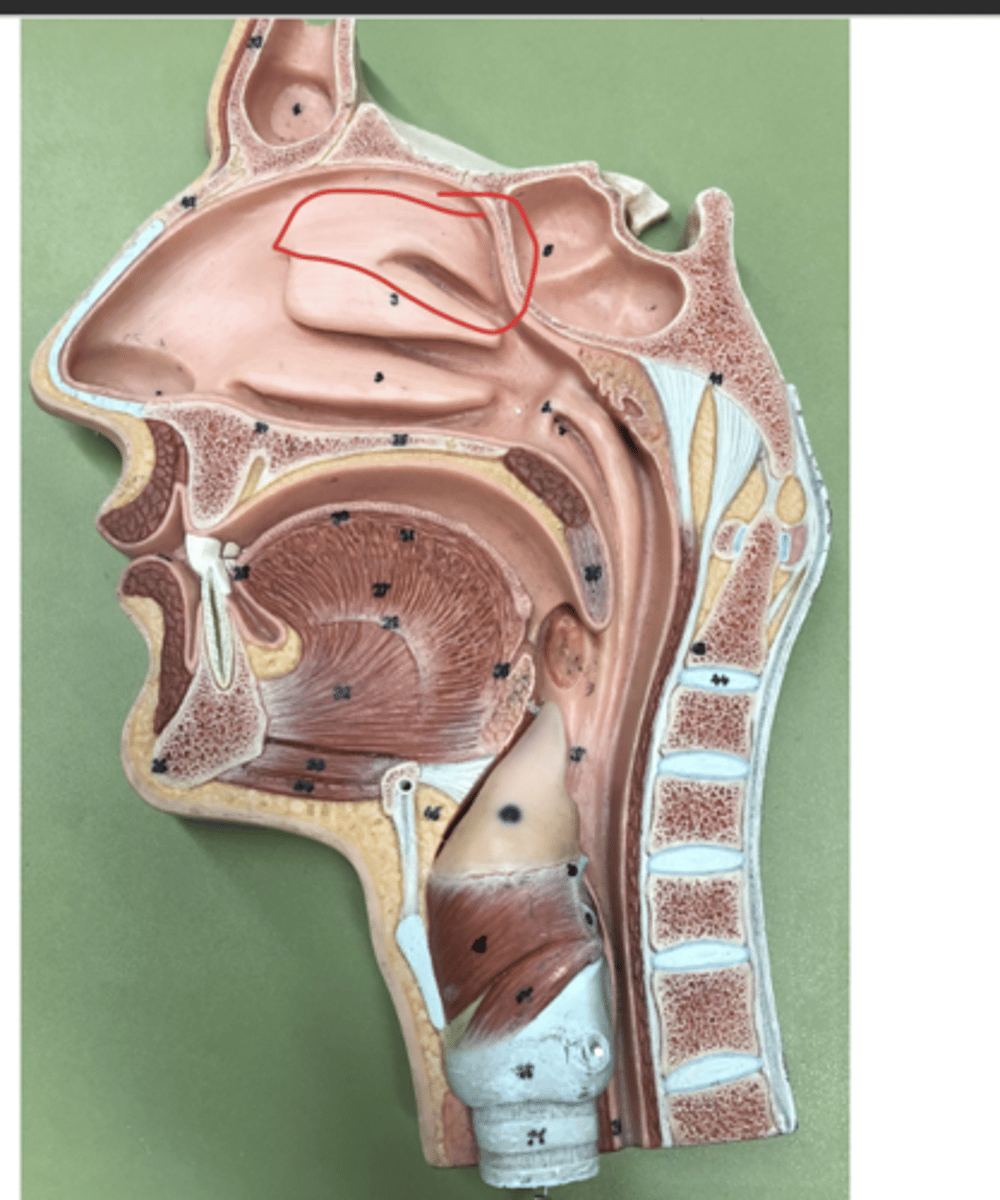
Inferior nasal conchae - The lowermost scroll-shaped bones on the sidewalls of the nasal cavity.
Hooks on either side of the vomer

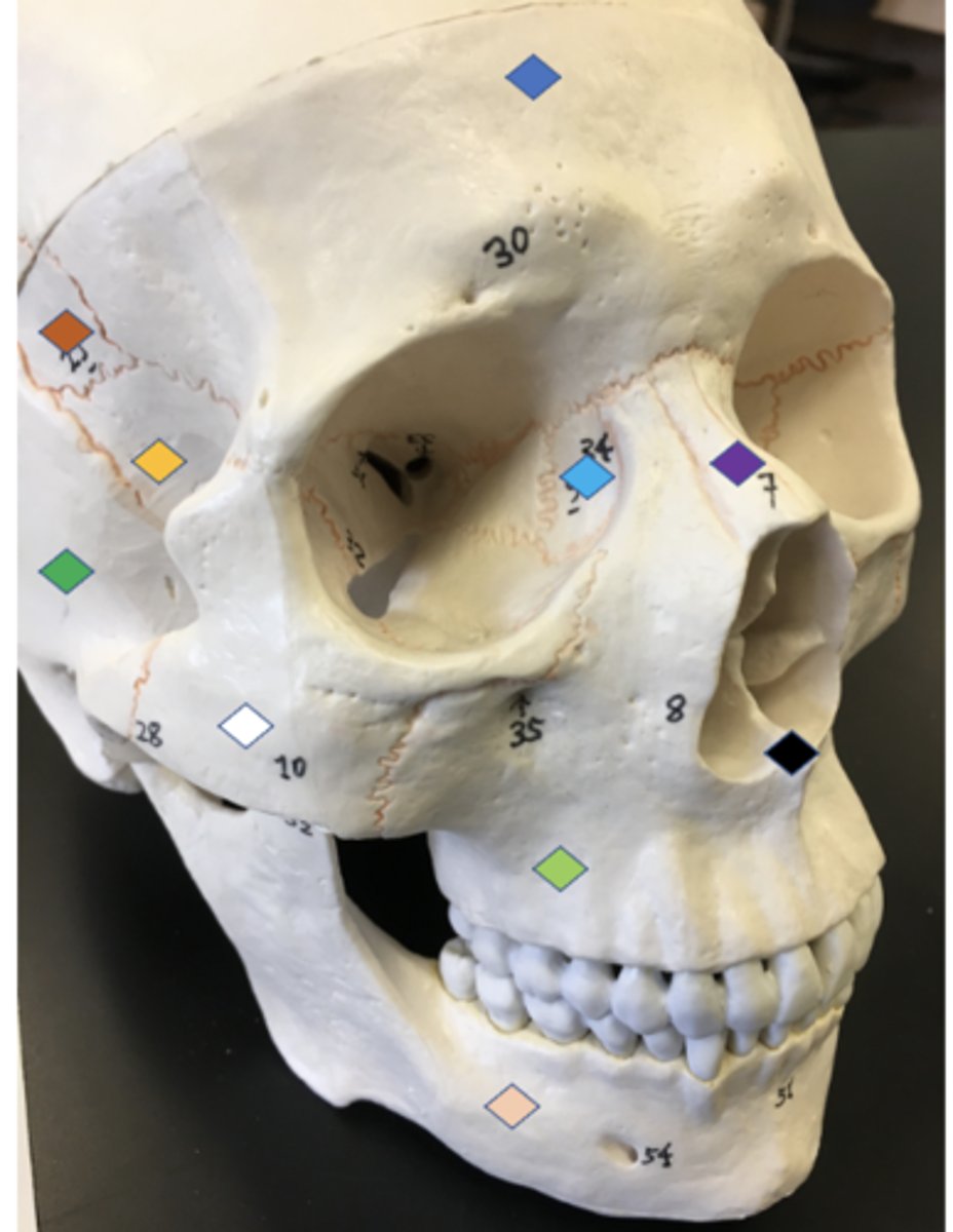
Zygomatic - Cheek bone
Green

Zygomatic arch - connects zygomatic bone to temporal bone
Bright green by #28 ( whole structure)
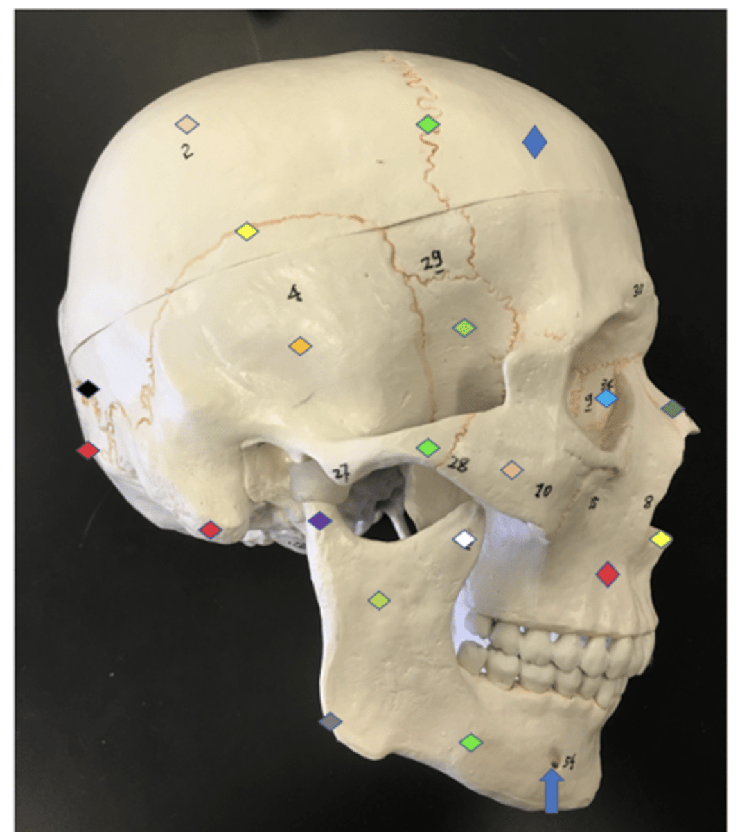
Temporal process - the zygomatic bone that connects the zygomatic to the temporal bone
#28
Lacrimal - Tear ducts
Light blue diamond
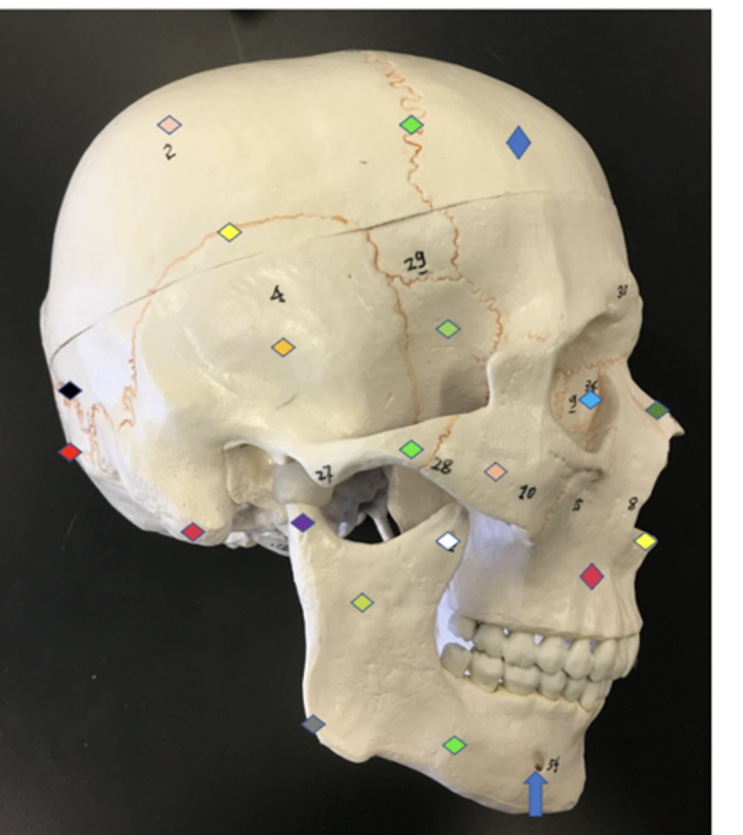
Lacrimal canal - hole where lacrimal is
Hole by light green
Vomer - nasal septum
White
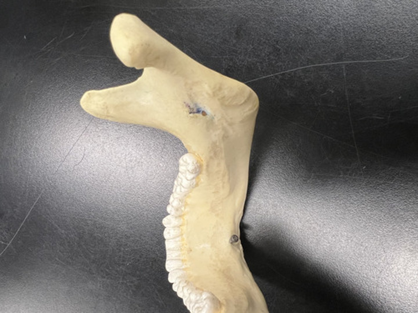
Mandible - Chin/whole lower jaw
Whole structure

Body - in between the angle and mental foramen
Light green section (solid line)

Ramus - section between the mandibular condyle and coronoid process
Red lines

Angle - point of the jawline
Red arrow

Mandibular condyle - outer hinge point of the jaw/ bigger notch
Dotted green circle

Coronoid process - in front of the mandibular condyle
Yellow dotted circle

Mental protuberance - point of the chin
Point at solid green line angle to the right by aquamarine arrow
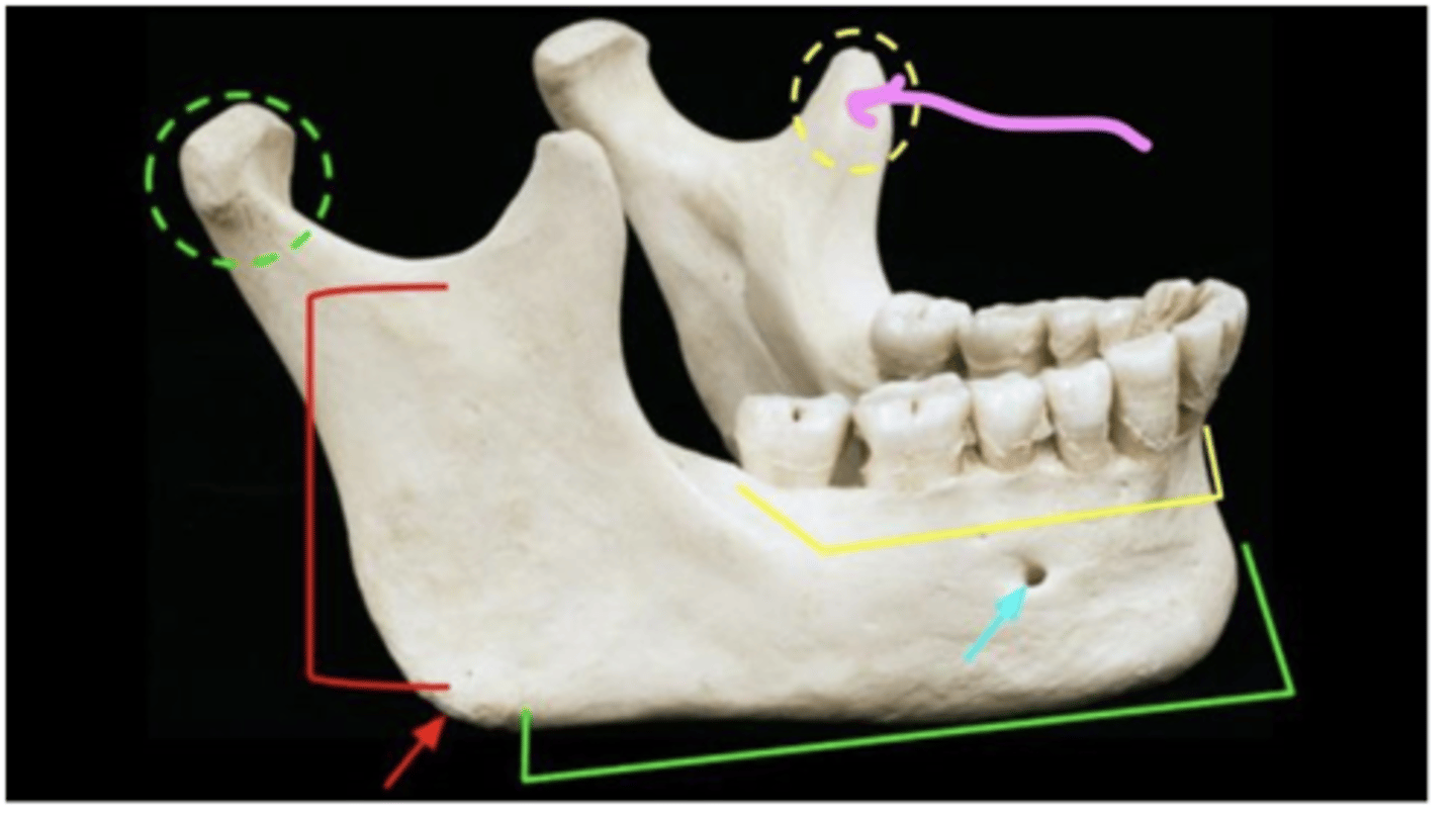
Mental foramen - hole on the chin
Aquamarine arrow
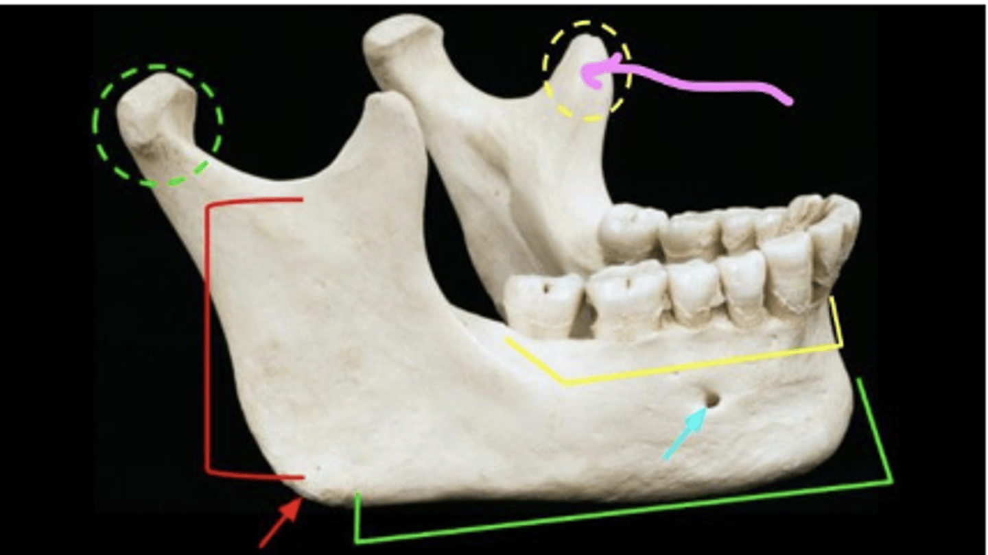
Mandibular foramen - hole on the inside of the mandible
Bluish marking
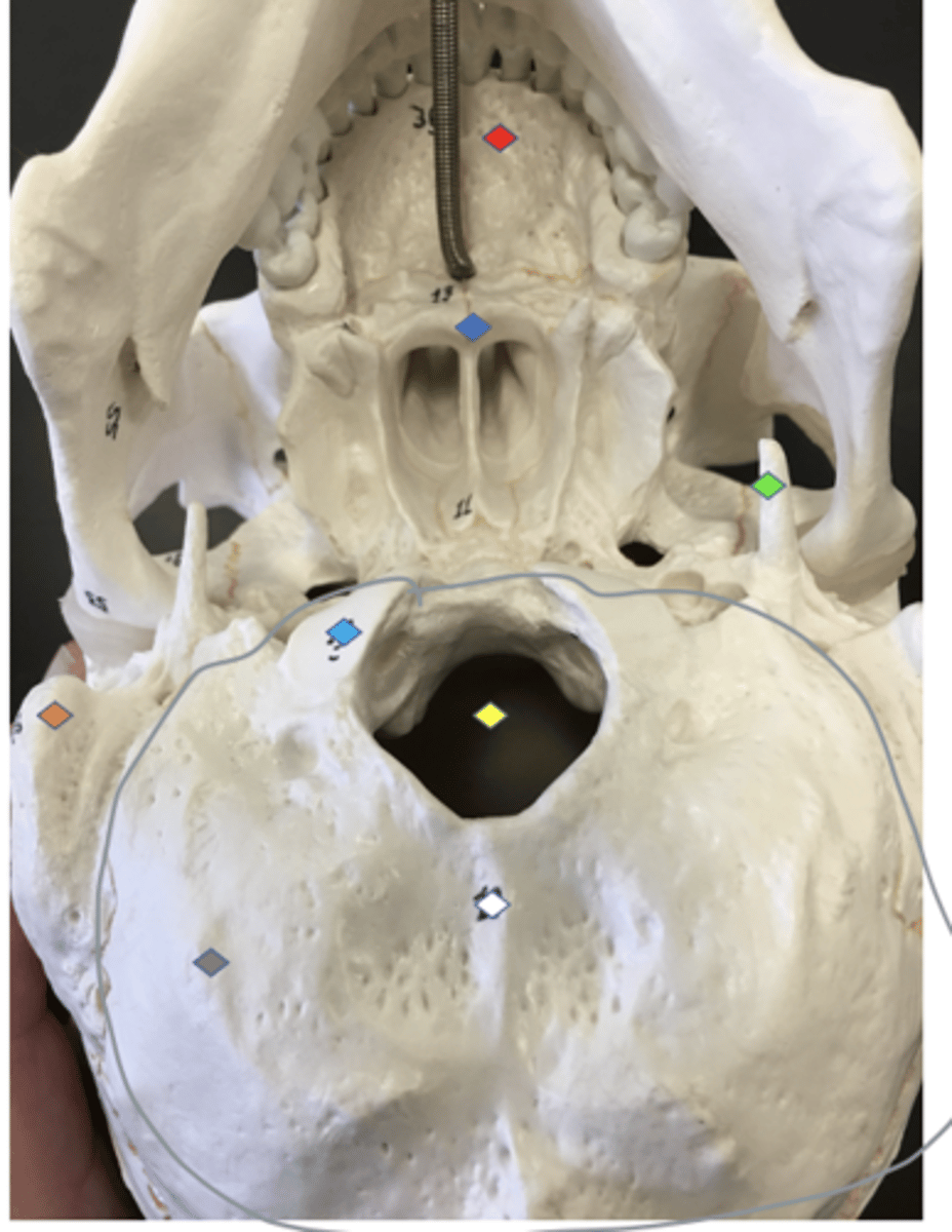
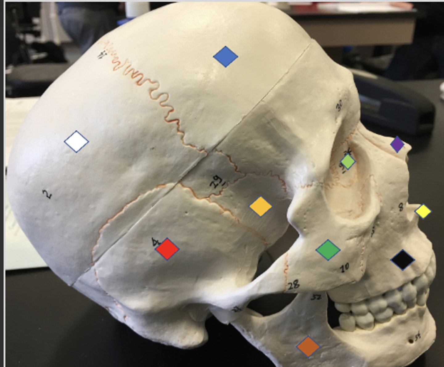
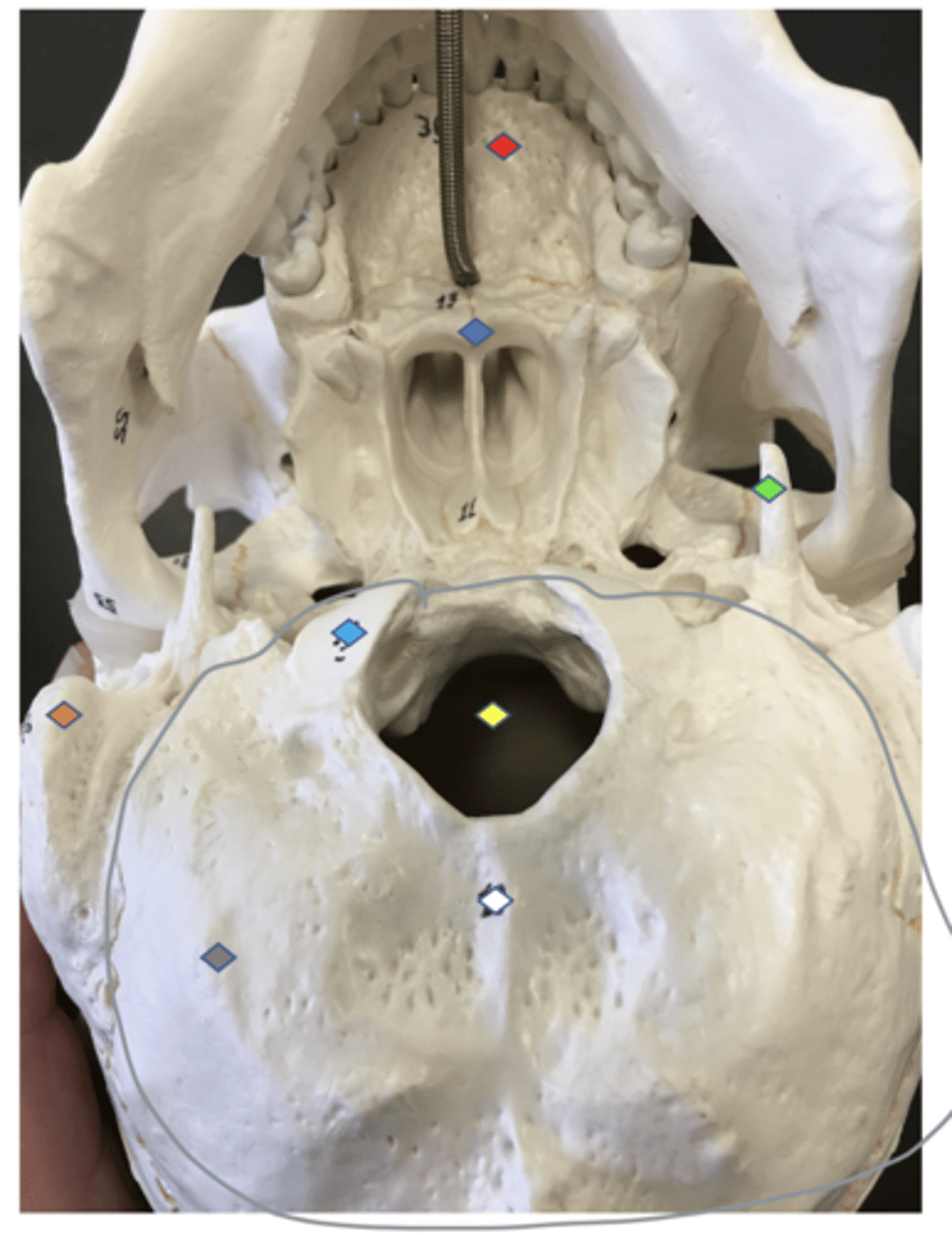
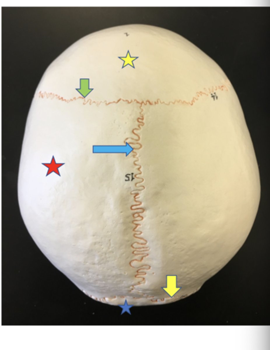
occipital - bone of the back of the skull
Grey circle
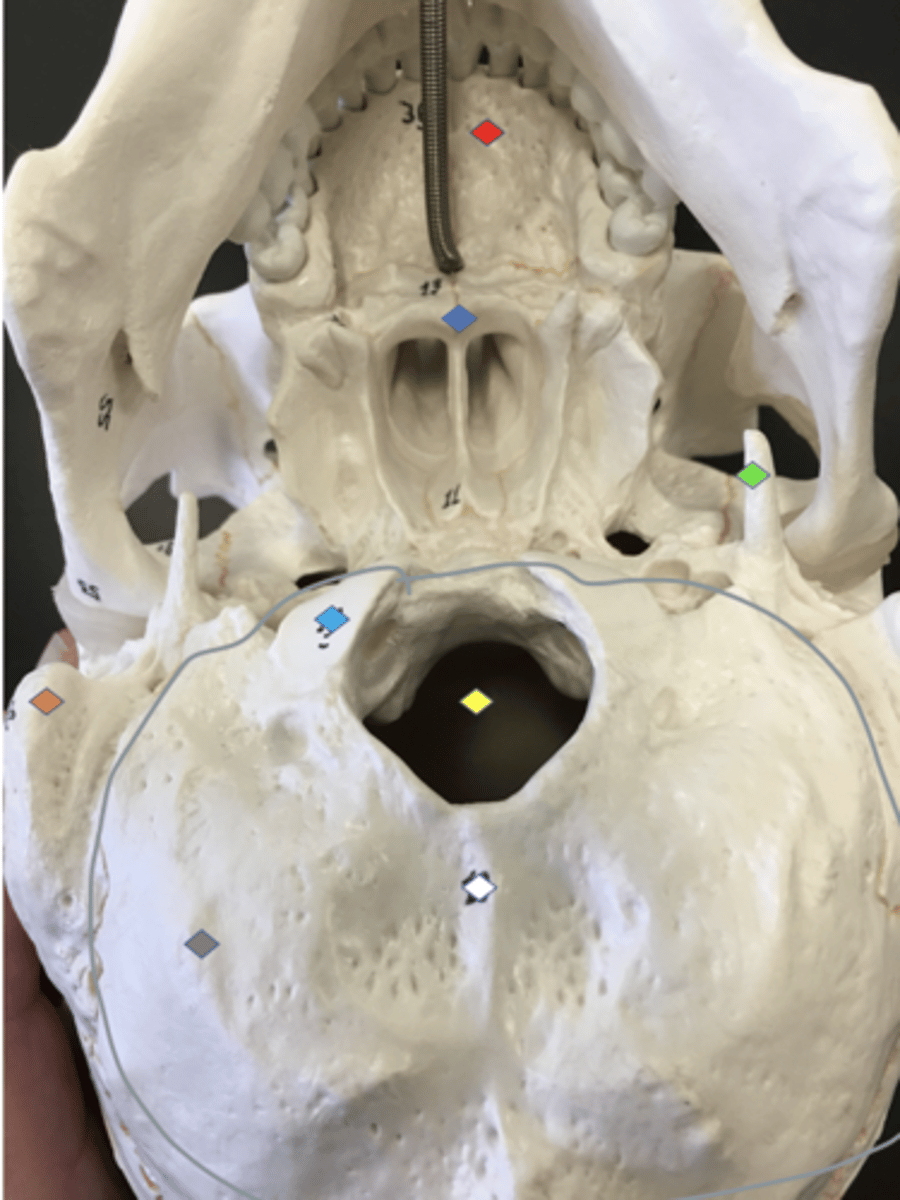
external occipital protuberance - Bump on the back of the head
White
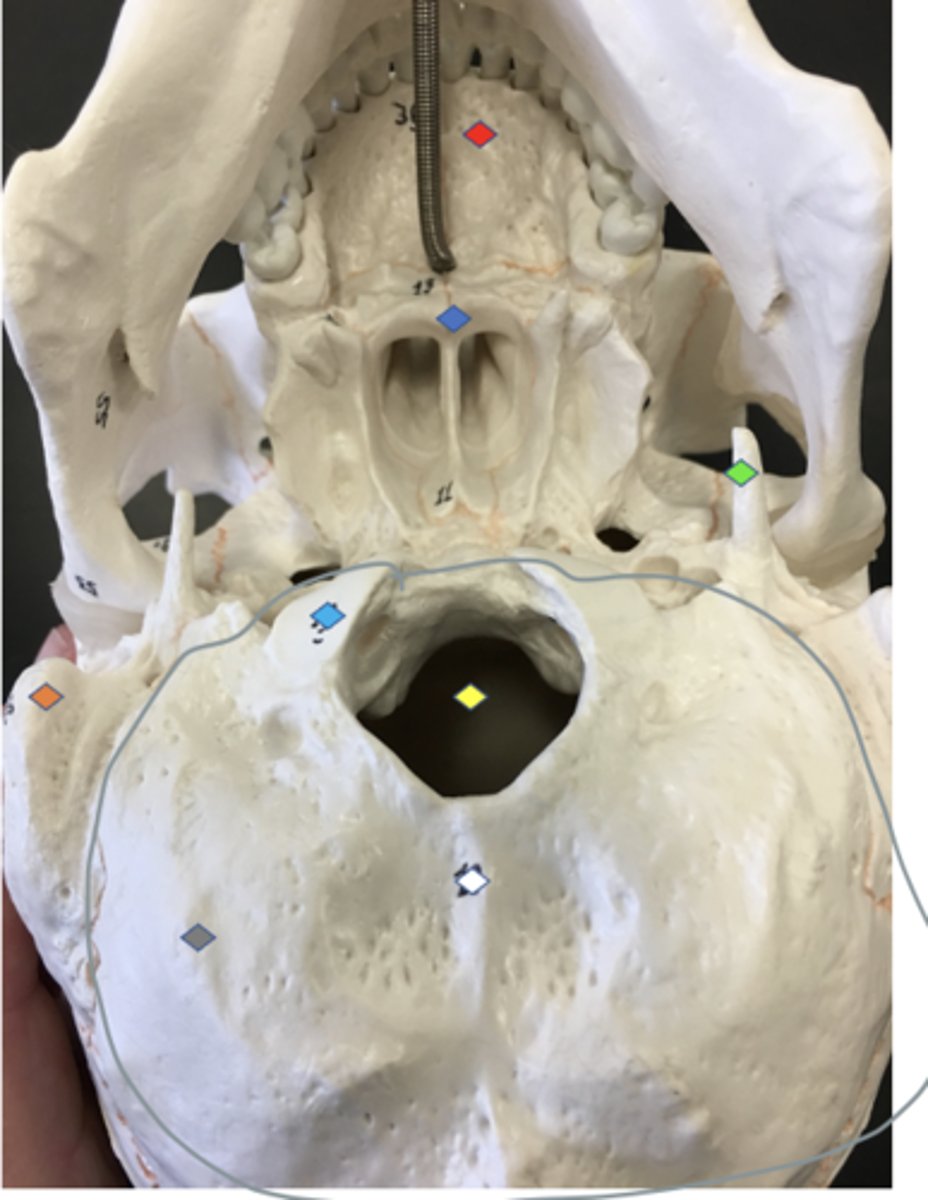
Foramen magnum - big hole in the occipital bone
Yellow

Hypoglossal canal - Tiny holes inside the foramen magnum
Structure inside foramen magnum
lambdoid suture - outlines the occipital in the back of the head
Yellow arrow
Occipital condyles - Rounded projections lateral to the foramen magnum that articulate with the first cervical vertebra (atlas)
Light blue
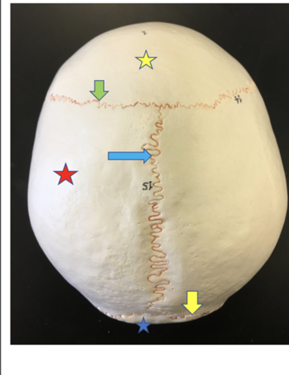
Parietal - side of the skull
Red star
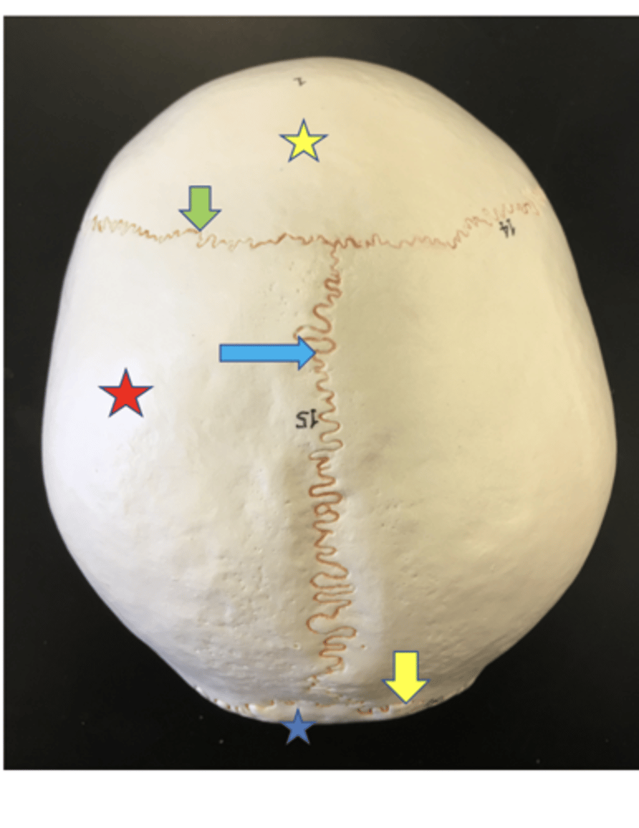
Sagittal suture - along the sagittal plane
Blue arrow

Frontal - forehead bone
yellow star

Coronal suture -along the frontal plane
green arrow
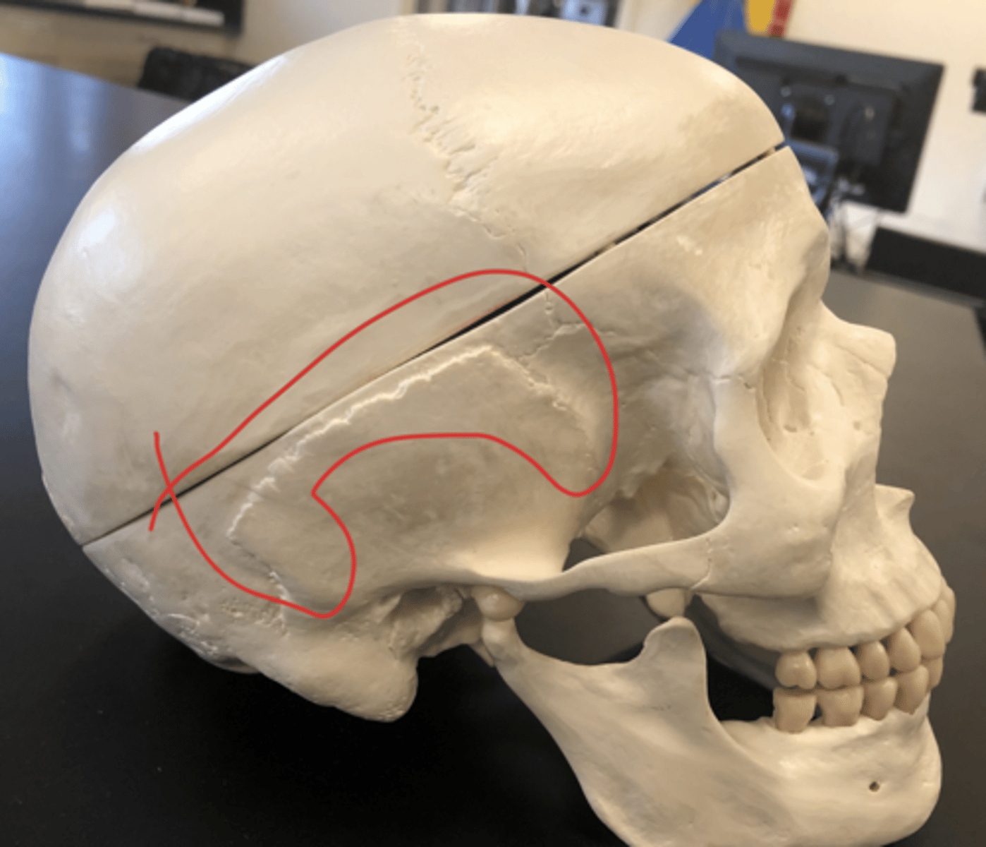
Temporal - temple bones
Red
Zygomatic process - the temporal bone that connects to the zygomatic bone on the zygomatic arch
#27
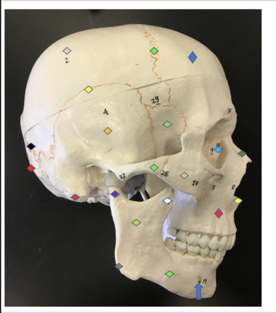
Styloid process - pointy processes on the side of the occipital bone and on the zygomatic bone
Green

Mastoid process - big bumps on the back of the zygomatic bone
Orange
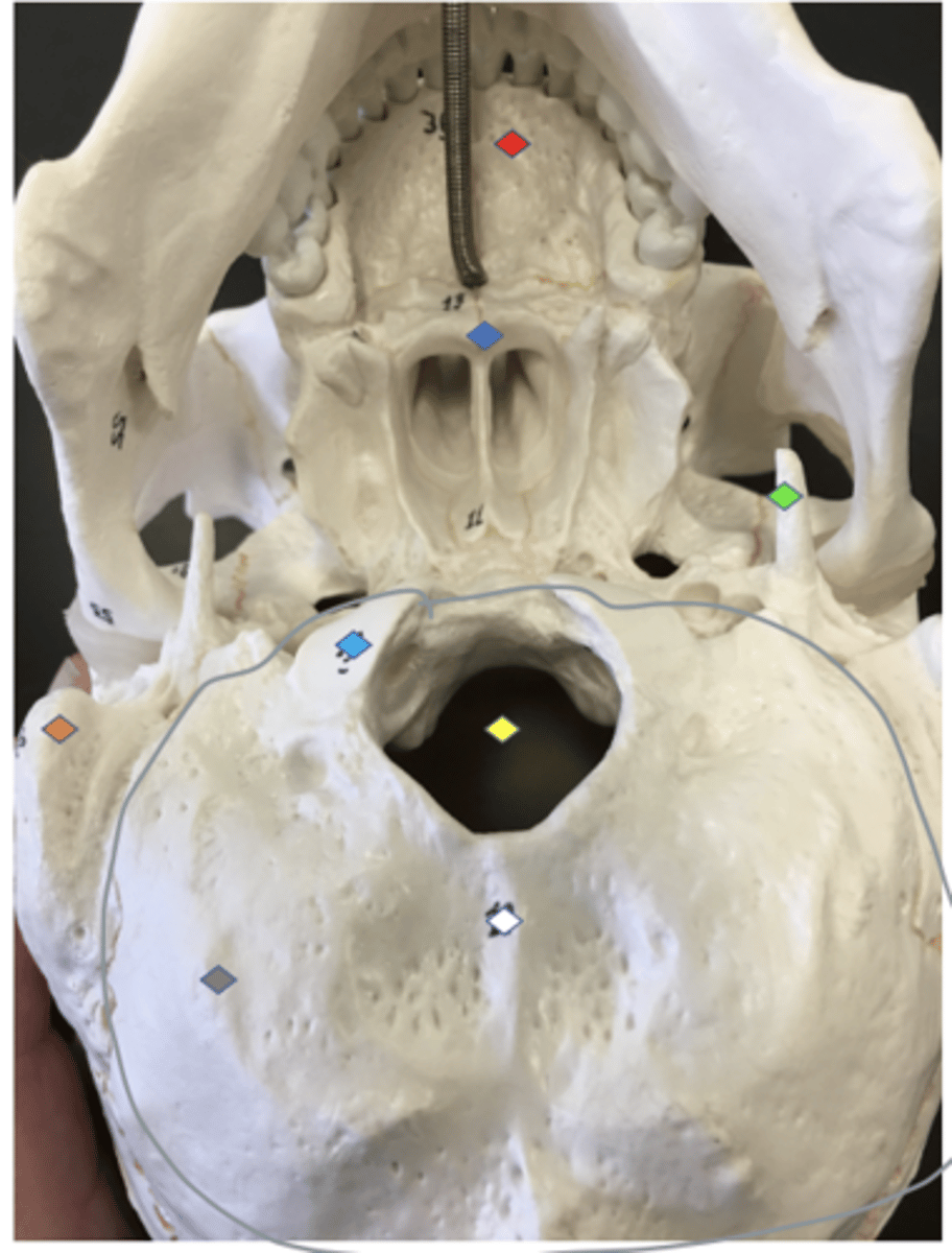
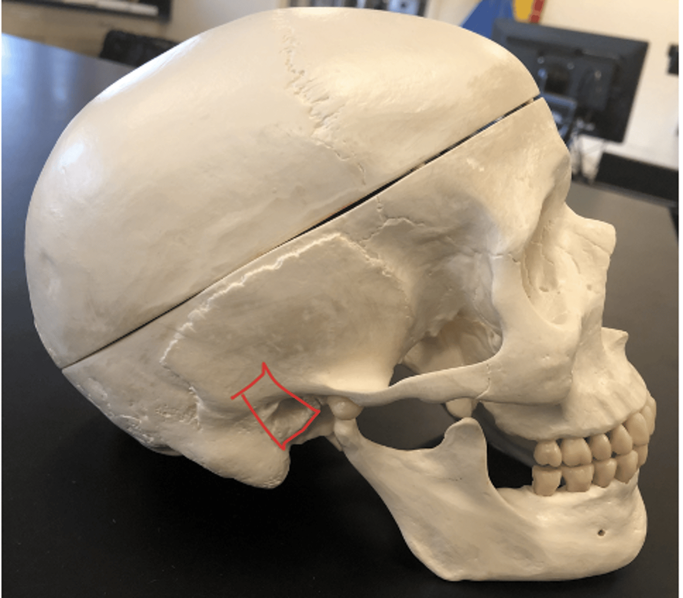
External acoustic meatus - Canal leading to eardrum and middle ear
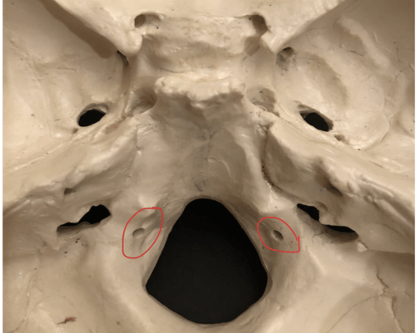
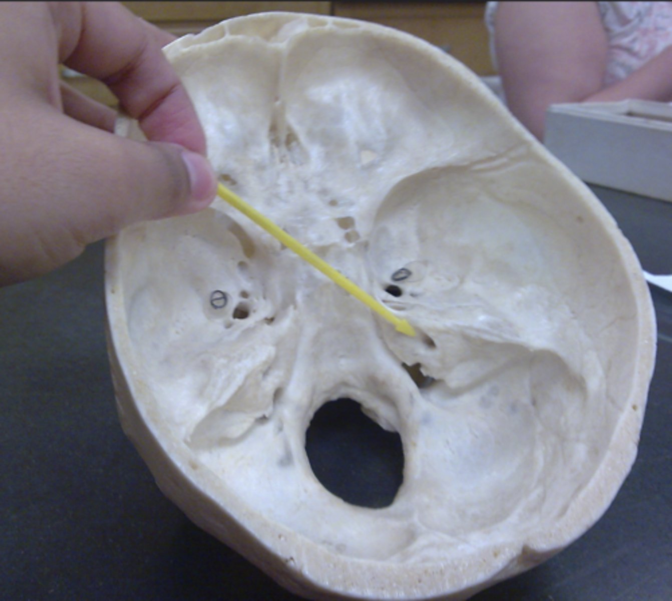
Internal acoustic meatus - socket below the sphenoid and to the sides of the foramen magnum
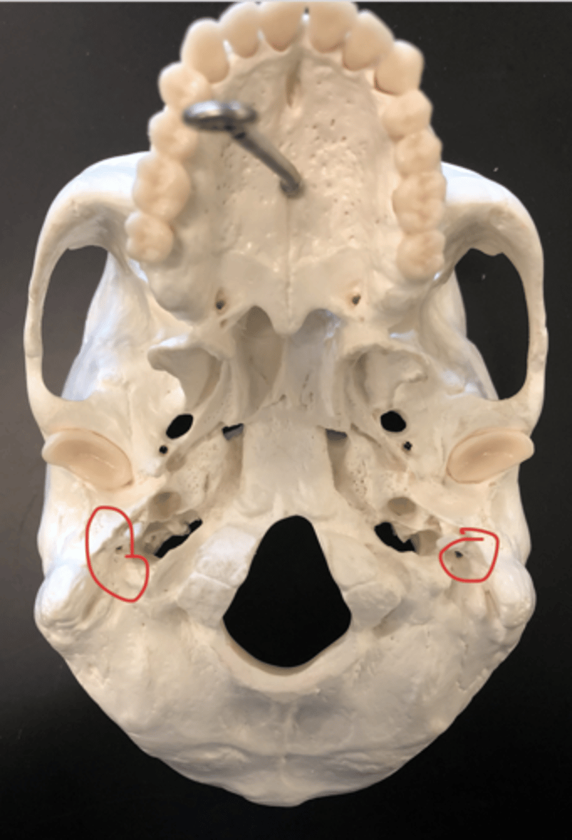
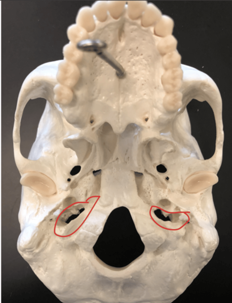
stylomastoid foramen - holes between the mastoid process and jugular foramen
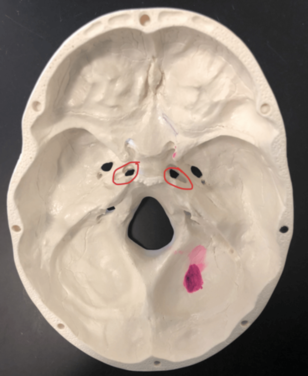
Carotid canal - above the jugular foramen; weird indentation; from the inferior view
#7
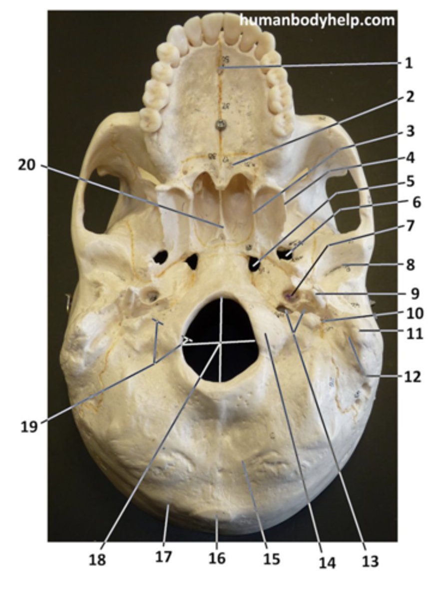
jugular foramen - J shaped hole near the mastoid process
Squamous suture - above the temporal bone
Sphenoid - butterfly structure above the occipital bone
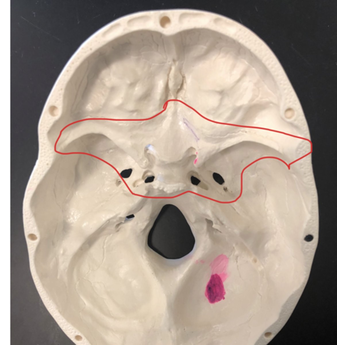
Sella turcica - in between lesser and greater wing; saddle shaped
Greater wing - bottom wing that's more indented in
Lesser wing - on top of the greater wing; looks like a bat
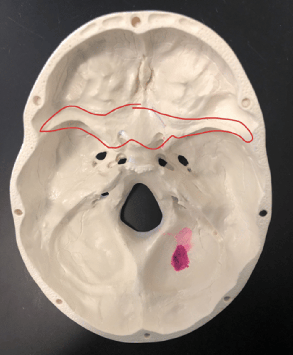
Pterygoid process - the legs of the goblin; under the palatine
"Legs of the goblin"
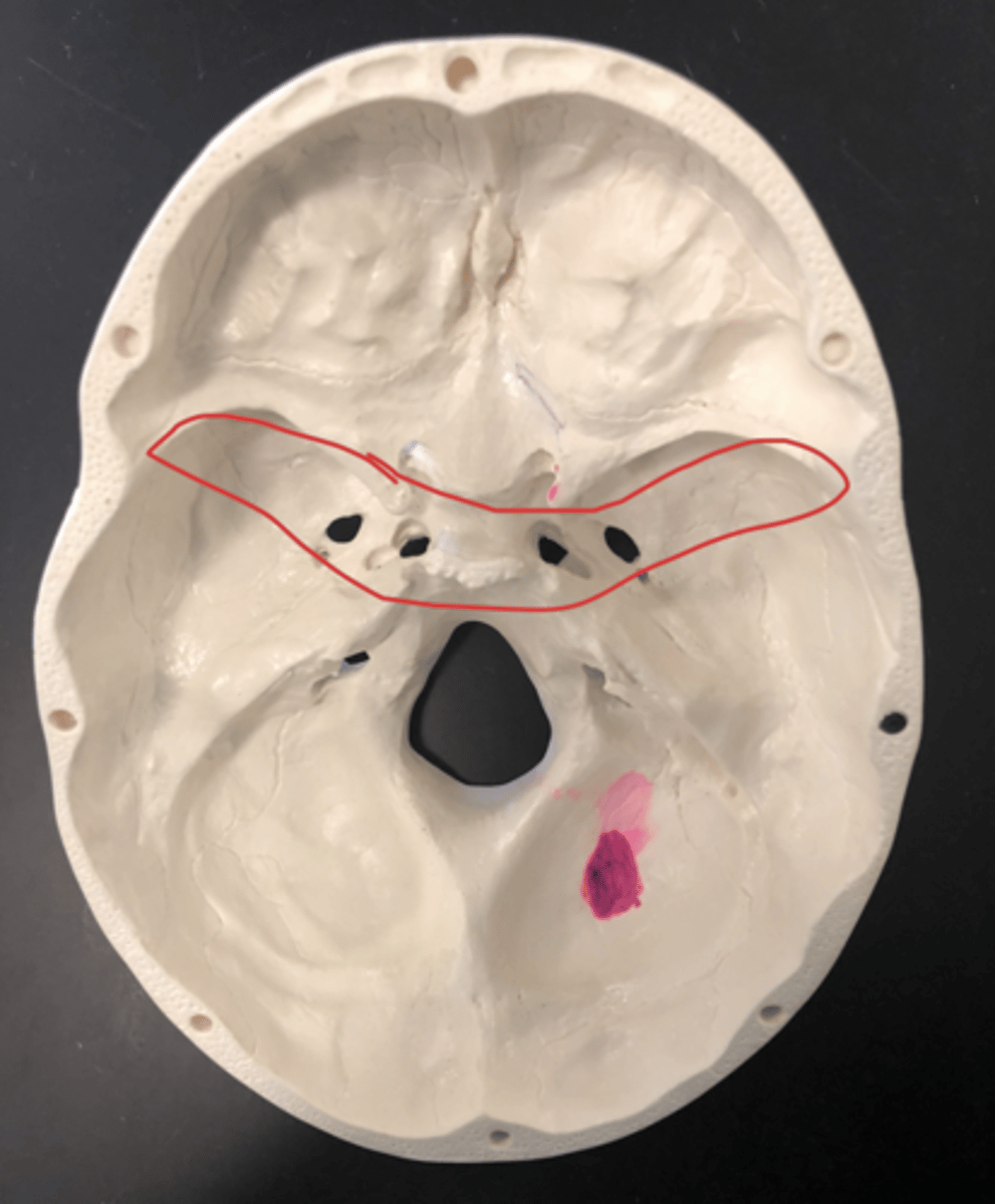
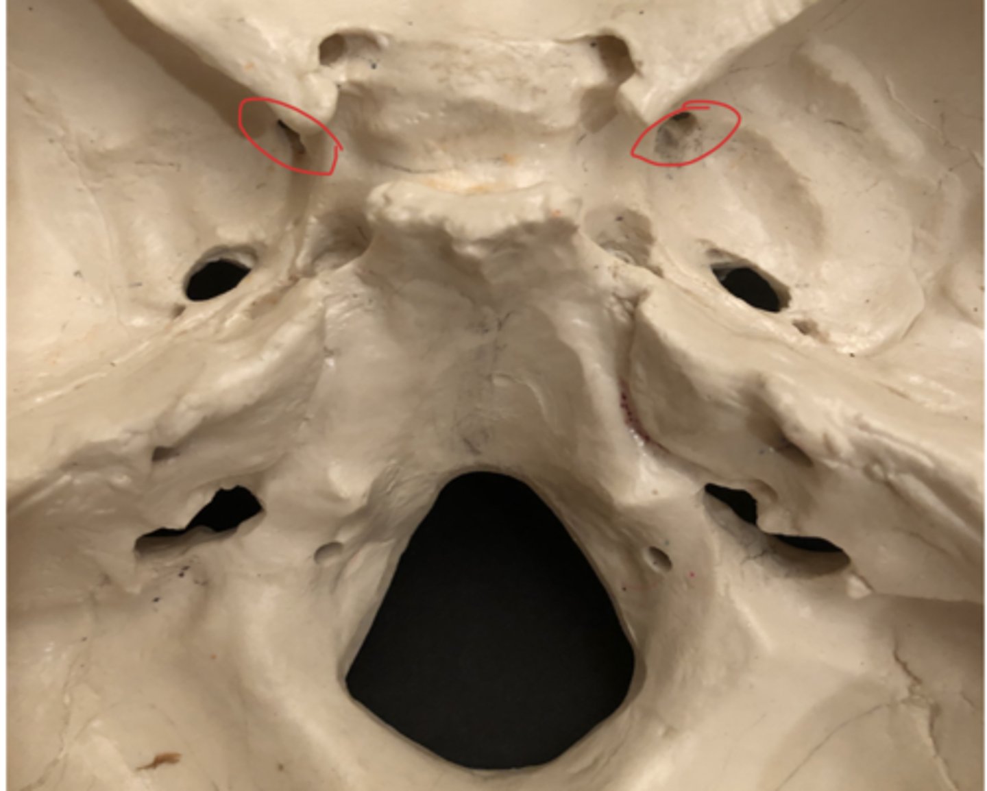
Foramen ovale - outer hole near lacerum, away from the sella turcica
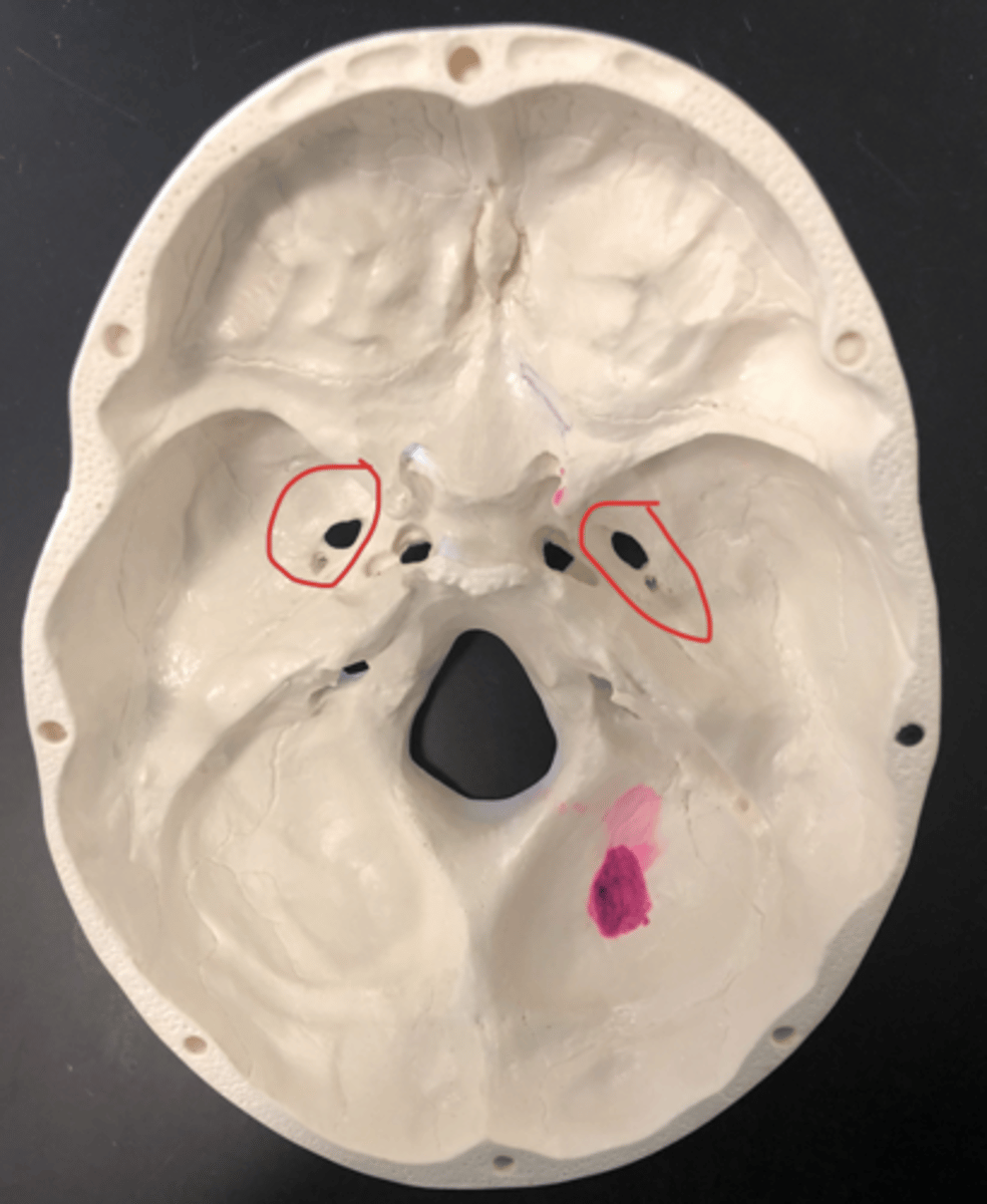
Foramen lacerum - first hole in inner greater wing
Optic canal - the eyes of the yoda in between the lesser wings
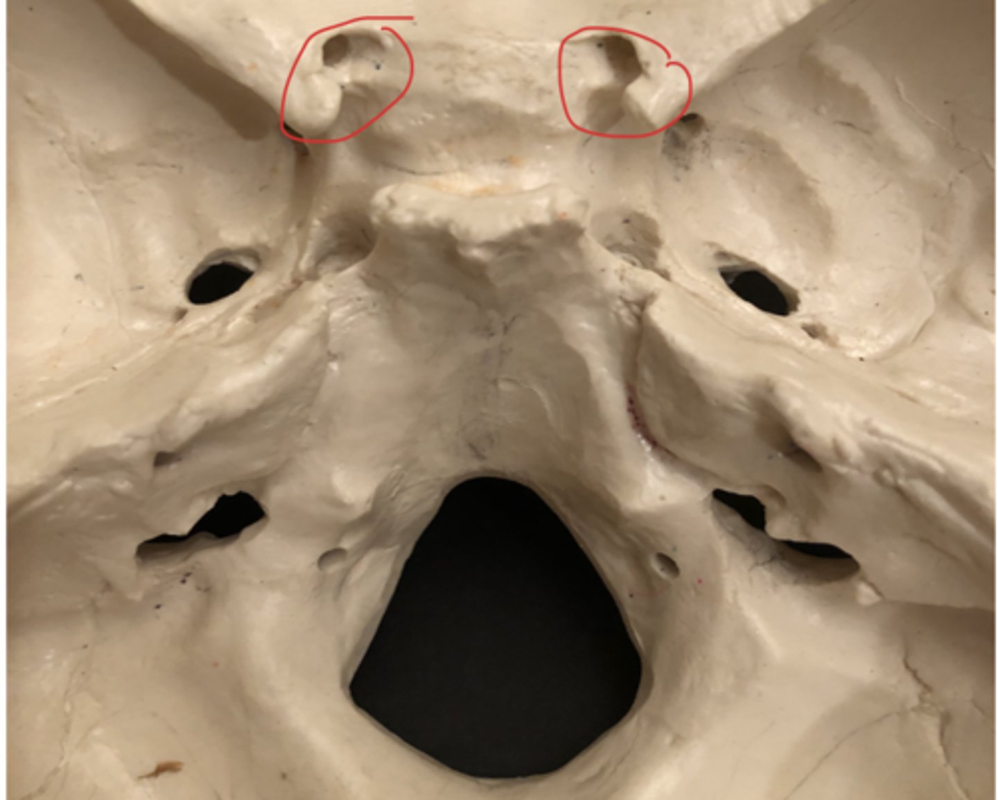
Foramen rotundum - superior to the lacerum; inside the greater wing
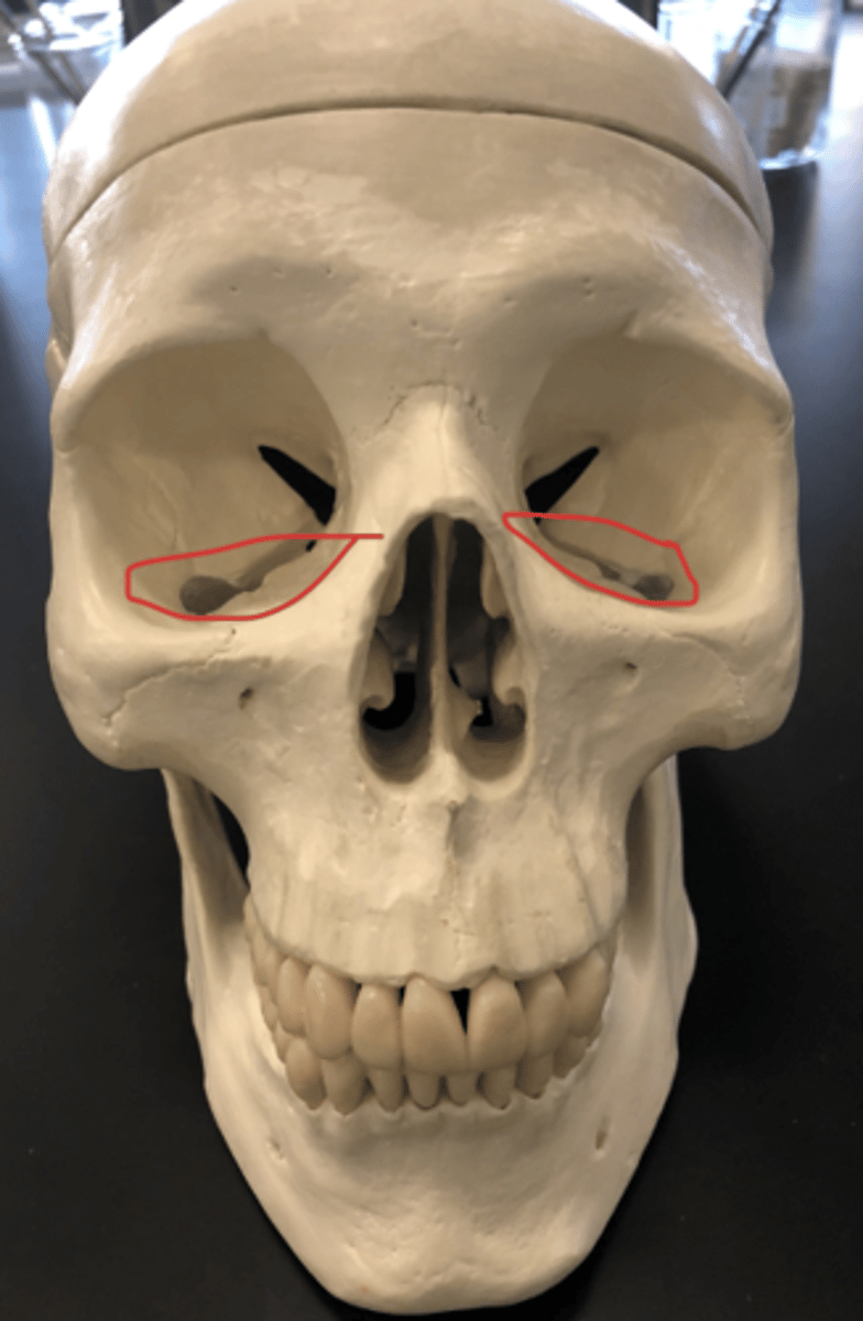
Superior orbital fissure - Face front: upper portion of the X hole in the eyes

Inferior orbital fissure - Face front; lower portion of the X hole in the eyes
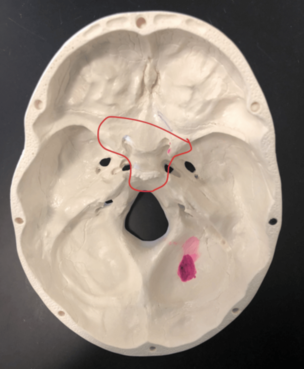
Ethmoid - bone that forms the back of the nose and encloses numerous air cells
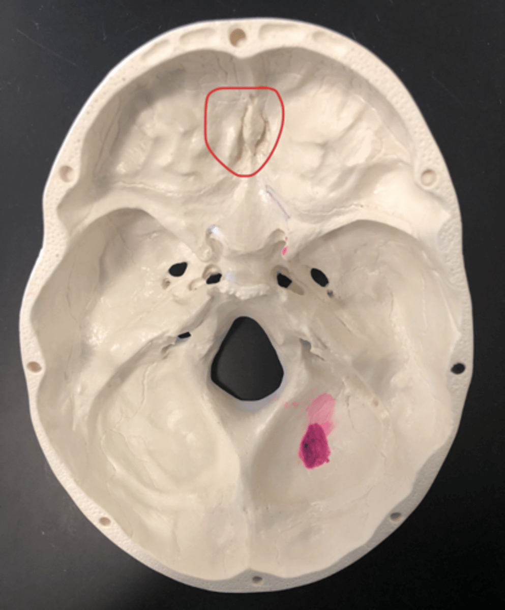
Crista Galli - protrusion on the ethmoid
Protrusion
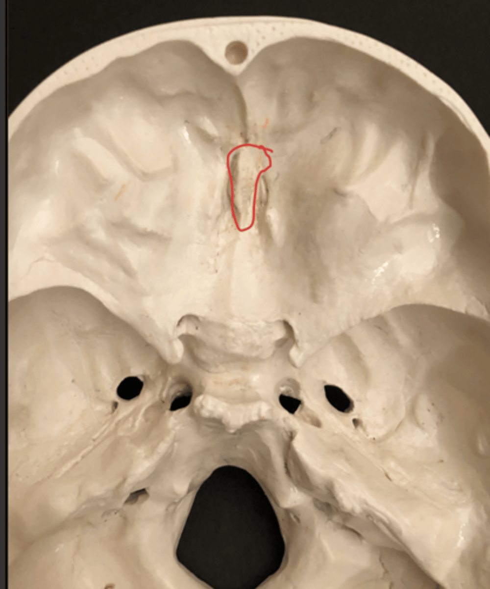
Cribriform plate - area around the crista galli; crib goes around the baby
Area Surrounding the protrusion

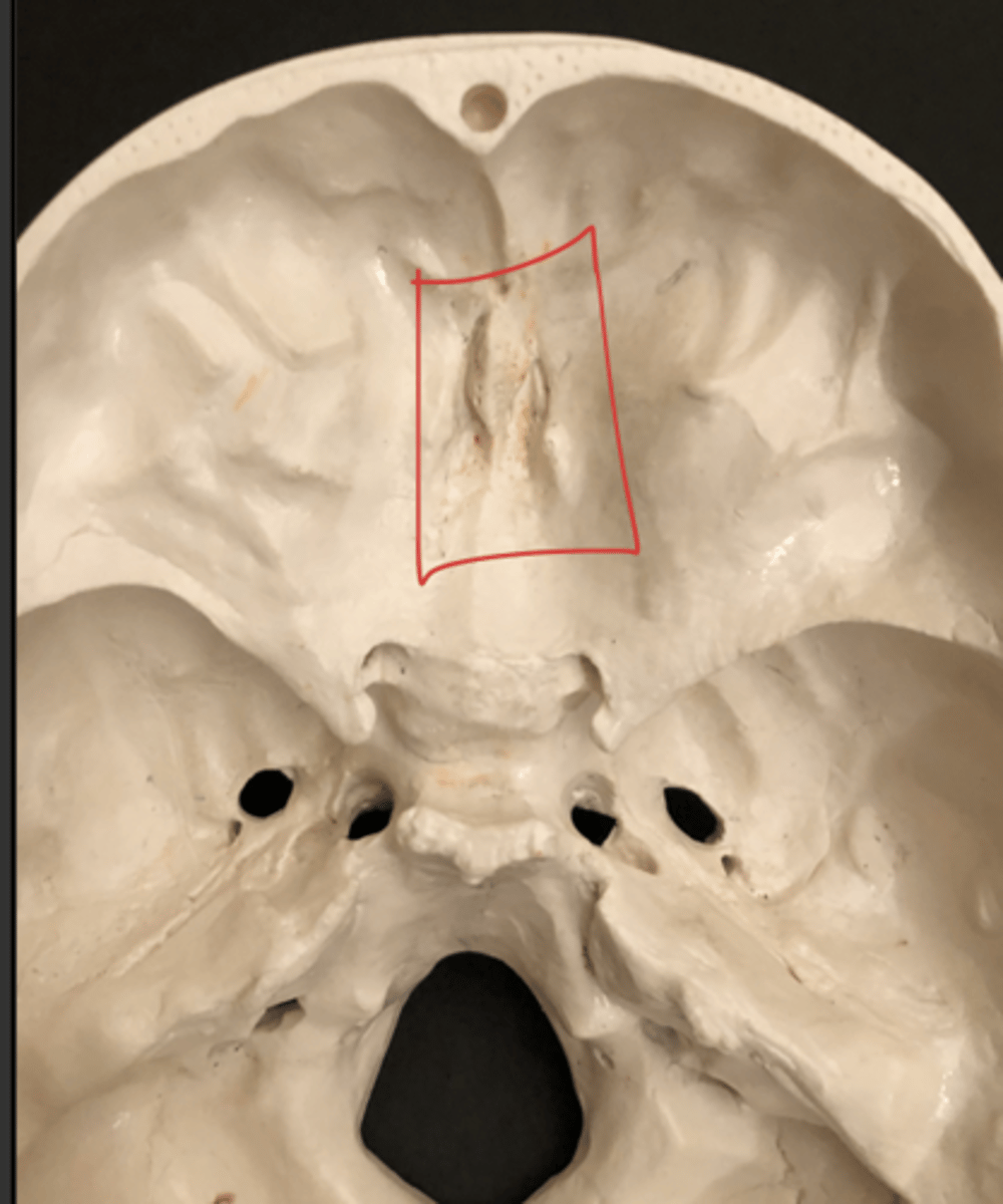
Olfactory Foramen - dots around the crista galli
Dots
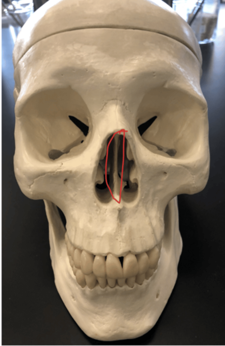
Perpendicular plate - same structure as the vomer but the top portion
Superior nasal conchae - bump on the inside of the nose; not really recognizable
Middle nasal conchae - short bump under the superior nasal conchae and above the inferior nasal conchae

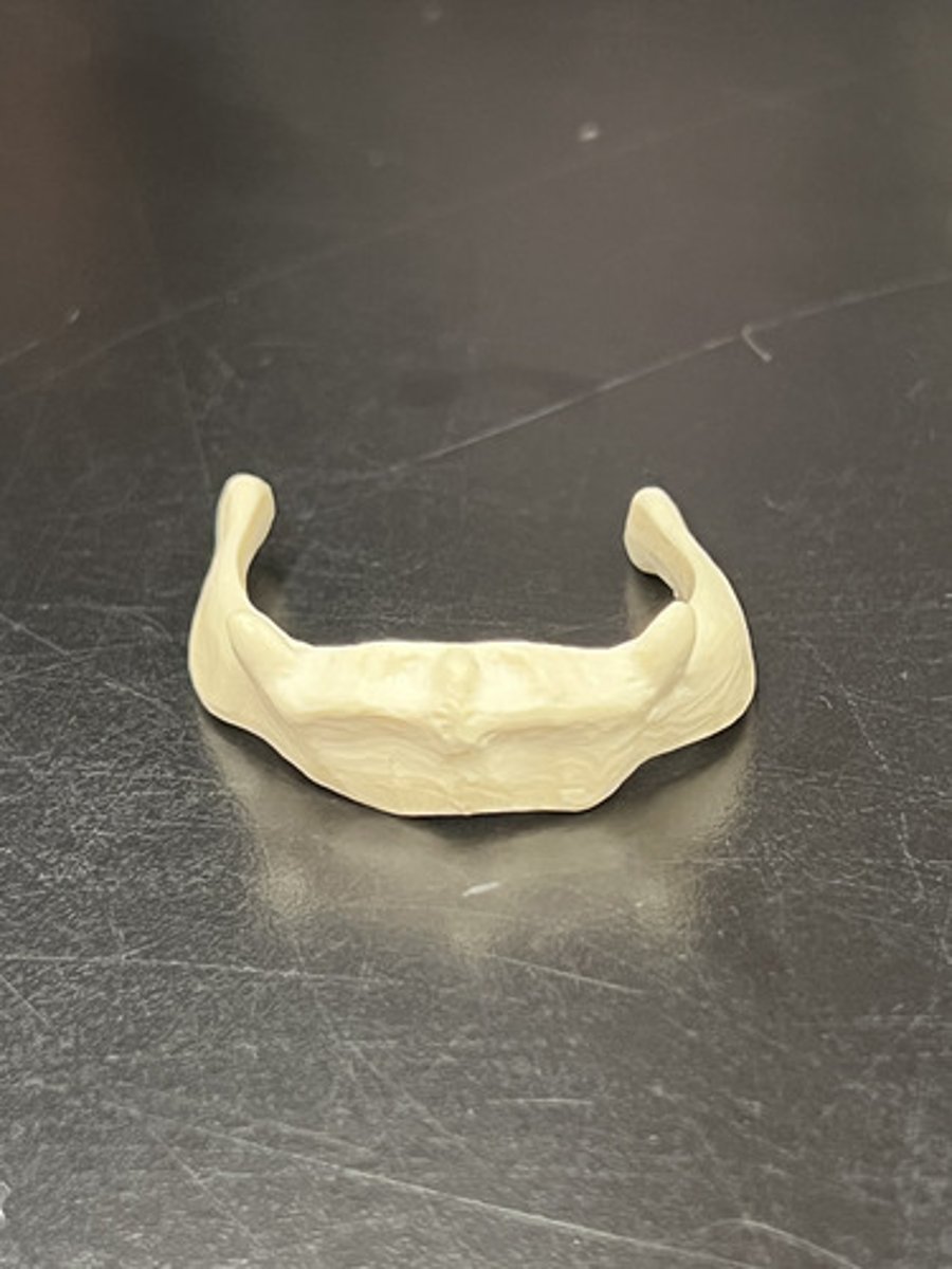
Hyoid - inside bone that supports the tongue; super small
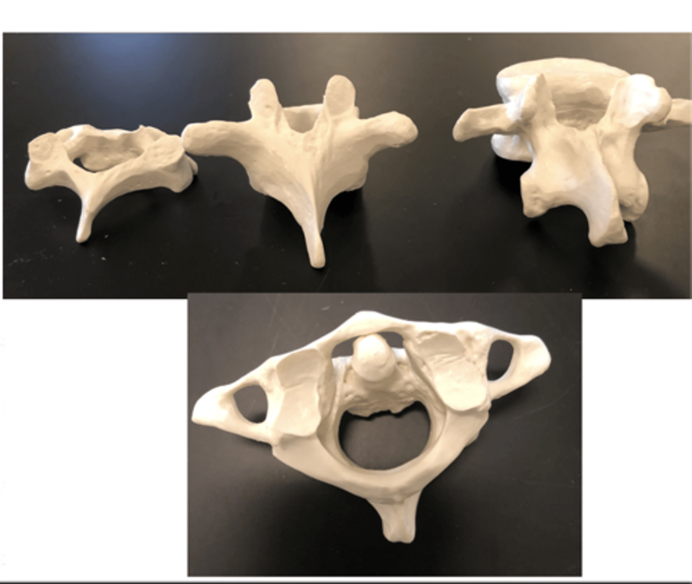
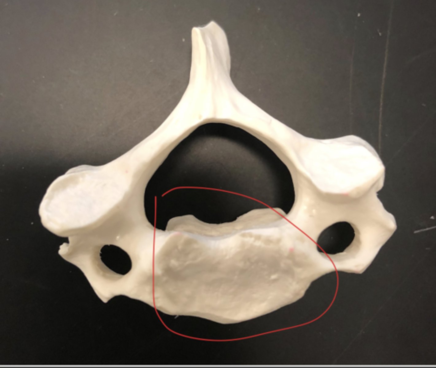
vertebrae - the bones forming the spinal column or backbone
vertebrae Body - space in between the transverse foramen
Spinous process - slender tip
Lamina - connects transverse process to spinous process
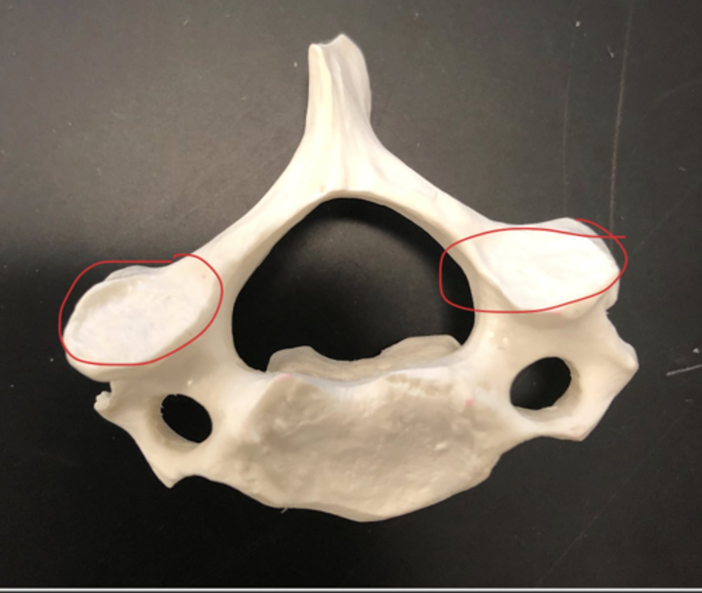
Transverse process - out to the side; two of them
Pedicle - connects the transverse process to the vertebral body
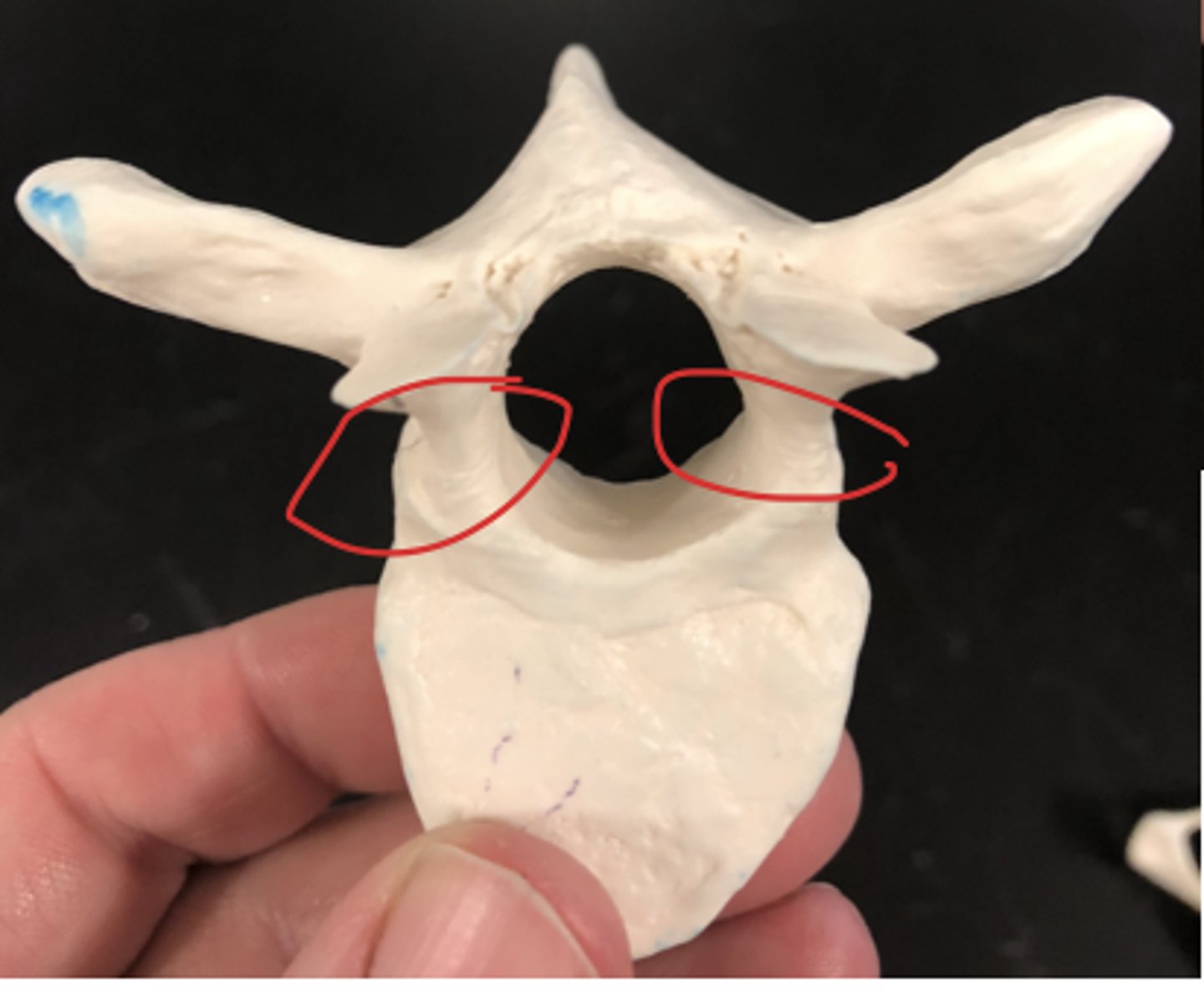
Articular facet - where we are forming a joint; flat surface
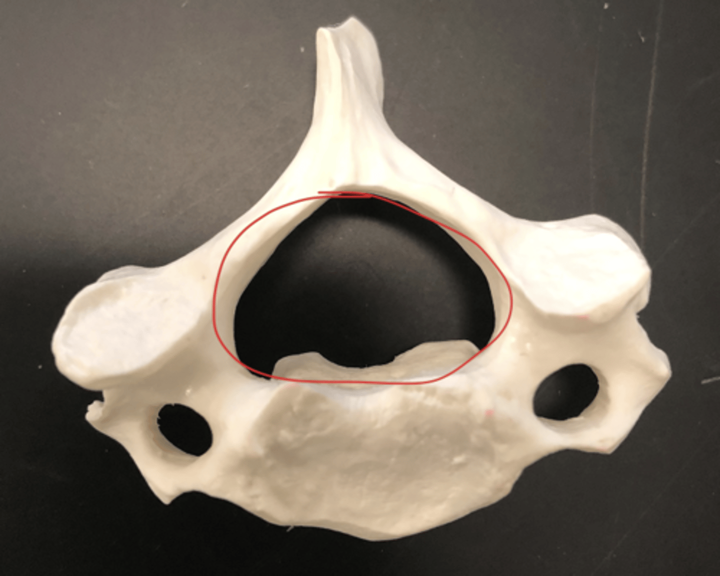
Vertebral canal - big hole in the middle
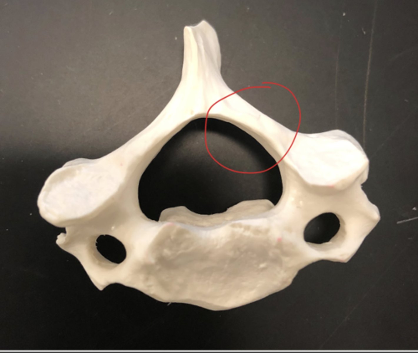
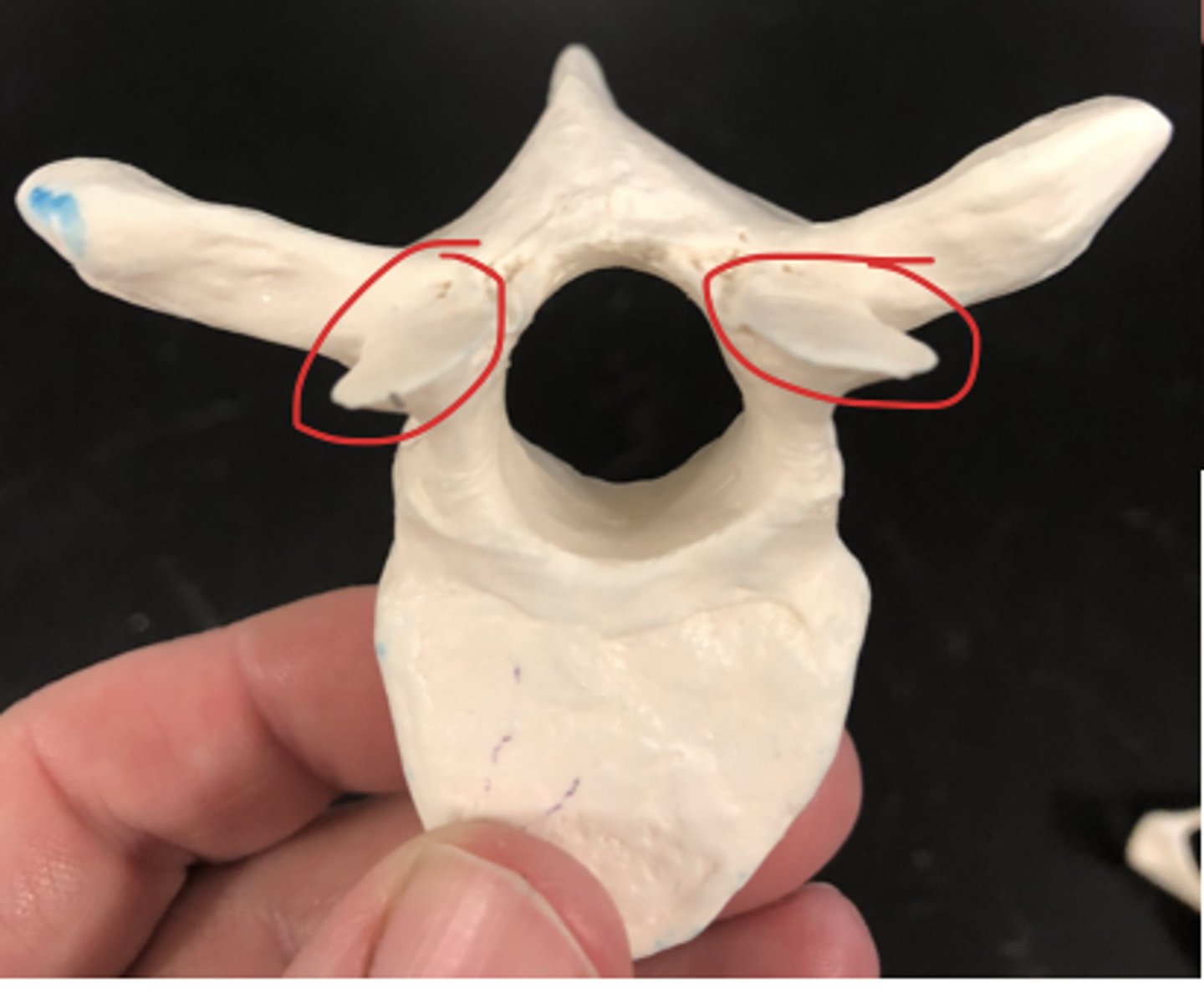
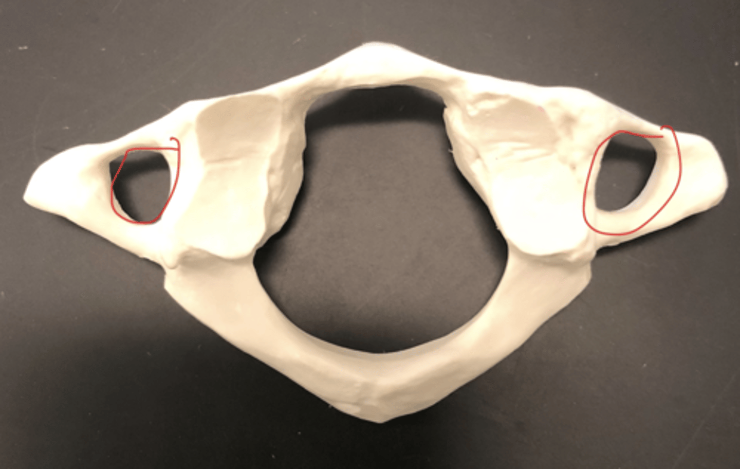
Cervical vertebrae (#7) - smaller holes on either side; transverse foramen (sloth face)
transverse foramen (cervical vertebrae)
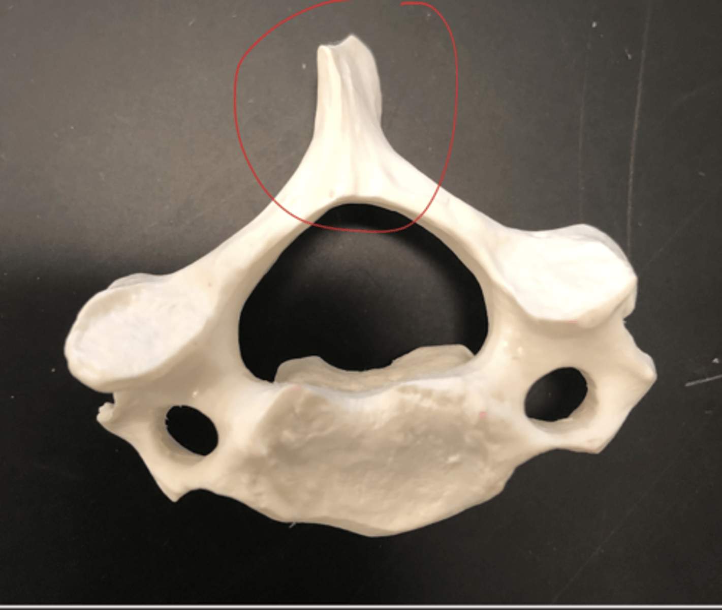
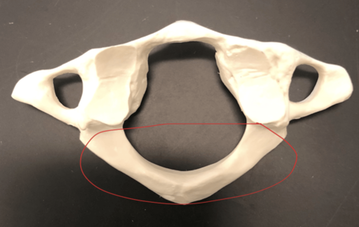
Atlas (C1) - sloth face ring

Anterior arch of the Atlas - less arched side, smooth
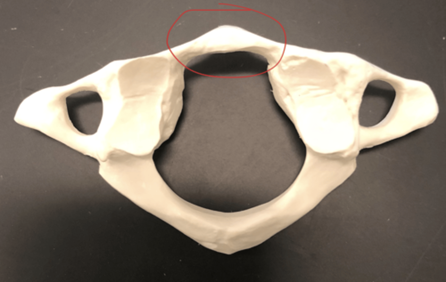
Posterior arch of the Atlas - spine on the back
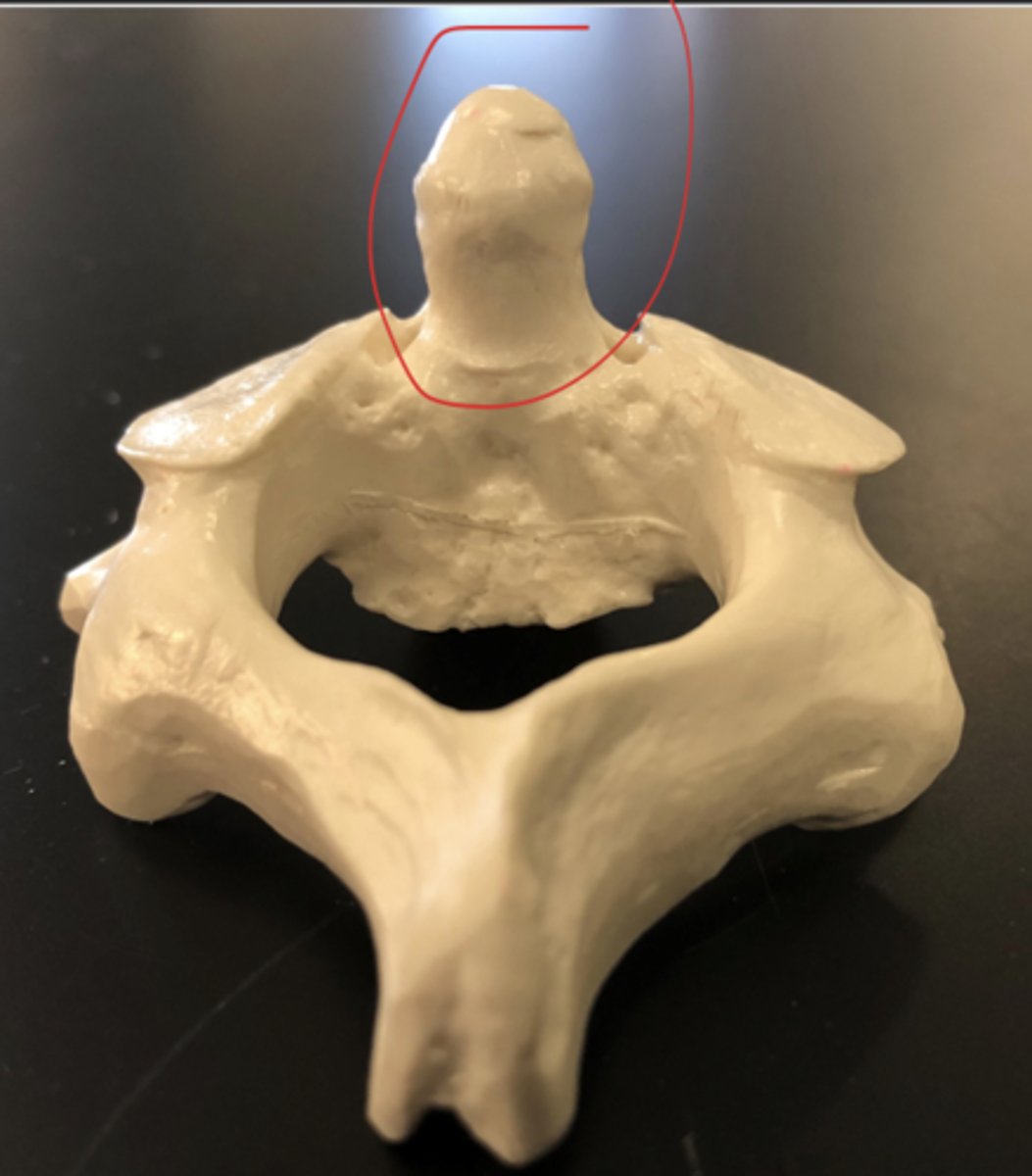
Axis (C2) - Rhino; what other bone spins on
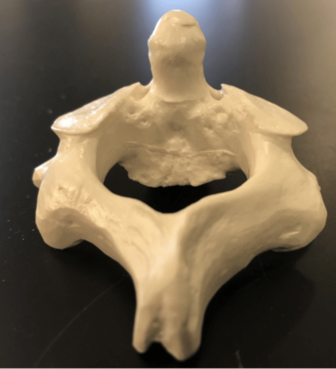
Dens - rhino horn
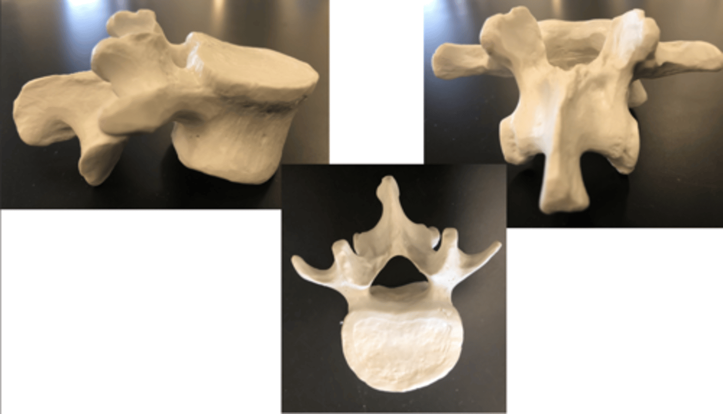
Thoracic vertebrae (#12) - looks like a giraffe with a downward spine
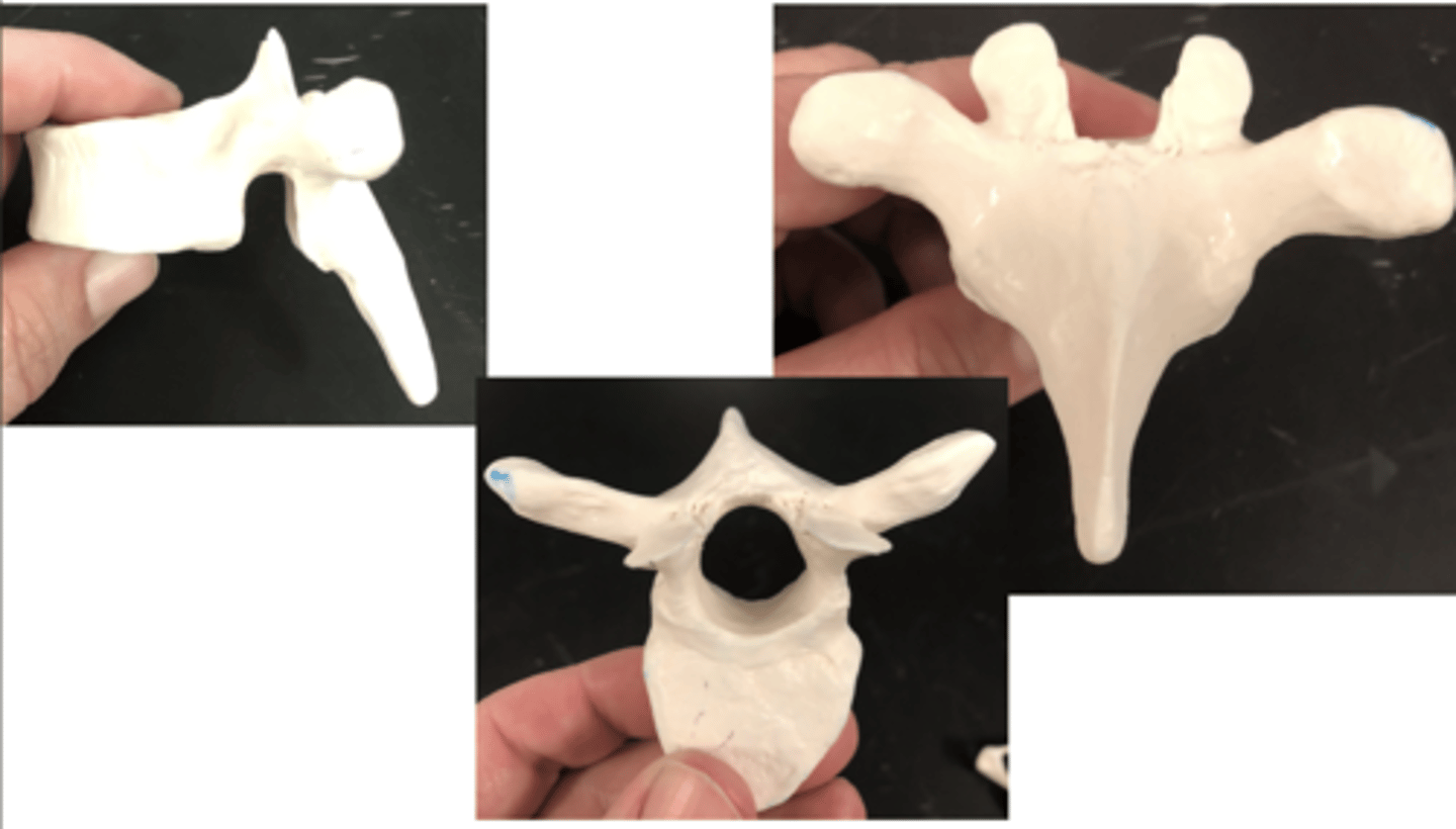
Costal facet of the thoracic vertebrae- behind the articular facet on the mouth of the spinous process

Lumbar vertebrae (#5).- big thick chunky clunky
Sacrum - elephant without the trunk
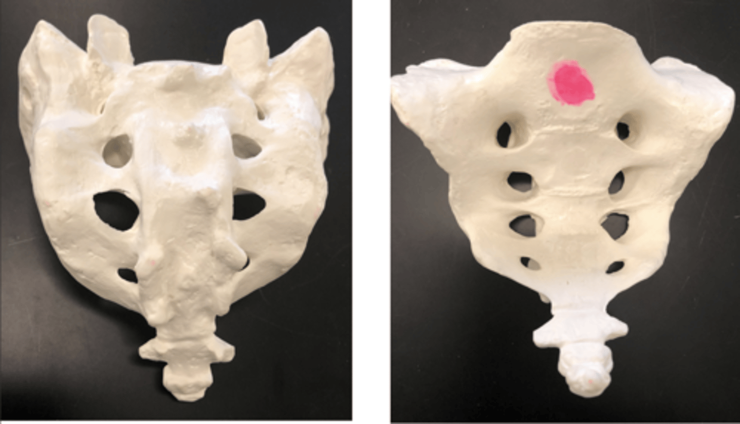
Sacrum base - widest part
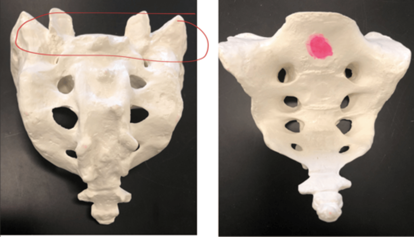
Sacrum apex - narrow part
Sacral foramina - all the holes
Holes

Median sacral crest - middle ridge down the trunk of the elephant
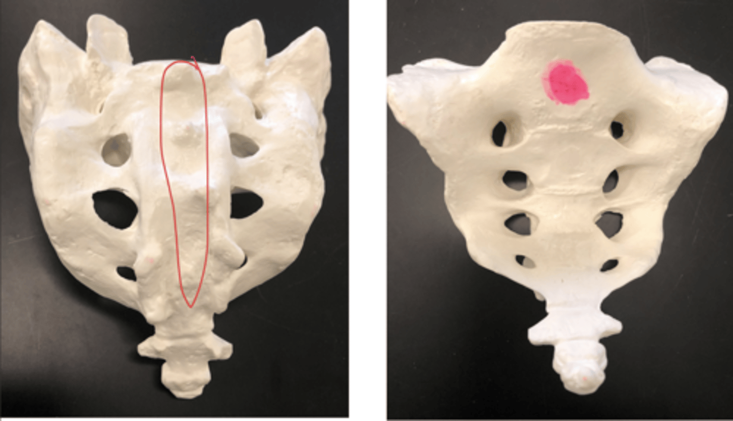
Lateral sacral crest - sides of the median sacral crest

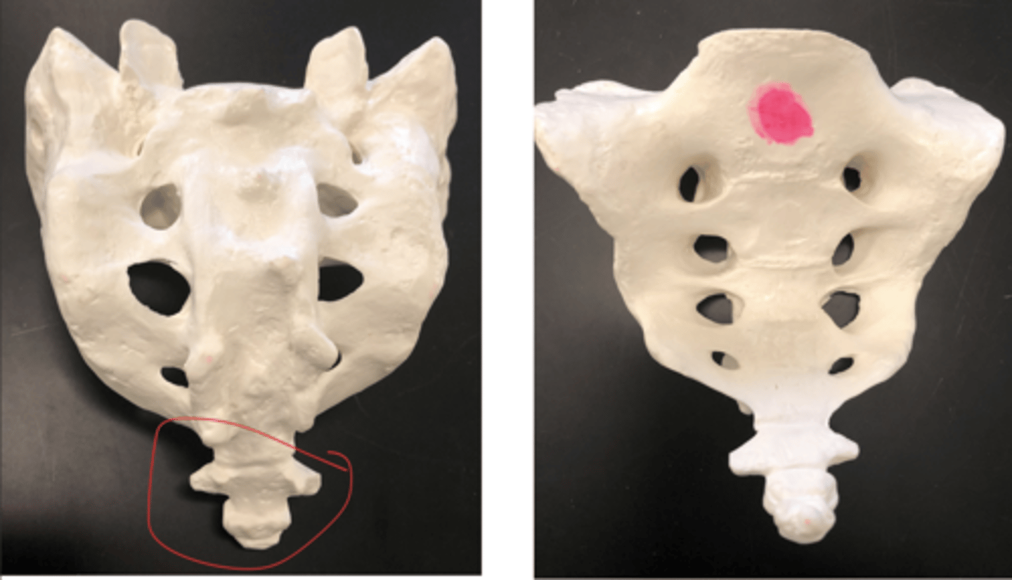
Coccyx - elephant trunk
Very end attachment

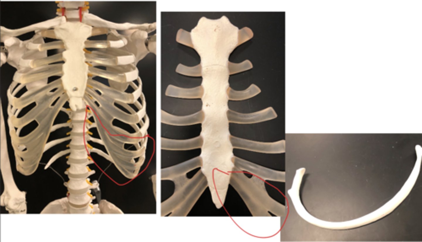
True ribs (1-7) - directly attached to sternum
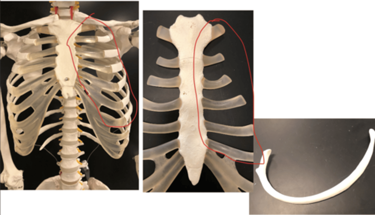
false ribs (8-12) - all come together into one cartilage fused together that is connected at one point in the sternum