lecture 4 equine pelvic limb part 2
1/90
Earn XP
Description and Tags
equine pelvic muscles- sparks
Name | Mastery | Learn | Test | Matching | Spaced | Call with Kai |
|---|
No analytics yet
Send a link to your students to track their progress
91 Terms
what do the gluteal muscles act to do
extend, abduct and medially rotate the limb at the coxal joint
what does the middle gluteal muscle have and what does this cause
a lumbar head which makes it an active participant in rearing
what types of headas does the caudal thigh have
vertebral heads
what do the caudal thigh muscles (hamstrings) act as
extension of coxal joint and flexion of stifle joint when non-weight bearing

what is A
middle gluteal

what is B
superficial gluteal

what is C
biceps femoris

what is D
semitendinosus
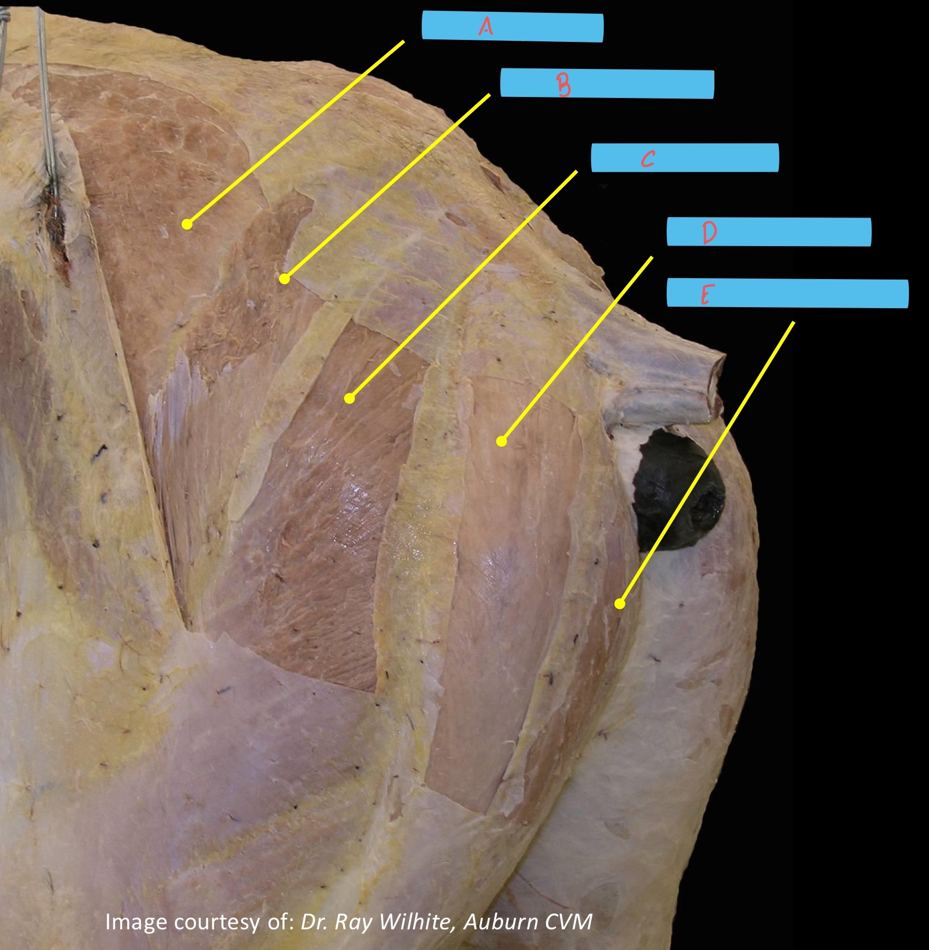
what is E
semimembranosus
origin of superficial gluteal m
tuber coxae and gluteal fascia
insertion of superficial gluteal m.
third trochanter
origin of middle gluteal m
Gluteal surface of ilium, tuber coxae, sacrosciatic ligament; Longissimus dorsi m. (vertebral head, as far cranially as cranial lumbar region
insertion of middle gluteal m.
Greater trochanter (caudal part); proximal femur between greater
and third trochanters
where tendon of middle gluteal m. insertion passes over cranial part of greater trochanter
trochanteric bursa
origin of deep gluteal m.
body of ilium, ischiatic spine
insertion of deep gluteal m.
greater trochanter (cranial part)
all parts of hamstring take partial origin from
tuber ischii but also have additional originswha
biceps femoris origin
sacrum and sacrosciatic ligament
origin of semitendinosus
sacrosciatic ligament and caudal vertebrae
origin of semimembranosus
caudal most edge of sacrosciatic ligament
biceps femoris has 3 parts what are their insertions
▪ Patella and lateral patellar ligament (1)
▪ Cranial border of the tibia (lateral aspect) (2)
▪ Crural fascia and tuber calcanei via common
calcanean tendon (3)
semitendinosus inserts on
cranial border of tibia (medial aspect) and tuber calcanei via common calcanean tendon
semimembranosus inserts on
medial epicondyle of femur and medial collateral ligament of stifle
what nerve innervates biceps femoris and semitendinosus
caudal gluteal
what are the pelvic heads of caudal thigh supplied by
sciatic n.
adductors of pelvic limb
sartorius
gracilis
adductor
pectineus
origin of sartorius
psoas fascia and tendon
insertion of sartorius
medial aspect of stifle
action of sartorius
flexes hip; flexes and extends (weight bearing)stifle joint
origin of gracilis
pelvic symphysis via symphyseal tendon
insertion of gracilis
medial aspect of stifle, cranial border of the tibia
origin of adductor
ventral surface of pubic and ischium, symphyseal tendon
insertion of adductor
caudal aspect of and medial epicondyle of femur
origin of pectineus
margin of pubis
insertion of pectineus
medial surface of femur
all adductor muslces provide motor innervation by obturator nerve except what
sartorius which is innervated by the saphenous nerve
the pectineus m. forms the caudal border of the
femoral triangle
what forms the cranial border of the femoral triangle
sartorius m.
where does the femoral nerve emerge from
ilioposoas m.
where does the obturator n. pass through
obturator foramen
lateral rotators of the hip
external obturator, internal obturator, gemelli, quadratus femoris
where do the lateral rotators of the hip insert
in trochanteric fossa and trochanteric crest of femur
where can be found as the pelvic floor muscle, dorsal to obturator foramen
internal obturator
what does the tendon of insertion of the internal obturator pass over
lesser ischiatic notch
gemelli origin
ischium ventral to lesser ishiatic notch
quadratus femoris origin
ventral aspect of ilium inserts on trochanteric crest of femur
quadriceps femoris group
rectus femoris
vastus lateralis
vastus medialis
vastus intermedius
where do quadriceps femoris insert
on the patella and tibial tuberosity via the patellar ligaments
where does rectus femoris originate
body of ilium craniodorsal to acetabulumwh
at is the only muscle of the quadriceps femoris to cross hip joint
rectus femoris
what does the rectus femoris do
flex the hip joint
what muscle of the crus is not present in horses
fibularis longus m.
what muscle of the crus is present in large animals and cat but not dogs and is a part of the common calcanean tendon
soleus m.
action of cranial muscles of the crus
flexion of hock

what is A
cranial tibial m
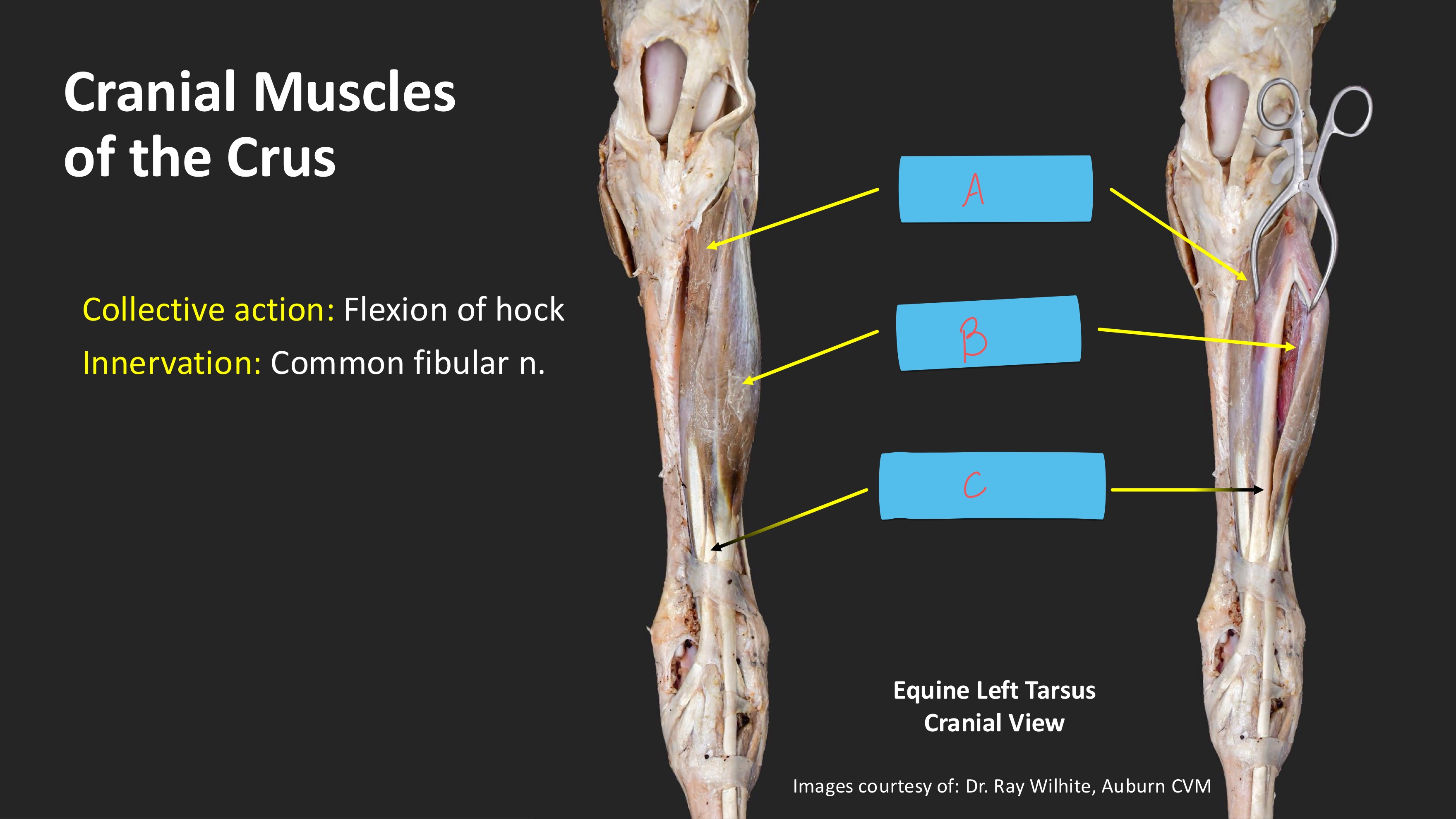
what is B
long digital extensor m.

what is C
peroneus tertius
origin of cranial tibial muscle
lateral condyle of tibia
insertions of cranial tibial muscles
▪ Dorsal tendon: Metatarsal tuberosity
▪ Medial tendon (a.k.a., cunean tendon): fused tarsal bones I & II
what is affiliated with the medial tendon of the cranial tibial m.
cunean bursa
origin of peroneus tertius
extensor fossa of femur in common with long digitala extensor m.
insertion of peroneus tertius
Dorsal tendon: Tarsal bone III, Metatarsal bone III
▪ Lateral tendon: Calcaneus, Tarsal bone IV
what does the peroneus tertius m. form
“tunnel” through which the tendon of the cranial tibial m. passes
origin of long digital extensor
extensor fossa of femur
origin of lateral digital extensor
lateral collateral ligament of stifel
where do the long and lateral digital extensors unite
at the level of the metatarsus, distal to the metatarsal (disatl) extensor retinaculum
where do the long and lateral digital extensors insert
extensor process of distal phalanx III
action of long and lateral digital extensor
flexes hock and extends digit
what makes up the extensor retinacula
crural extensor retinaculum
tarsal extensor retinaculum
metatarsal extensor retinaculum
collective a ction of the caudal muscles of the crus
extension of hock
triceps surae muscle group
gastrocnemius and soleus muscle
origin of triceps surae muscle group
Medial and lateral supracondylar tuberosities of femur (lateral and medial heads of gastrocnemius); head of fibula (soleus)
insertion of triceps surae group
calcaneal tuber
origin of superficial digital flexor m
supracondylar fossa of femur
insertion of superficial digital flexor muscle
Calcaneal tuber; eminences
(medial and lateral) on palmar and
distal aspect of proximal phalanx and
proximal aspect of middle phalanx
action of superficial digital flexor muscle
Extension of hock; flexion
of fetlock and pastern joints;
counteracts flexion (buckling
forward) of pastern joint when
weight bearing
origin of deep digital flexor m
Lateral condyle of tibia and caudal
proximal surface of tibia
insertion of deep digital flexor
flexor surface of distal phalanx
action of deep digital flexor
extension of hock and flexion of all digital joints
tell me about the calcanean bursae
Subcutaneous
between skin and SDF tendon;
subtendinous located deep to the
SDF tendon where it passes over
the point of the hock.
Surrounds the
lateral digital flexor tendon from
~2-3 inches proximal to the
medial malleolus to a quarter of
the way down the metatarsus.
tarsal sheath
digital sheath
surrounds the tendons of the SDF and DDF proximal and distal to the fetlock
major components of reciprocal apparatus
superficial digital flexor m
peroneus tertius m
what does the reciprocal apparatus do
turns the limb in a pantograph
femur and metatarsus move in parallel
the reciprocal apparatus links what
the action between the stifle and the hock
how to diagnose peroneus tertius rupture
ability to extend the hock when the stifle is flexed
animal is lame but can usually bear weight on the limb
if the stifle can be locked in extension what will this allow
lock the hock in extension as well
through patellar locking mechanism
explain the patellar locking mechanism
during patellar locking mechanism what maintains extension of tarsus
SDF
clinical condition in which the patella becomes locked in position on the trochlear resting surface commonly known as stifled horse
upward fixation of patella
how does upward fixation of patella most likely occur
by a neuromuscular disorder or spasm of the medial thigh muscles