In-Depth Study of Medical Language: Chapter 6 - Key Terms and Definitions
1/136
There's no tags or description
Looks like no tags are added yet.
Name | Mastery | Learn | Test | Matching | Spaced | Call with Kai |
|---|
No analytics yet
Send a link to your students to track their progress
137 Terms
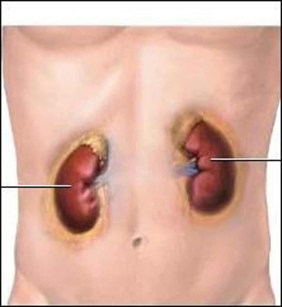
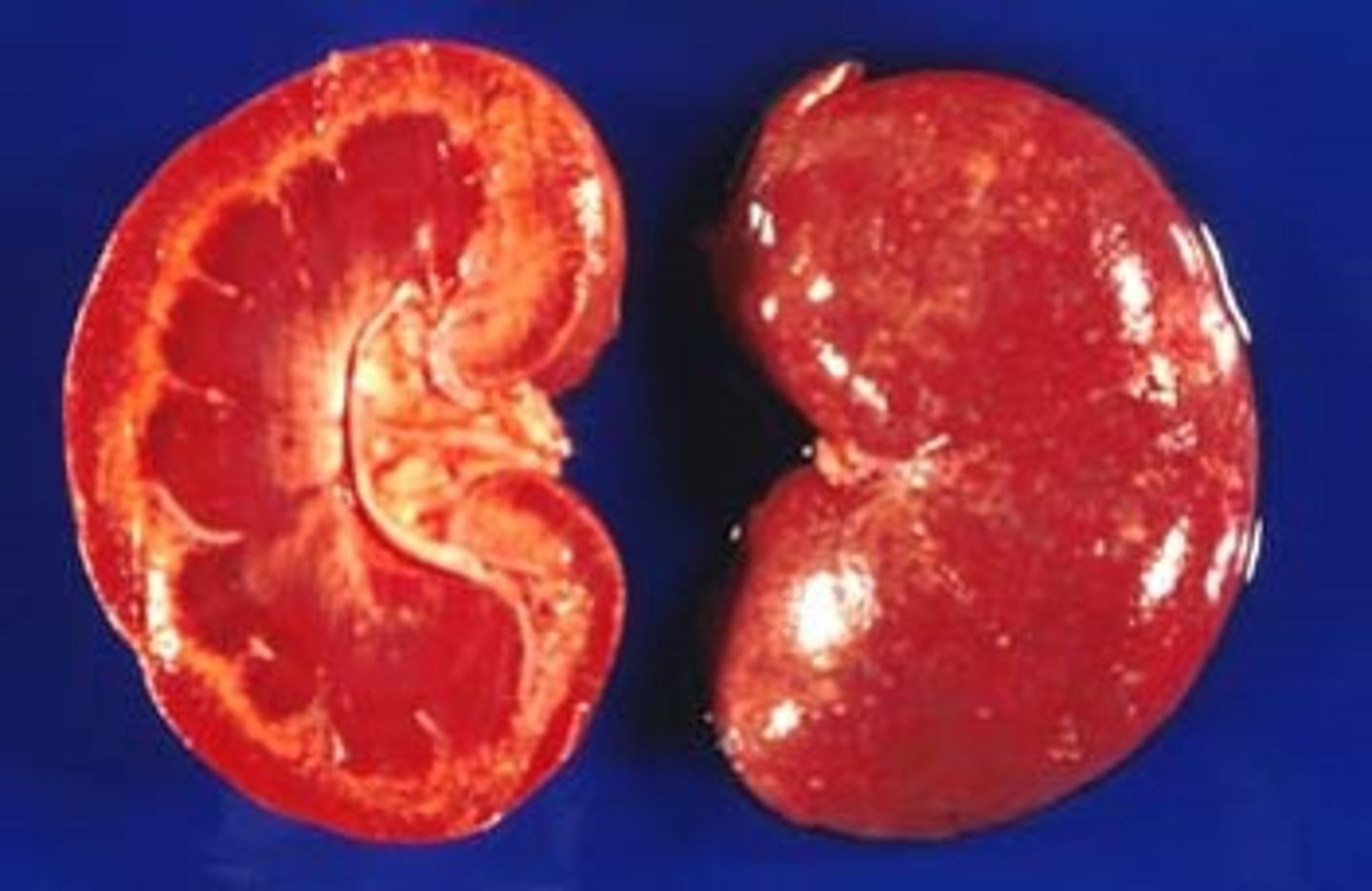
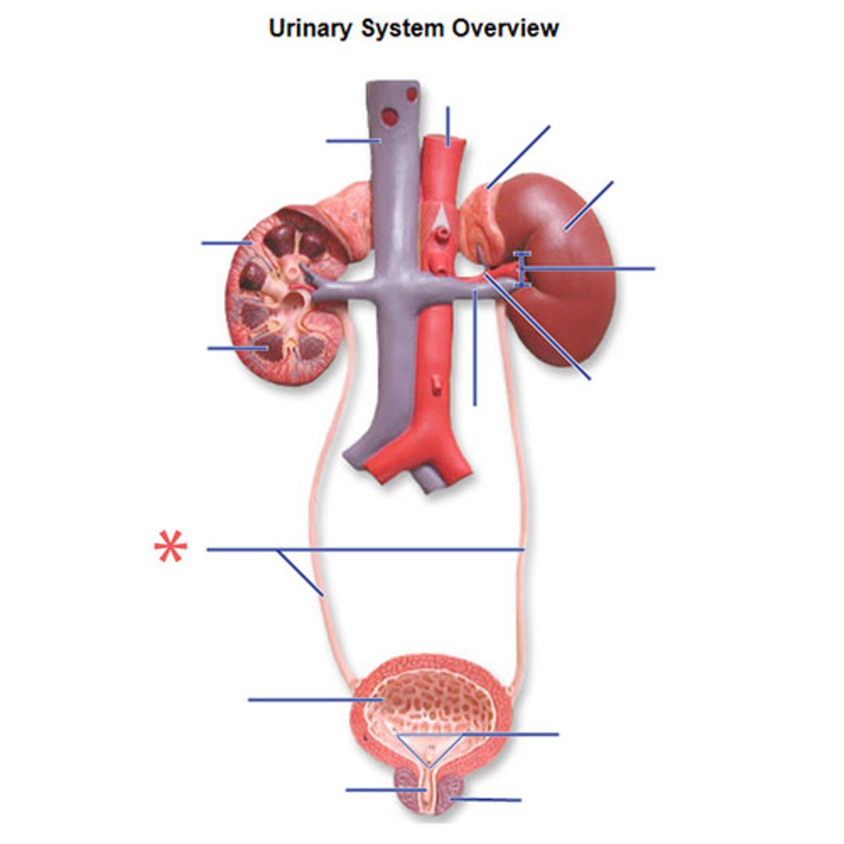
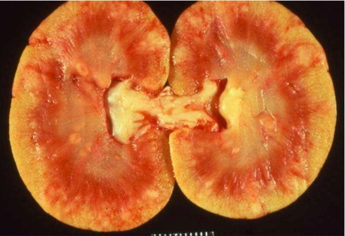
kidneys
two bean-shaped organs located on each side of the vertebral column on the posterior wall of the abdominal cavity behind the parietal peritoneum. Their function is to remove waste products from the blood and to aid in the maintaining water and electrolyte balances
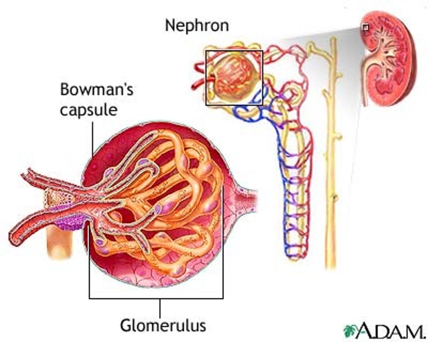
nephron
urine-producing microscopic structure. Approximately 1 million nephrons are located in each kidney
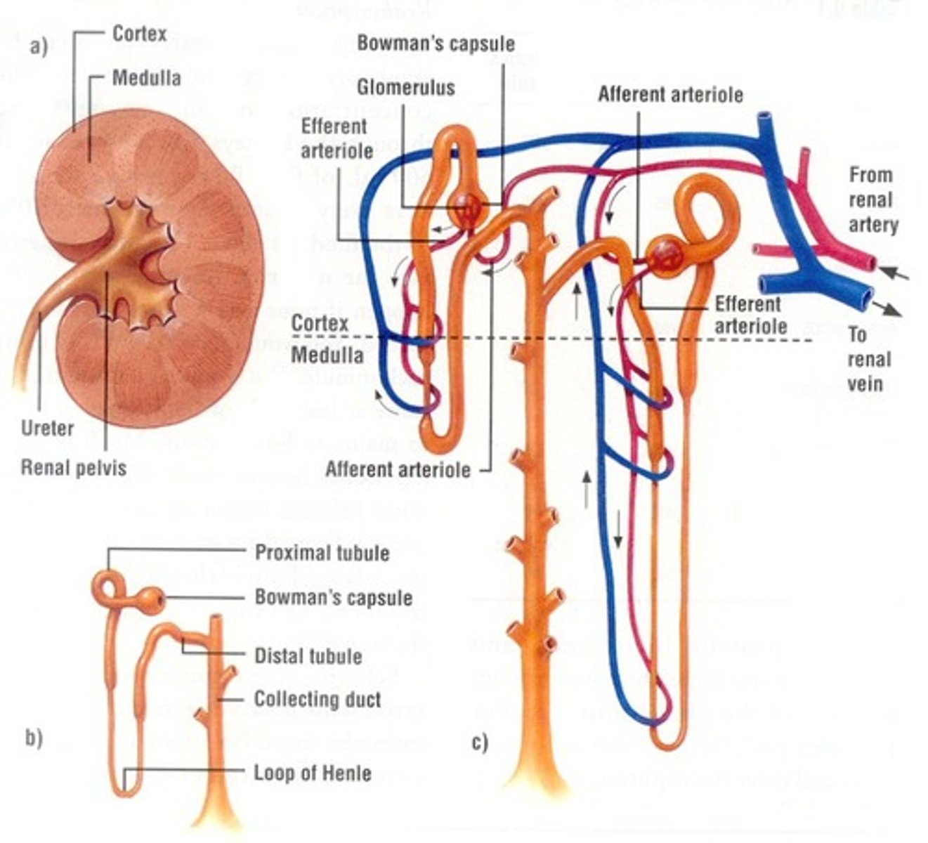
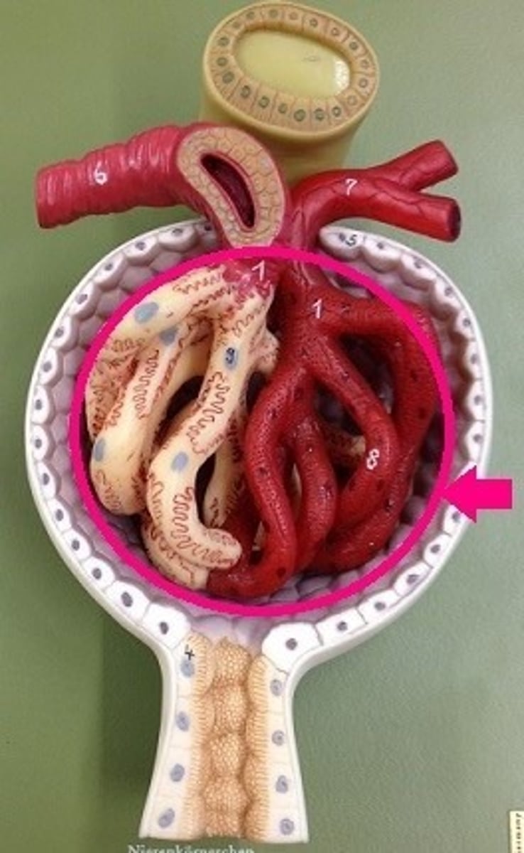
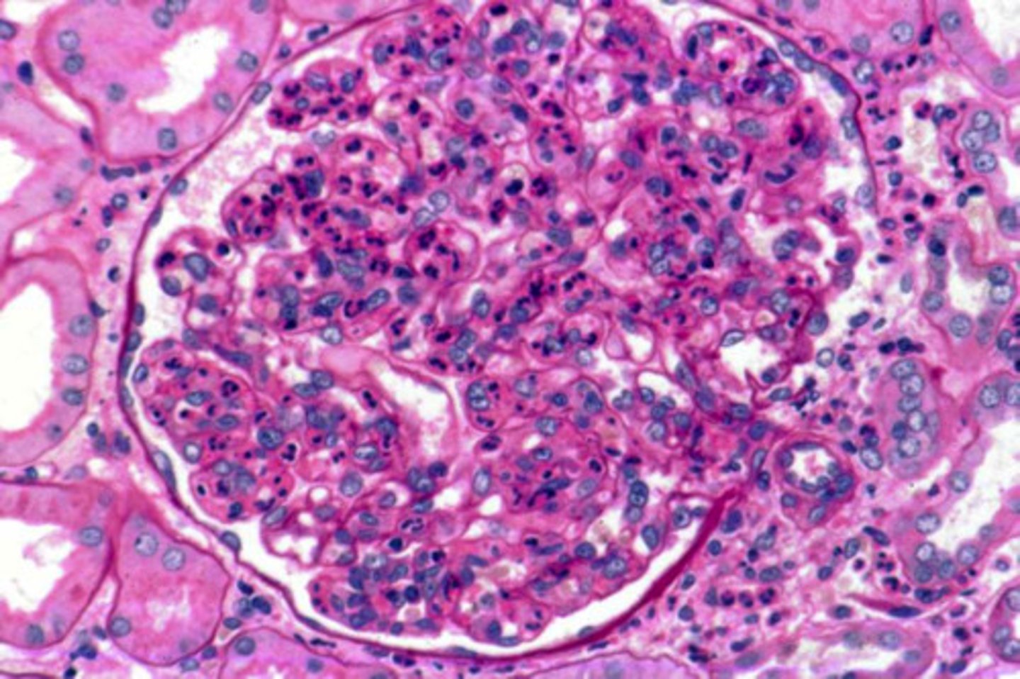
glomerulus
cluster of capillaries at the entrance of the nephron. The process of filtering the blood, thereby forming urine, begins here
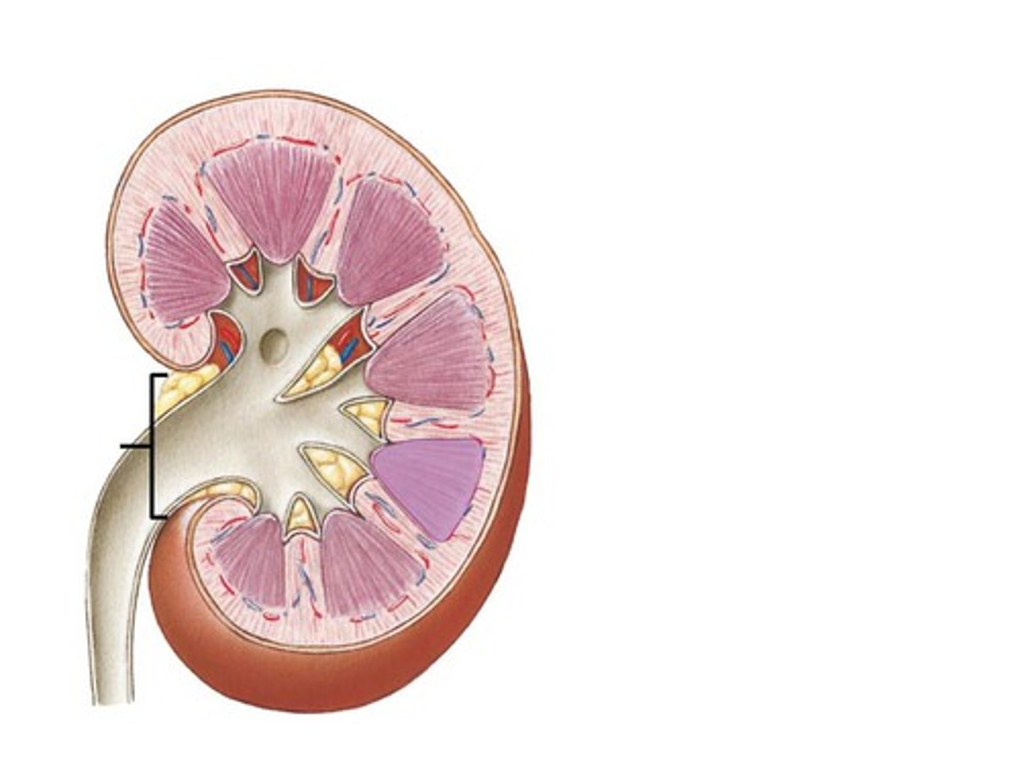
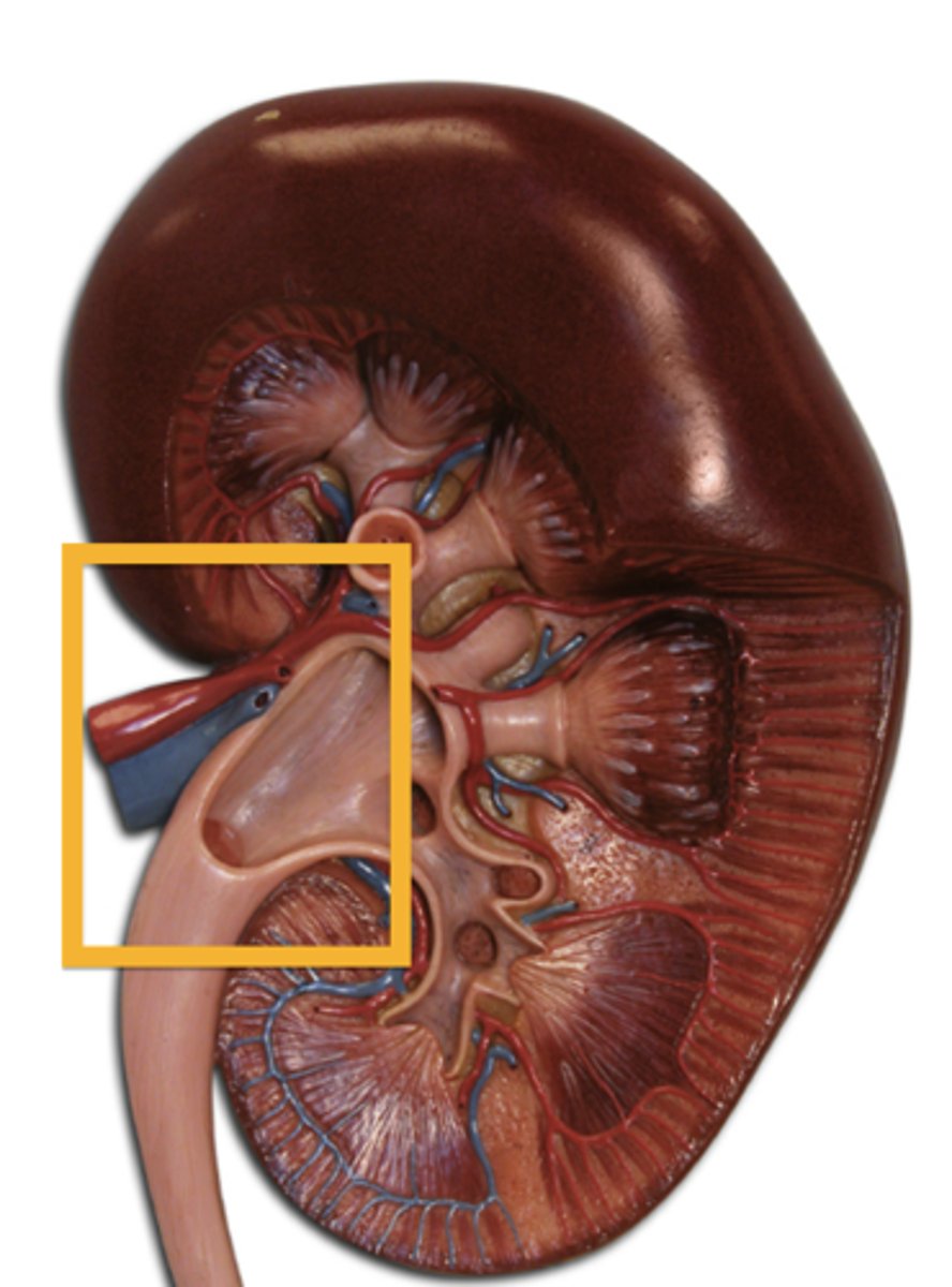
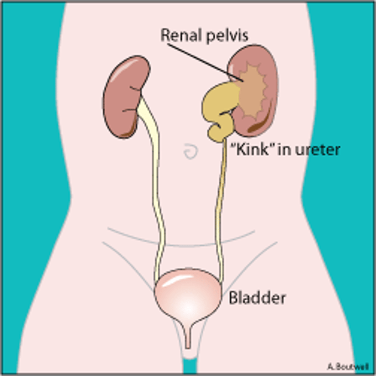
renal pelvis
funnel-shaped reservoir that collects the urine and passes it to the ureter

hilum
indentation on the medial side of the kidney where the ureter leaves the kidney
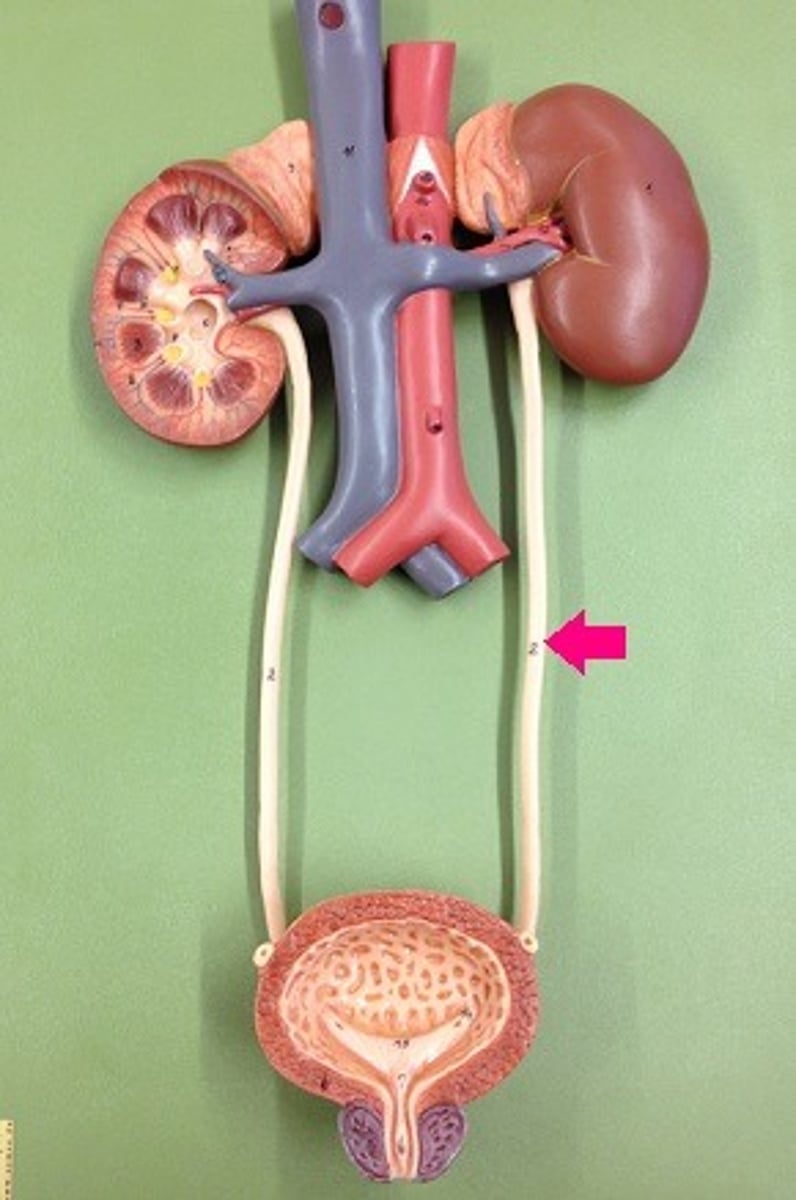
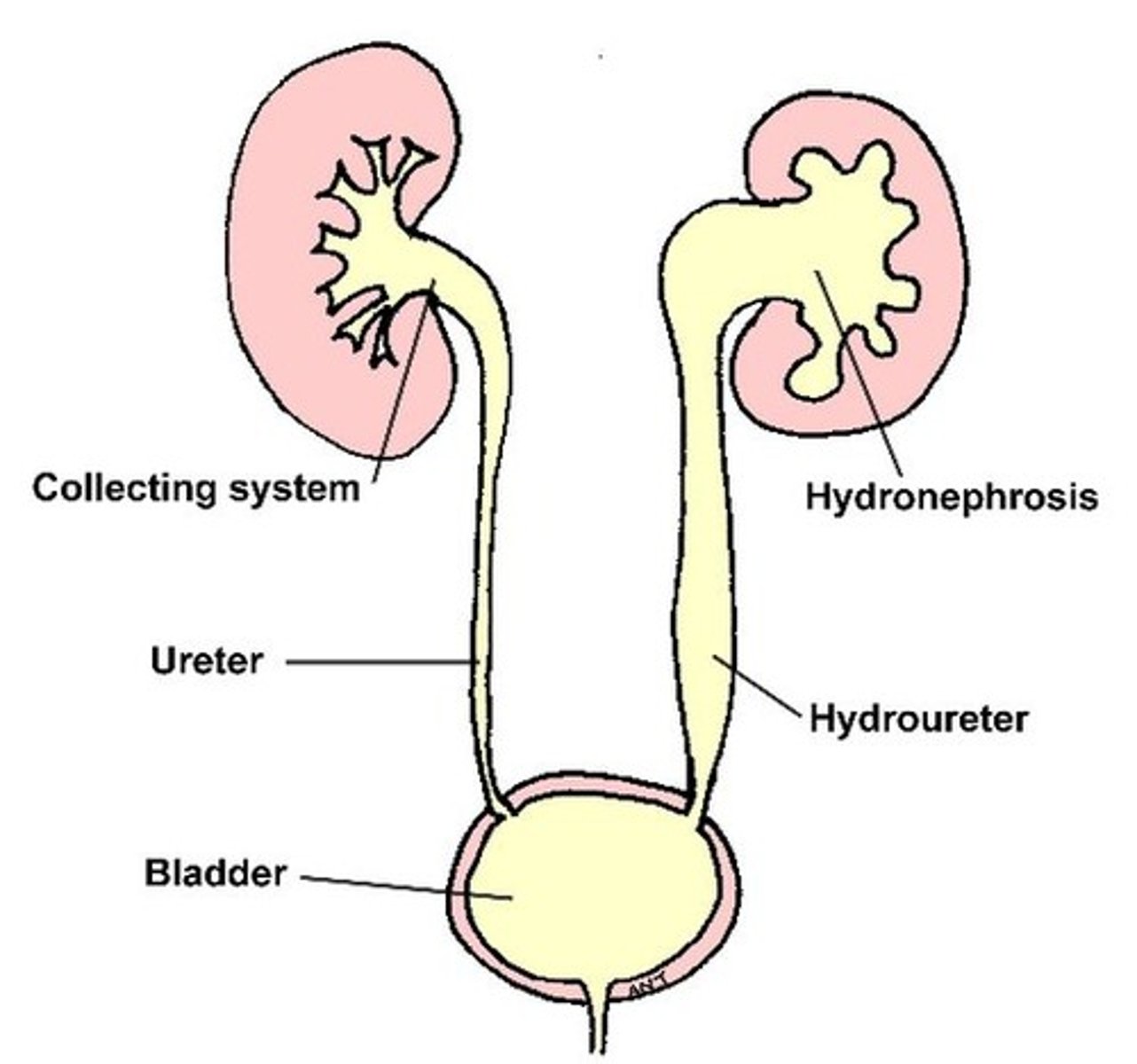
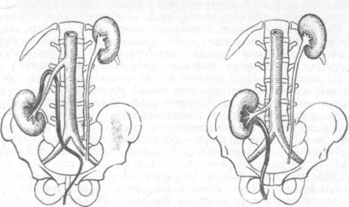
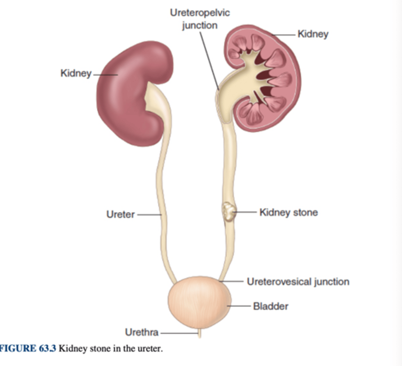
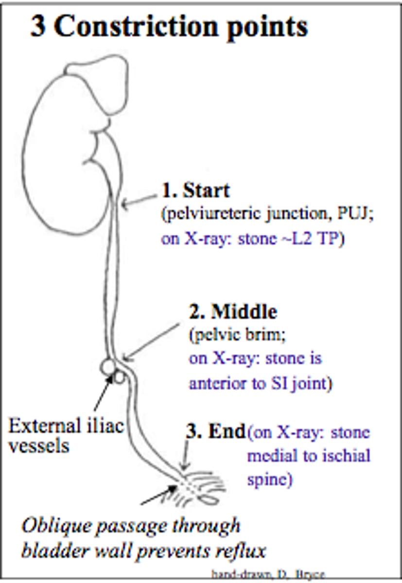
ureters
two slender tubes, approximately 10 to 13 inches (26 to 33 cm) long, that receive the urine from the kidneys and carry it to the posterior portion of the bladder
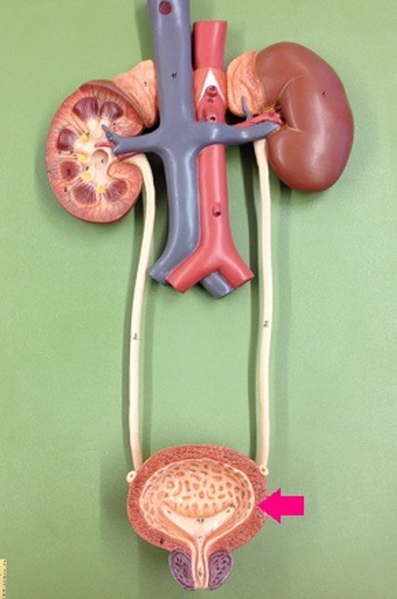
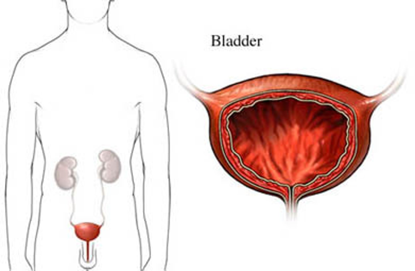
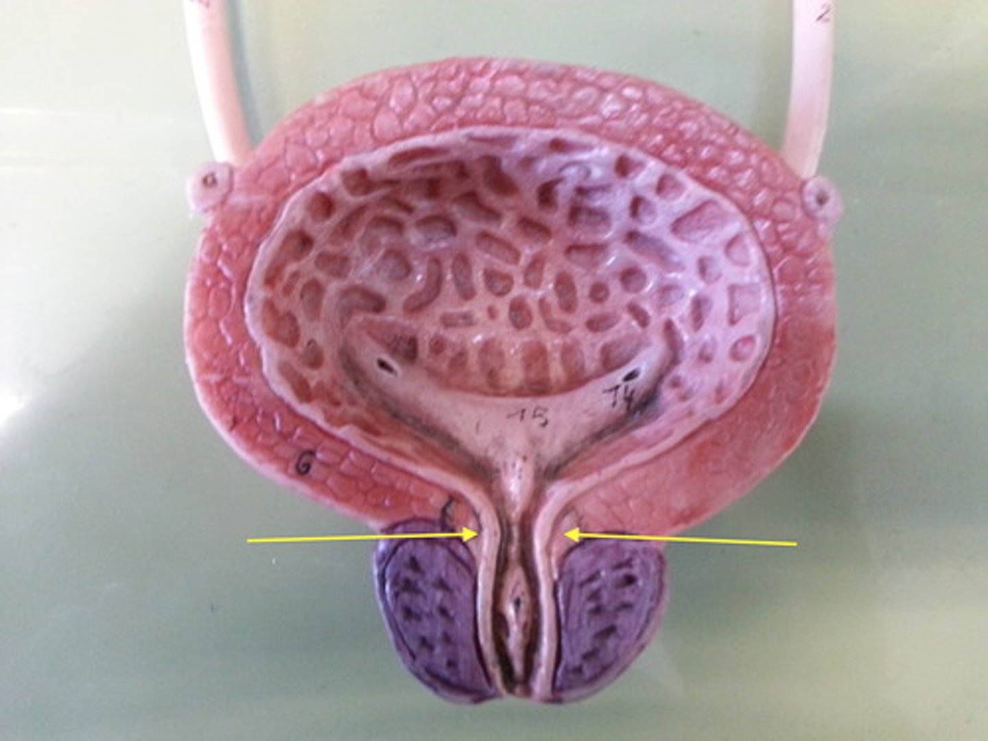
urinary bladder
muscular, hollow organ that temporarily holds the urine. As it fills, the thick, muscular wall becomes thinner and the organ increases in the size
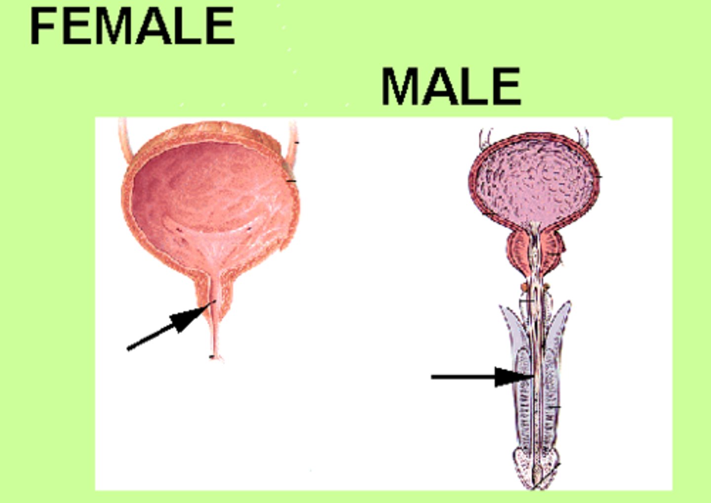
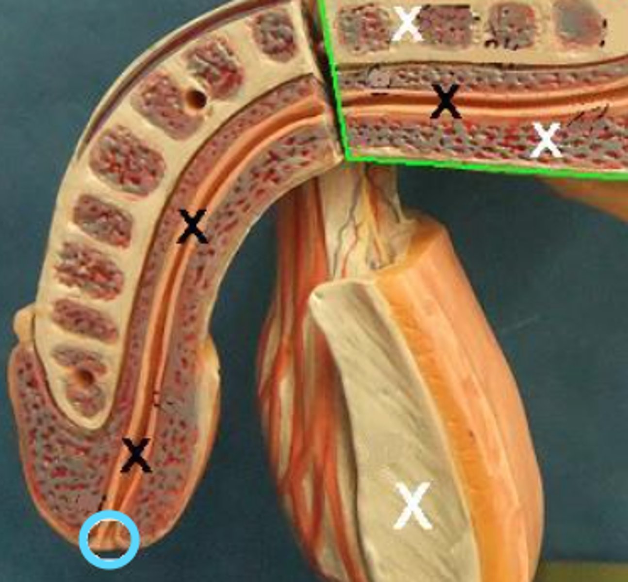
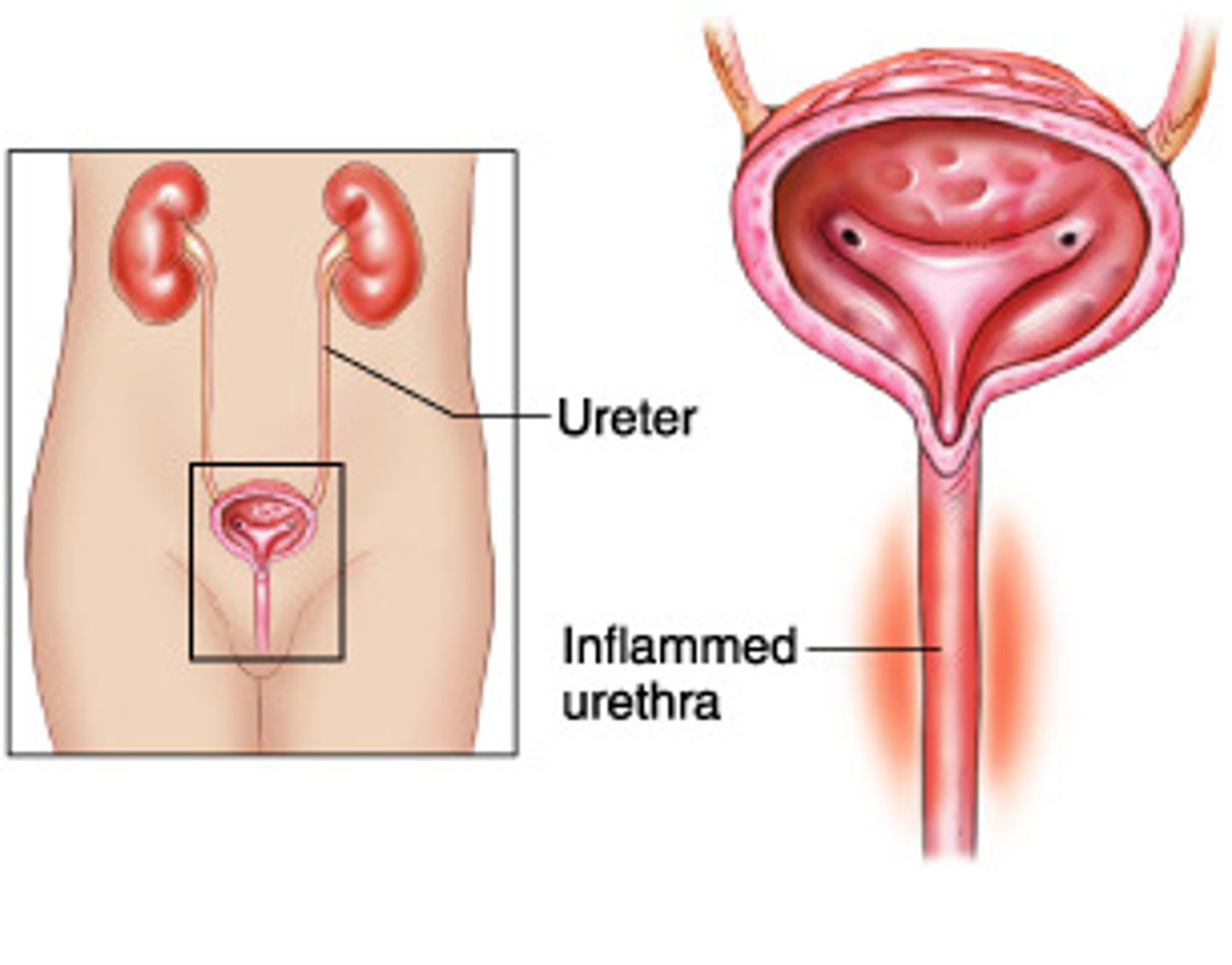
urethra
lowest part of the urinary tract, through which the urine passes from the urinary bladder to the outside of the body. This narrow tube caries in length by sex. It is approximately 1.5 inches (3.8cm) long in the female and approximately 8 inches (20cm) in the male, in whom it is also part of the reproductive system. It carries seminal fluid (semen) at the time of ejaculation
urinary meatus
opening through which the urine passes to the outside
cyst/o
vesic/o
bladder, sac
glomerul/o
glomerulus
meat/o
meatus (opening)
nephr/o
ren/o
kidney
pyel/o
renal pelvis
ureter/o
ureter
urethr/o
urethra
albumin/o
albumin

azot/o
urea, nitrogen

blast/o
developing cell, germ cell
glyc/o
glycos/o
sugar

hydr/o
water

lith/o
stone, calculus

noct/i
night

olig/o
scanty, few
urin/o
ur/o
urine, urinary tract
-iasis
-esis
condition
-lysis
loosening, dissolution, separating
-ptosis
drooping, sagging, prolapse

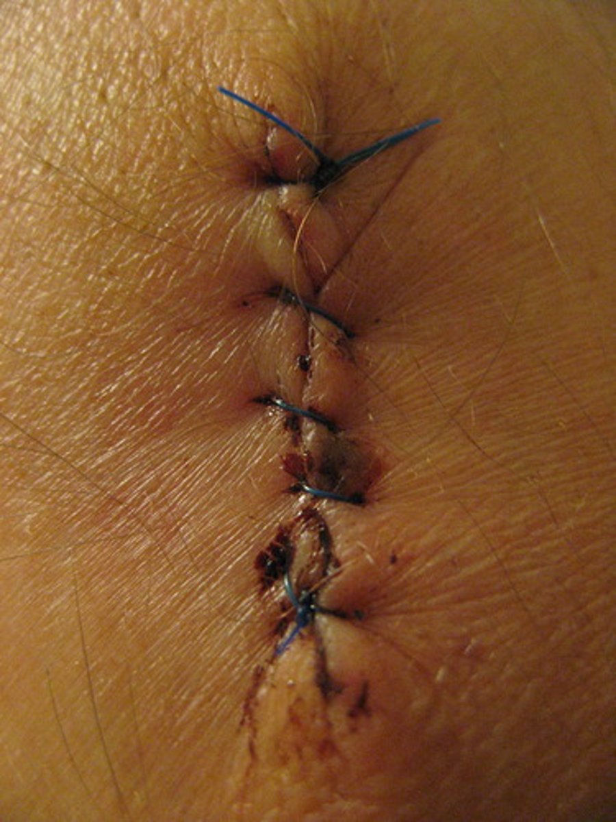
-rrhaphy
suturing, repairing
-tripsy
surgical crushing
-trophy
nourishment, development
-uria
urine, urination
azotemia
urea in the blood (a toxic condition) also called uremia
cystitis
inflammation of the bladder

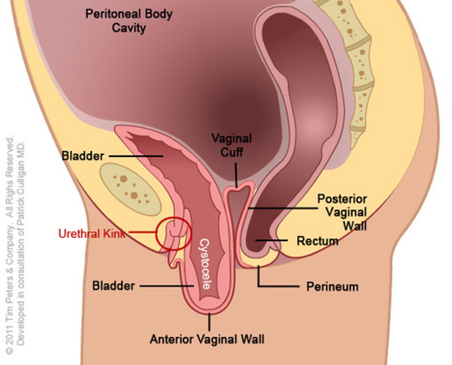
cystocele
protrusion of the bladder
cystolith
stone in the bladder
glomerulonephritis
inflammation of the glomeruli of the kidney
hydronephrosis
abnormal condition of water in the kidney
nephritis
inflammation of a kidney
nephrblastoma
kidney tumor containing developing cell
nephrohyptertrophy
excessive development (increase in size) of the kidney
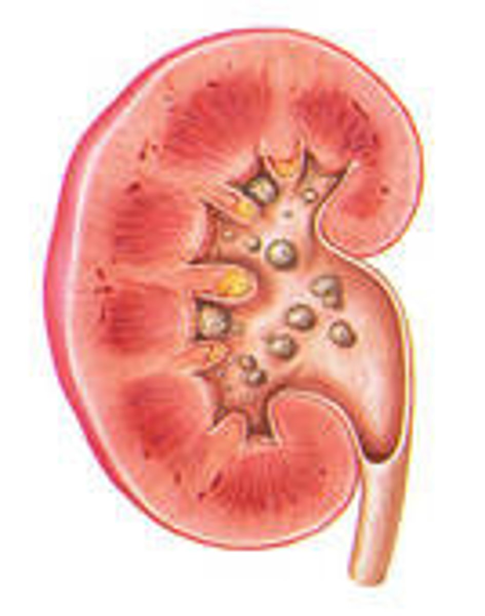
nephrolithiases
condition of stone(s) in the kidney
nephroma
tumor of the kidney
nephromegaly
enlargement of a kidney
nephroptosis
drooping kidney
pyelitis
inflammation of the renal pelvis
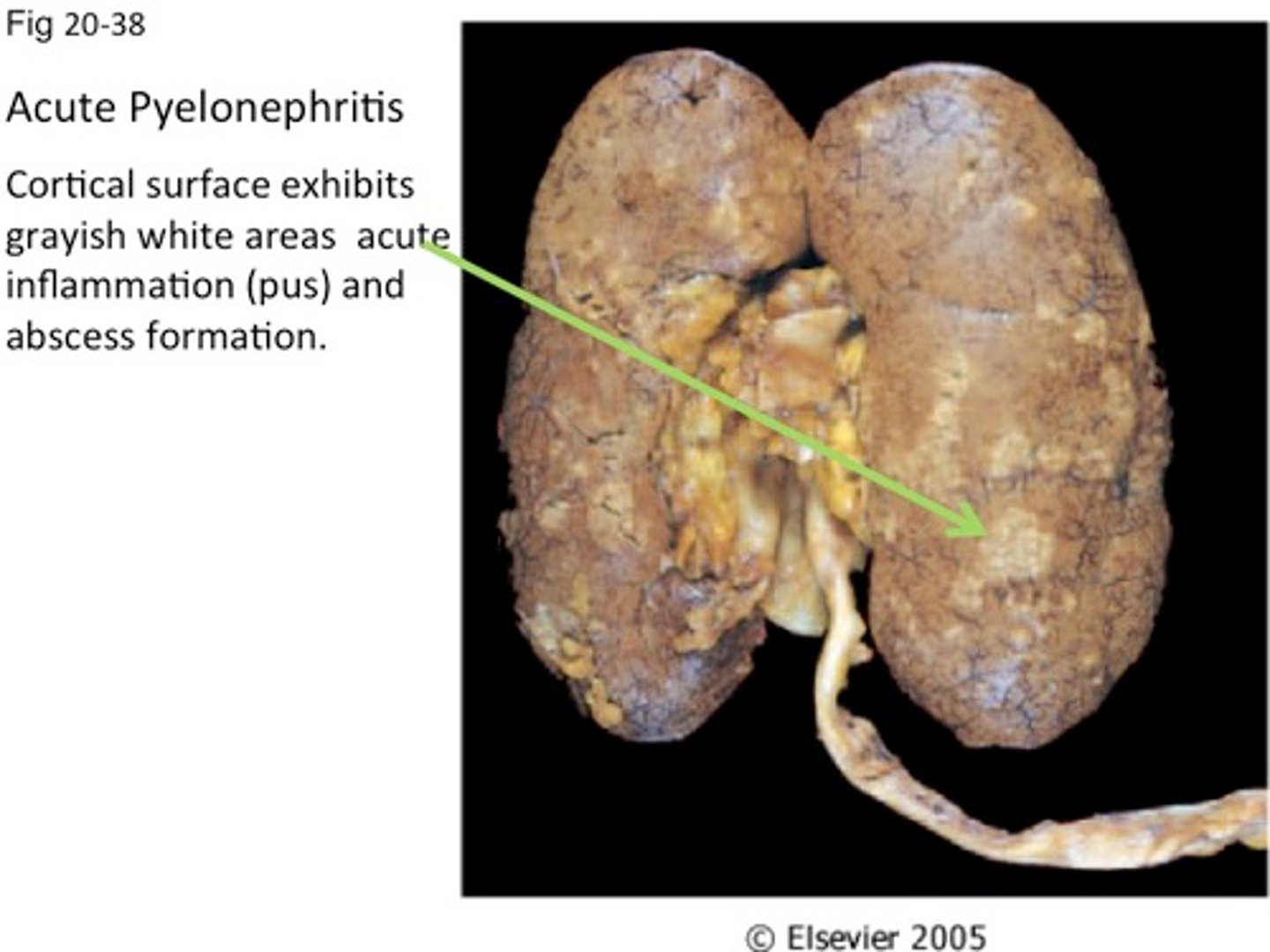
pyelonephritis
inflammation of the renal pelvis and the kidney
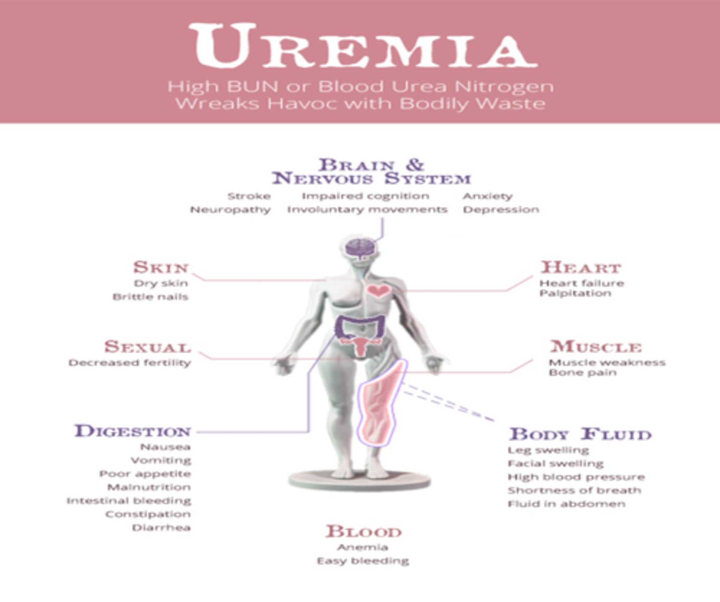
uremia
urine (urea Nitrogen) in the blood (refers to the presence of azotemia and a wide range of signs and symptoms associated with chronic kidney disease, including polyuria (excessive urination) polydipsia (excessive thirst), vomiting and weight loss; associated with renal failure) also called uremic syndrome)
ureteritis
inflammation of a ureter
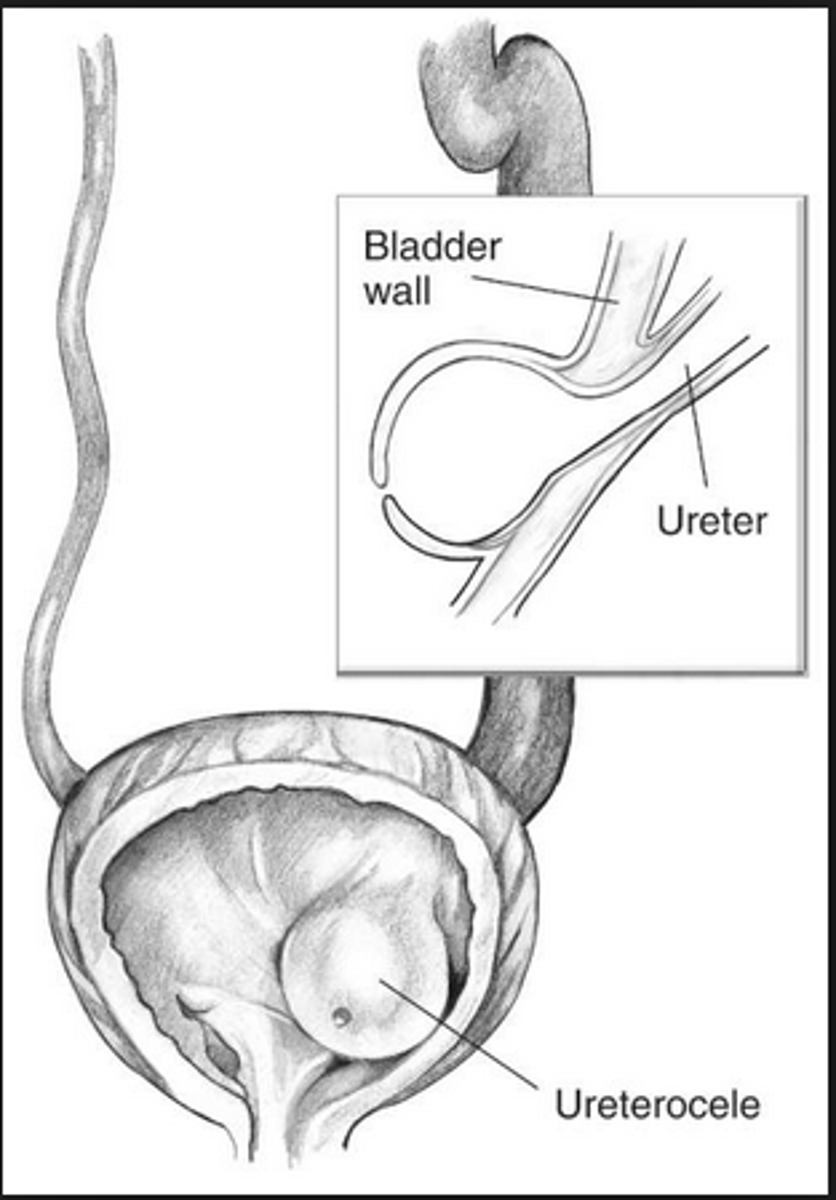
ureterocele
protrusion of a ureter
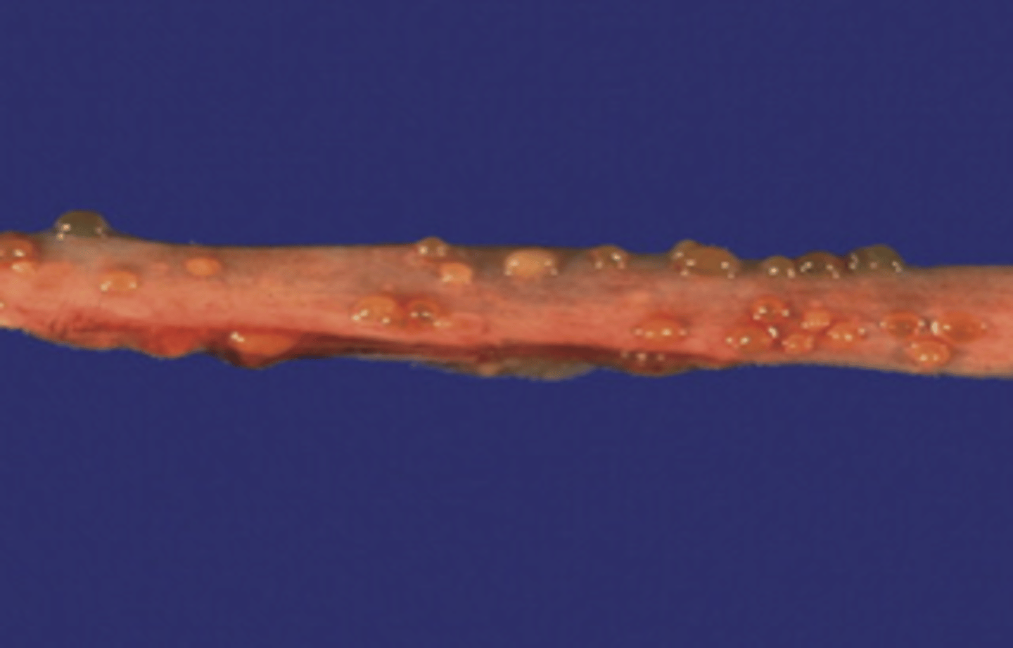
ureterolithiasis
condition of stones in the ureters
urethritis
inflammation of the urethra
ureterostenosis
narrowing of the ureter
urethrocystitis
inflammation of the urethra and the bladder

vesicoureteral reflux (VUR)
reflux pertaining to the bladder and ureter (condition in which urine flows backward towards the kidneys. May occur in up to 10% of children, and in some adults)
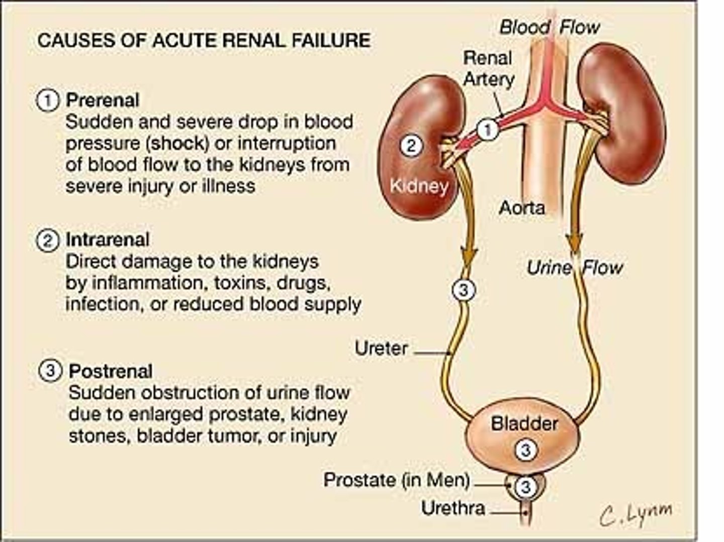
Acute Kidney Injury (AKI)
abrupt decline in kidney function that occurs over hours to days and is usually reversible (also called acute renal failure (ARF))
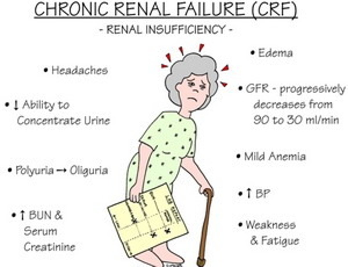
Chronic Kidney Disease (CKD)
progressive, irreversible loss of kidney function (also called chronic renal failure [CRF])
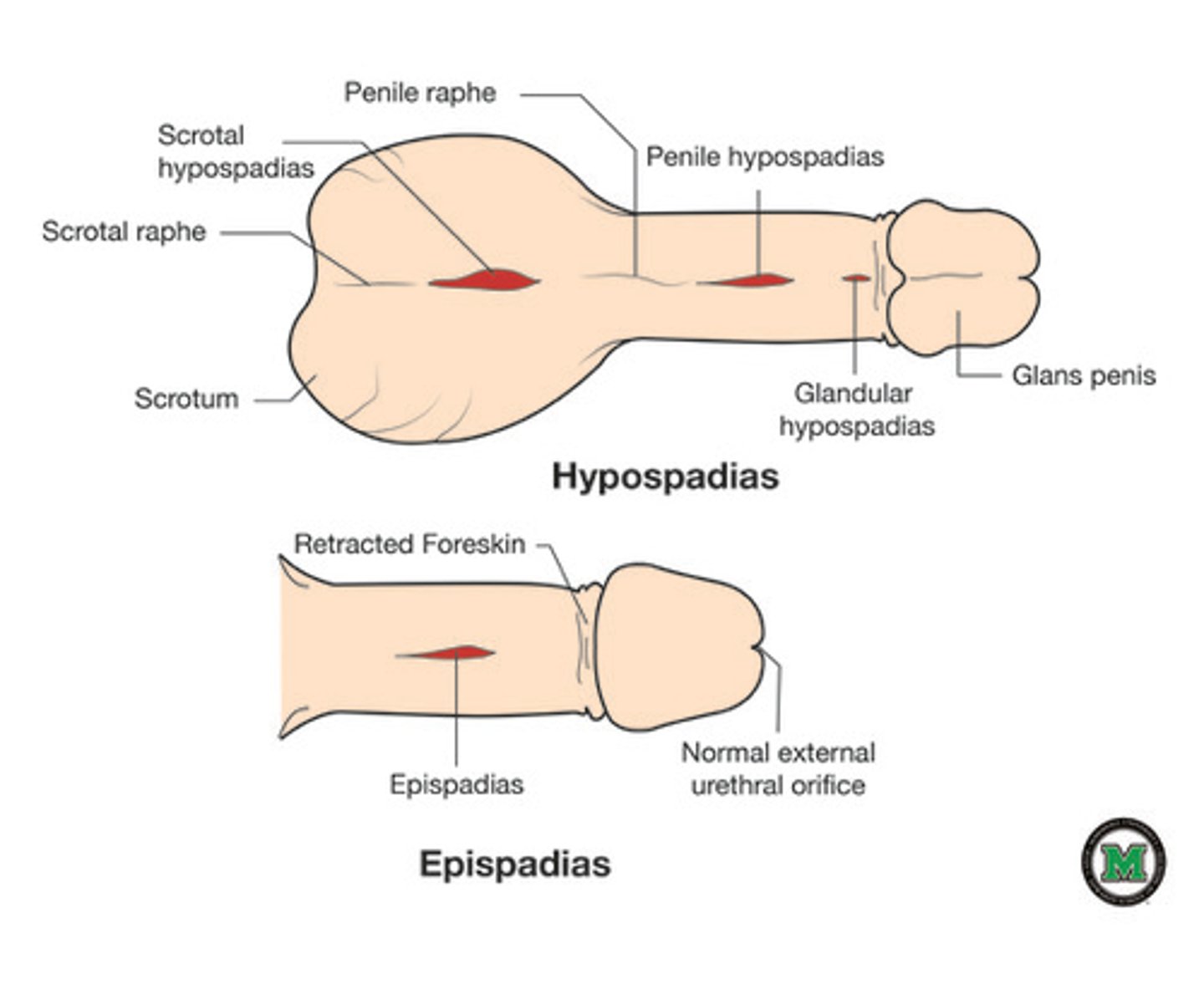
epispadias
congenital defect in which the urinary meatus is located on the upper surface of the penis
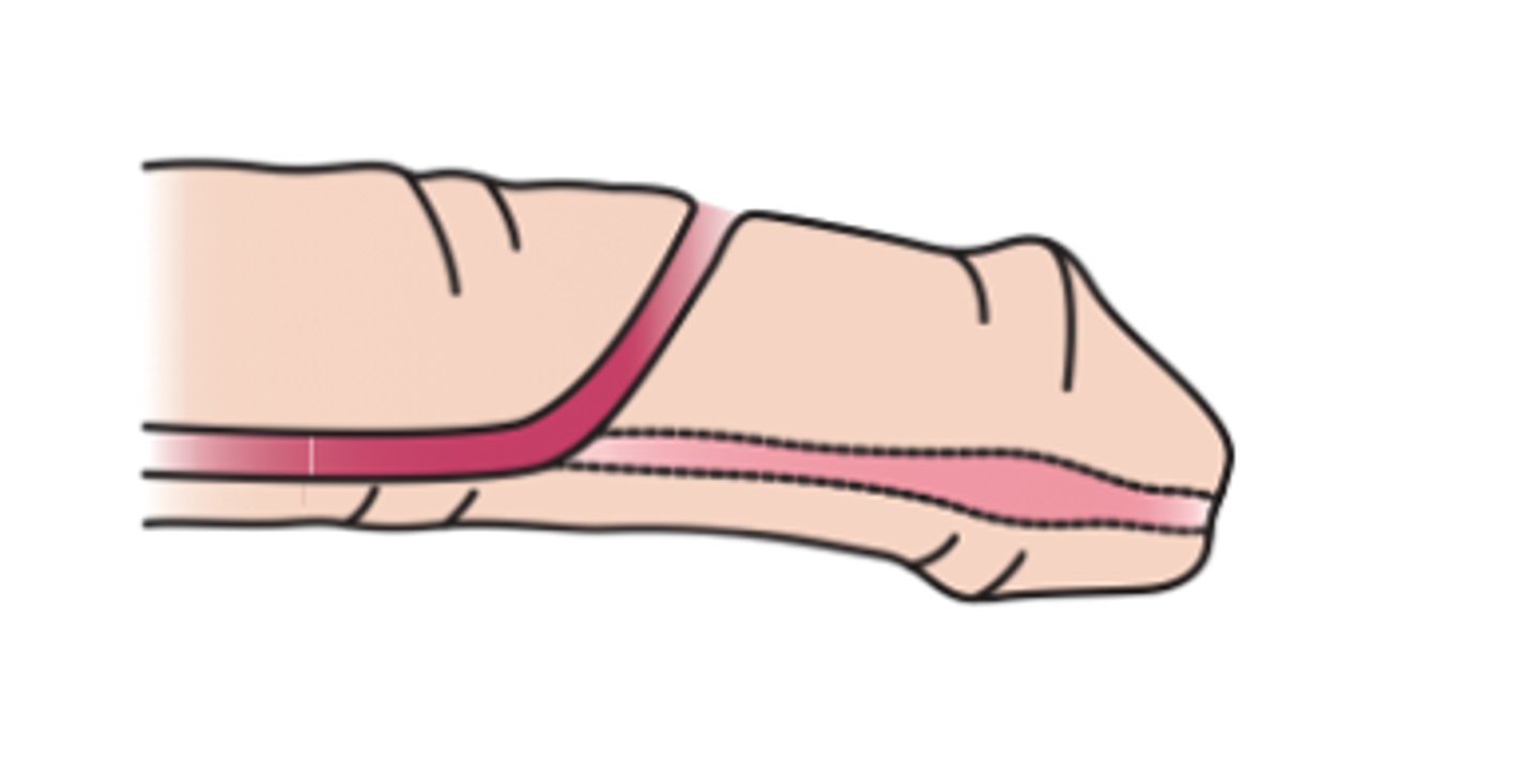
hypospadias
congenital defect in which the urinary meatus is located on the underside of the penis; a similar defect can occur in the female
polycystic kidney diesase (PKD)
condition in which the kidney contains many cysts and is enlarged
renal calculus
stone in the kidney

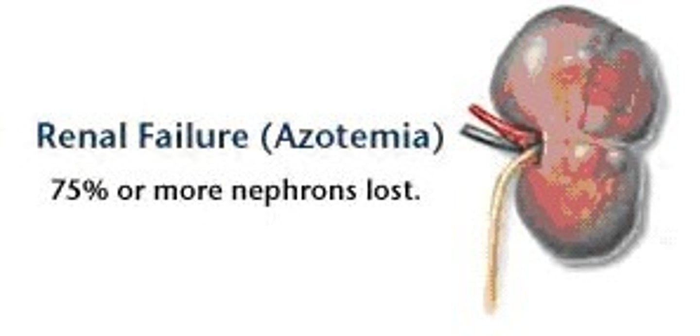
renal failure
loss of kidney function resulting in its inability to remove waste products from the body and maintain electrolyte balance
renal hypertension
elevated blood pressure resulting from kidney disease

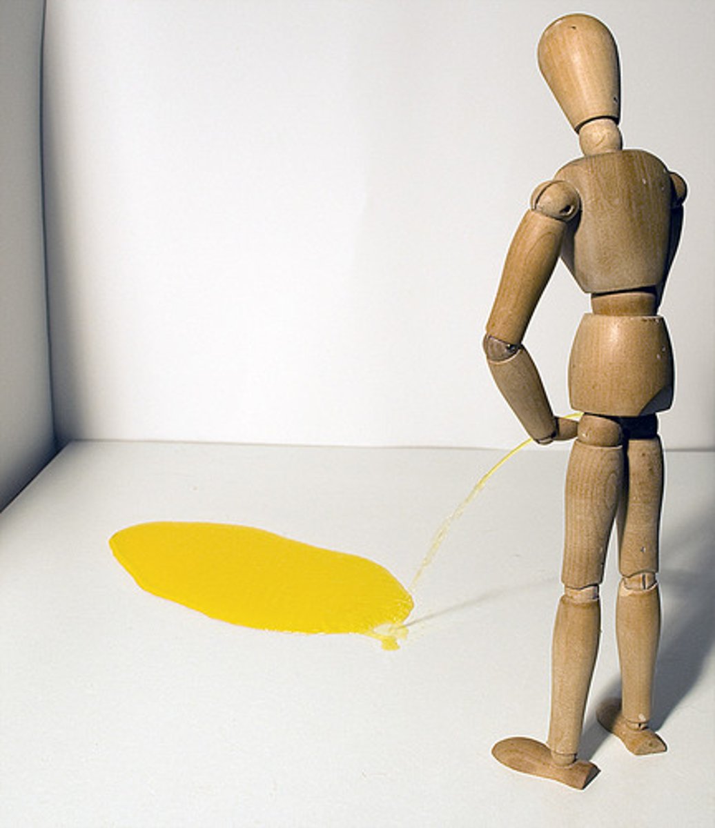
urinary retention
abnormal accumulation of urine in the bladder because of an inability to urinate
urinary suppression
sudden stoppage of urine formation
urinary tract infection (UTI)
infection of one or more organs of the urinary tract
cystectomy
excision of the bladder
cystolithotomy
incision of the bladder to remove a stone
cystorrhaphy
suturing the bladder
cystostomy
creating an artificial opening into the bladder
custotomy, vesicotomy
incision of the bladder
lithtripsy
surgical crushing of a stone
meatotomy
incision of the meatus
nephrectomy
excision of a kidney
nephropyelolithotomy
incision through the kidney to remove a stone
nephrolithotripsy
surgical crushing of stones in the kidney
neprholysis
separating the kidney (from other body structures)
nephropexy
surgical fixation of the kidney
nephrostomy
creation of an artificial opening into the kidney
pyelolithotomy
incision of the renal pelvis to remove a stone
pyeloplasty
surgical repair of the renal pelvis
ureterectomy
excision of a ureter
ureterostomy
creation of an artificial opening into the ureter
urethroplasty
surgical repair of the urethra
vesicourethral suspension
suspension pertaining to the bladder and urethra
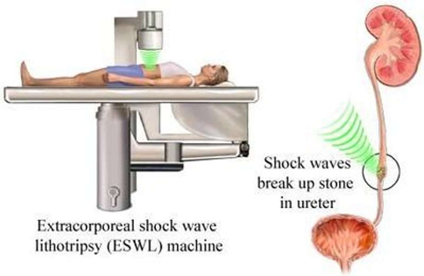
extracorporeal shock wave lithotripsy
a noninvasive treatment for removal of a kidney or ureteral stone(s). By using ultrasound and fluoroscopic imaging, the stone is positioned at the focal point. Repeated firing of shock waves renders the body in the urine
fulfuration
destruction of living tissue with an electric spark
renal transplant
surgical implantation of a donor kidney to replace a nonfunctioning kidney
cystogram
radiographic image of the bladder
cystography
radiographic imaging of the bladder
intravenous urogram (IVU)
radiographic image of the urinary tract
nephrography
radiographic imaging of the kidney
nephrosonography
process of recording the kidney using sound (ultrasonography)
nephrotomogram
sectional radiographic image of the kidney
renogram
(graphic) record of the kidney
retrograde urogram
radiographic image of the urinary tract (retrograde means to move in a direction opposite from normal) with contrast medium instilled through urethral catheters by a cystoscope
voiding cystourethrography (VCUG)
radiographic imaging of the bladder and the urethra. Radiopaque dye is instilled in the bladder. Radiographic images called cystourethrograms are taken of the bladder and during urination of the dye
cystoscope
instrument used for visual examination of the bladder
custoscopy
visual examination of the bladder
nephroscopy
visual examination of the kidney