ch. 3/4 parisitology nematodes
 Call Kai
 Learn
Learn Practice Test
 Spaced Repetition
 Match
Match Flashcards
 Knowt Play
1/48

There's no tags or description
Looks like no tags are added yet.
Name | Mastery | Learn | Test | Matching | Spaced | Call with Kai |
|---|
No analytics yet
Send a link to your students to track their progress
49 Terms
types of nematodes
free living (in water and soil)
those that parasitize plants
those that parasitize domesticated/wild animals and humans
nematode anatomy
spicules
unsegmented
elongated
round on both ends
bilaterally symmetric
2mm to 100 cm in size
dioecious (separate sexes)
Spirocerca lupi
GI tract; esophageal worm
forms nodule in esophageal wall
eggs in feces and vomit
paper clip shape
30 - 37 micrometers
6 month prepatent period
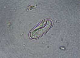
spirocerca lupi
Ollulanus tricuspis
common name: feline trichostrongyle
GI tract; in stomach
identify in cat vomit/fecal float
females = larviparous (bear live larva)
L3 infective stage
ascarids
large roundworms
host: canine, feline, raccoon
location: small intestines
route of infection: ingestion of larvated eggs, contaminated environment, transplacental, transmammary
Toxocara canis, Toxocara cati, Toxascaris Leonina, Baylisascaris procyonis
detect with fecal flotation
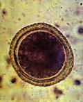
ascarid egg
transplacental migration of ascarids
larvae migrate through liver and lungs, get coughed up, get swallowed, then develop in small intestine
Toxocara adults
3 - 18 cm
tan
3 prominent lips of anterior end
cati has smaller, arrowhead appearance
swim to stay in intestines and sometimes stomach
ascarid diseases
V/D
pot belly
poor hair coat
abdominal pain
gastroenteritis-inflammation, hypersensitivity
zoonotic ascarids
visceral larval migrans
ocular larval migrans
neural larval migrans
ascarid life cycle
adults mate in small intestine
eggs passed in the host’s feces
become stage 2 larvae on ground
ingested by a host and the larvae migrate to grow
migrate to lungs, coughed up, swallowed, restart cycle
ascarid treatment
pyrantel pamoate (strongid): vermifuge; paralyzes worms that are passed in feces
fenbendazole (panacur): vermicide; kills parasites and they are broken down by body
Baylisascaris procynosis
mostly from small intestine of raccoons
can attack brain in humans
neurological larval migrans can lead to neurologic signs
use monthly heartworm prevention
corticosteroids as supportive care
hookworms
location: small intestine
route of infection: ingestion of 3rd stage larvae, contaminated environment, larval penetration of skin, transplacental migration, transmammary transmission
attach to small intestine; do not swim; suck blood
fecal flotation to detect
Ancylostoma: caninum, tubaeforme, braziliense
Unicinaria stenocephala
hookworm life cycle
adults attach to small intestines
voracious feeding and secondary hemorrhage cause anemia in host
eggs passed through host’s feces
eggs mature to L1 larvae in external
L1 hatch, feed, grow, molt to L2
L2 do same thing to L3 infective stage
L3 can be ingested or penetrate through skin, migrate to lungs, coughed up, swallowed, start process over
hookworm diseases
severe and more common in puppies and kittens
dark/tarry diarrhea
anemia/pale mm
weight gain or weight loss
zoonotic hookworms
cutaneous larval migrans; “creeping eruptions”
not transmitted person to person
hookworm treatment
pyrantel (monthly)
fenbendazole (panacur): vermicide; kills parasites, broken down by body
Strongyloides stercoralis
intestinal threadworm
in mucosa of dogs, cats, humans
only female is parasitic
route of infection: penetrate skin or oral ingestion
partheogenesis: reproduction without fertilization from male
zoonotic by fecal-oral
facultative parasites
Strongyloides stercoralis (intestinal threadworm) life cycle
eggs laid in mucosa
eggs hatch into rhabditiform larvae (non infective)
larvae penetrate glandular epithelium and pass to lumen
pass out of feces
Strongyloides stercoralis diseases
bloody mucoid diarrhea
dehydration
stunted growth
diagnosis/treatment of Strongyloides stercoralis
use Baermann technique for diagnostic
treat with ivermectin or fenbendazole
whipworms
location: cecum and colon
route of infection: ingestion of eggs, contaminated environment
canine and feline
suck blood
larvated eggs can be viable for several years
fecal flotation for detection
Trichuris: vulpis, campanula, serrata
whipworm eggs
barrel shaped
yellow/brown
bipolar end plugs / operculum on ends where larvae come out
smooth shell
unembryonated when laid
intermittently shed
whipworm diseases
diarrhea
mucous coated stool
anemia
some may be asymptomatic or subclinical
whipworm treatment
fenbendazole (panacur)
mebendazole
vermicide: kill parasite and are broken down by body
Enterobius vermicularis
human pinworm
in human cecum and colon
NO dogs or cats; omnivores and herbivores
can be found in primates
centrifugation or cellophane tape
Oxyuris equi
common name: equine pinworm
location: large intestine
clinical signs: pruritus in anal region, tail rubbing, behavior
diagnosis: cellophane tape
treatment: moxidectin, piperazine, pyrantel
Dirofilaria immitis
common name: canine heartworm
canine, feline, ferrets, sealions
location: right ventricle and pulmonary arteries
dioecious
born live; no egg
can cause pulmonary “coin lesions” with humans
heartworm life cycle
infection from a bite of infected female mosquito
need intermediate host
definitive host: dogs
microfilaria
baby heartworm
in peripheral/circulating blood
6 months to become adult and get to heart
heartworm diseases
exercise introlerance
right side heart enlargement
ascites (fluid in abdominal cavity)
fainting
coughing
heartworm diagnosis
detect microfilariae
direct smear
hematocrit test (above buffy coat)
modified knott’s test
filter
antigen test (for adult female) ELISA
radiographs/ultrasound
modified knott’s test
1 mL EDTA mixed with 9 mL 2% formalin
invert tube
centrifuge at 1100 to 1500 rpm for 5 to 8 min
pour liquid off
add drop of methylene blue
place on slide with coverslip
10x or 40x
differentiate between Dirofilaria immitis and Acanthocheilonema reconditum
filter test
1 mL blood into EDTA or heparin tube
add 9 mL lysing solution and mix
attach to filter unit
remove filter from holder and place on microscope
add methylene blue stain and add cover glass
feline heartworm disease
resistant; takes more exposure to induce disease
can clear L3 to L5 larva
respiratory symptoms
feline heartworm diagnosis
antigen test: active infection
antibody test: indicate exposure
heartworm treatment
adult stage: malarsomine (immiticide)
microfilariae: ivermectin or milbemycin
cage rest for 1 month
no treatment for cats
macrolitic lactones
against microfilariae, L3, L4, young adults
preventatives
ivermectin, selamectin, moxidectin, milbemycin oxime
effective for 1 month
CAUTION with collies and MDR1 deficient dogs
Wolbachia spp.
intracellular gram neg rickettsiae
immune complex disease
treat with doxycycline
caval syndrome
hundreds of heartworms
sudden onset of severe lethargy, weakness, anemia, hemoglobinemia, hemoglobinuria
Aelurostrongylus abstrusus
common name: feline lungworm
first stage larvae in cat feces
definitive host: cats
transport/reservoir host: rodents, frogs/toads, reptiles, birds
feline lungworm diagnosis
Baermann technique on feces or sputum
treatment: fenbendazole
Oslerus osleri
location: trachea, lungs parenchyma, bronchioles
common name: canine lung worm
can transmit from mother to pups from licking them
fecal float or baermann technique
Dioctophyme renale
common name: giant kidney worm
location: kidney and peritoneal cavity of canines
affects/destroys right kidney
signs: hematuria and dysuria
no treatment other than surgery
intermediate host: earthworm, frogs, fish, crayfish
Pearsonema Plica
Pearsonema feliscati
bladder worms in dogs and cats
signs: asymptomatic or cystitis
diagnosis: eggs in urine sediment, bipolar plugs
intermediate/transport host: earthworm
treatment: fenbendazole or ivermectin
Dracunnulus insignis
location: subcutaneous tissue on limbs
affects raccoons
signs: chronic skin ulcer and secondary bacterial infection on limbs
diagnosis: exam of skin lesion
intermediate hosts: water fleas (cyclops)/copepod
ingestion of contaminated water, tadpoles, frogs
Thelazia spp.
common name: eye worm
location: conjunctival duct and lacrimal duct
cattle, dogs, horses
intermediate host: muscid flies (face fly) transmit L3 into tears
signs: